. 2020 Feb 12;11(10):2834-2842.
doi: 10.1039/c9sc05381f. eCollection 2020 Mar 14.

Wavelength-gated photoreversible polymerization and topology control

Affiliations

Wavelength-gated photoreversible polymerization and topology control

Hendrik Frisch et al. Chem Sci. .

Abstract

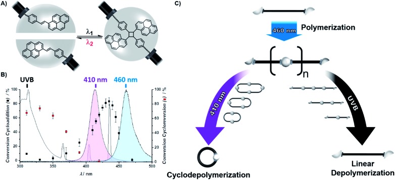
We exploit the wavelength dependence of [2 + 2] photocycloadditions and -reversions of styrylpyrene to exert unprecedented control over the photoreversible polymerization and topology of telechelic building blocks. Blue light (λ max = 460 nm) initiates a catalyst-free polymerization yielding high molar mass polymers (M n = 60 000 g mol-1), which are stable at wavelengths exceeding 430 nm, yet highly responsive to shorter wavelengths. UVB irradiation (λ max = 330 nm) induces a rapid depolymerization affording linear oligomers, whereas violet light (λ max = 410 nm) generates cyclic entities. Thus, different colors of light allow switching between a depolymerization that either proceeds through cyclic or linear topologies. The light-controlled topology formation was evidenced by correlation of mass spectrometry (MS) with size exclusion chromatography (SEC) and ion mobility data. Critically, the color-guided topology control was also possible with ambient laboratory light affording cyclic oligomers, while sunlight activated the linear depolymerization pathway. These findings suggest that light not only induces polymerization and depolymerization but that its color can control the topological outcomes.


Figures

Scheme 1
Scheme 1. (A) Chemical structure of the reversible [2 + 2] photocycloaddition of styrylpyrene used as a wavelength gated binding site of telechelic polymers. (B) Action plot for the conversion of the photocycloaddition (black cubes) and the photocycloreversion of the cycloadduct (red cubes) after irradiation with the same number of photons at different wavelengths, as previously reported for the styrylpyrene derivative hydroxyl-styrylpyrene. The normalized emission spectra of the herein utilized UVB lamp (grey) and LEDs (violet, λmax = 410 nm and blue, λmax = 460 nm) indicate which reaction is triggered in the respective photonic field. (C) Schematic representation of the polymerization and depolymerization of L1 comprising two terminal styrylpyrene units. Irradiation of L1 with blue light induces its photopolymerization yielding P1, which can be either cyclodepolymerized under violet light or linearly depolymerized under UVB light.
Fig. 1
Fig. 1. (A) SEC trace of the cyclization of L1 (black) into the macrocycle C1 (blue) using blue light irradiation. (B) Cycloreversion of C1 (grey) to L1 (black) under UVB irradiation.
Fig. 2
Fig. 2. (A) SEC trace of the photopolymerization of L1 (grey) to P1 (black, Mn = 60 kDa, Đ = 2, DP = 38), cyclic oligomers (DP = 1–5) denoted by black arrows. The different cyclic oligomers were identified by SEC-ESI-MS and the recorded mass spectra are plotted along with the simulated isotopic patterns of the respective sum formula. (B) Mass spectrum of the first eluting species assigned to a monomeric species (m/z of the most abundant ion = 1626.76763, simulated for C92H114O24Na1+ = 1626.76233, Δm/z = 3.26 ppm). (C) Mass spectrum of the second eluting species assigned to a dimeric species (m/z of the most abundant ion = 1626.76917, simulated for C184H228O48Na22+ = 1626.76233, Δm/z = 4.20 ppm). (D) Mass spectrum of the third eluting species assigned to a trimeric species (m/z of the most abundant ion = 1626.77647, simulated for C276H342O72Na33+ = 1626.76233, Δm/z = 8.69 ppm). (E) Mass spectrum of the fourth eluting species assigned to a tetrameric species (m/z of the most abundant ion = 2161.37567, simulated for C368H456O96Na33+ = 2161.35383, Δm/z = 10.10 ppm). (F) Mass spectrum of the fifth eluting species assigned to a pentameric species (m/z of the most abundant ion = 1374.16201, simulated for C464H578O122Na66+ = 1374.14239, Δm/z = 11.38 ppm).
Fig. 3
Fig. 3. Depolymerization of P1 under UVB (left columns) and cyclodepolymerization under violet light (right columns). Selected SEC traces of the depolymerization of P1 after different irradiation times, with highlighted characteristic molar mass ranges of the cyclic and linear oligomers (DP = 1–6). Refer to chapter 1.5 and 1.6 of the ESI for the full depolymerization kinetics. XIC traces of the oligomers (DP = 1–5) extracted from the SEC-ESI-MS (bottom). Refer to the ESI chapter 1.10 for the full mass spectra of the oligomers and the selected ions.
Fig. 4
Fig. 4. Depolymerization of P1 (black) into linear oligomers (highlighted in red) within 4 min of sunlight irradiation.

References

    1. Staudinger H. Ber. Dtsch. Chem. Ges. A. 1920;53(6):1073–1085. doi: 10.1002/cber.19200530627. - DOI
    1. Szwarc M. Nature. 1956;178(4543):1168–1169. doi: 10.1038/1781168a0. - DOI
    1. Moad G., Rizzardo E., Thang S. H. Polymer. 2008;49(5):1079–1131. doi: 10.1016/j.polymer.2007.11.020. - DOI
    1. Kamigaito M., Ando T., Sawamoto M. Chem. Rev. 2001;101(12):3689–3746. doi: 10.1021/cr9901182. - DOI - PubMed
    1. Hawker C. J., Bosman A. W., Harth E. Chem. Rev. 2001;101(12):3661–3688. doi: 10.1021/cr990119u. - DOI - PubMed