

Photobiomodulation of lymphatic drainage and clearance: perspective strategy for augmentation of meningeal lymphatic functions
- PMID: 32206394
- PMCID: PMC7041454
- DOI: 10.1364/BOE.383390
Photobiomodulation of lymphatic drainage and clearance: perspective strategy for augmentation of meningeal lymphatic functions
Abstract
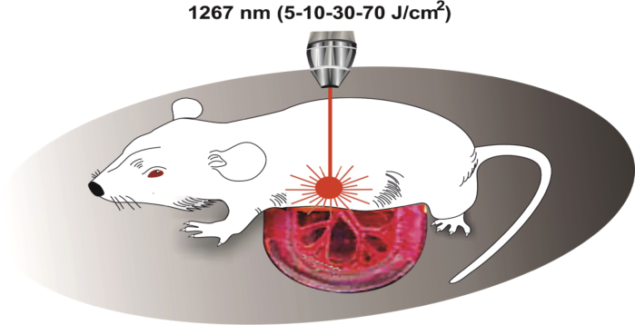
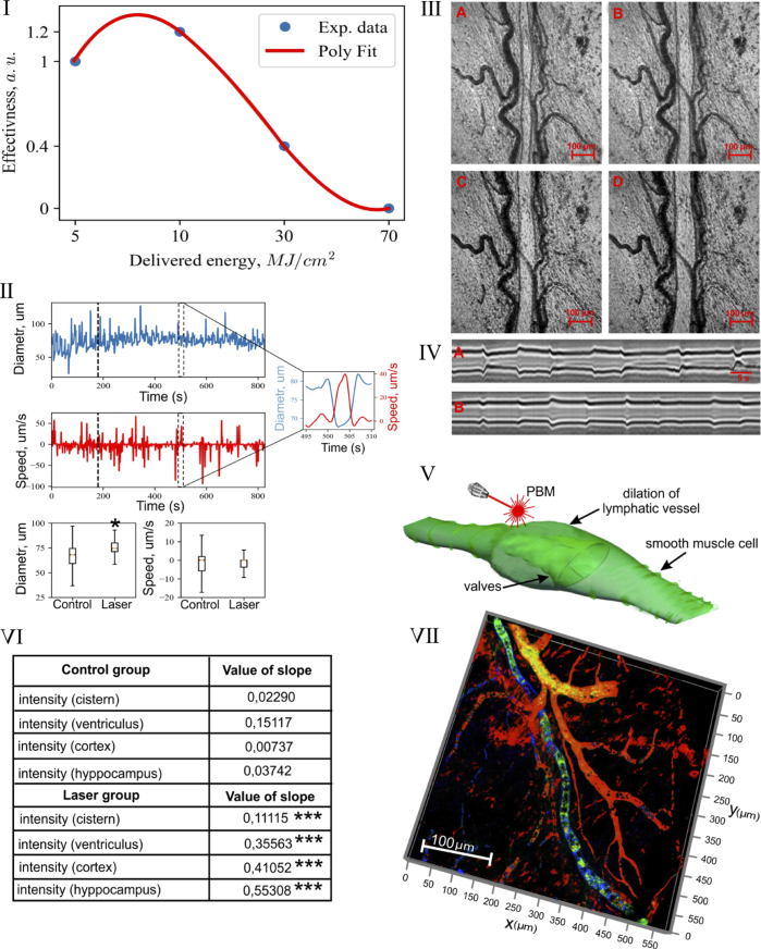
There is a hypothesis that augmentation of the drainage and clearing function of the meningeal lymphatic vessels (MLVs) might be a promising therapeutic target for preventing neurological diseases. Here we investigate mechanisms of photobiomodulation (PBM, 1267 nm) of lymphatic drainage and clearance. Our results obtained at optical coherence tomography (OCT) give strong evidence that low PBM doses (5 and 10 J/cm2) stimulate drainage function of the lymphatic vessels via vasodilation (OCT data on the mesenteric lymphatics) and stimulation of lymphatic clearance (OCT data on clearance of gold nanorods from the brain) that was supported by confocal imaging of clearance of FITC-dextran from the cortex via MLVs. We assume that PBM-mediated relaxation of the lymphatic vessels can be possible mechanisms underlying increasing the permeability of the lymphatic endothelium that allows molecules transported by the lymphatic vessels and explain PBM stimulation of lymphatic drainage and clearance. These findings open new strategies for the stimulation of MLVs functions and non-pharmacological therapy of brain diseases.
© 2020 Optical Society of America under the terms of the OSA Open Access Publishing Agreement.
Conflict of interest statement
The authors declare that there are no conflicts of interest related to this article.
Figures
References
-
- Absinta M., Ha S.-K., Nair G., Satil P., Luciano N. J., Palisoc M., Louveau A., Zaghloul K. A., Pittaluga S., Kipnis J., Reich D. S., “Human and nonhuman primate meninges harbor lymphatic vessels that can be visualized noninvasively by MRI,” eLife 6, e29738 (2017).10.7554/eLife.29738 - DOI - PMC - PubMed
-
- Semyachkina-Glushkovskaya O., Abdurashitov A., Dubrovsky A., Bragin D., Bragina O., Shushunova N., Maslyakova G., Navolokin N., Bucharskaya A., Tuchin V., Kurths J., Shirokov A., “Application of optical coherence tomography for in vivo monitoring of the meningeal lymphatic vessels during opening of blood-brain barrier: mechanisms of brain clearing,” J. Biomed. Opt. 22(12), 1–9 (2017).10.1117/1.JBO.22.12.121719 - DOI - PMC - PubMed
LinkOut - more resources
Full Text Sources
Other Literature Sources