. 2020 Mar 18;6(12):eaaz1170.
doi: 10.1126/sciadv.aaz1170. eCollection 2020 Mar.

10,000 social brains: Sex differentiation in human brain anatomy

Affiliations

10,000 social brains: Sex differentiation in human brain anatomy

Hannah Kiesow et al. Sci Adv. .

Abstract

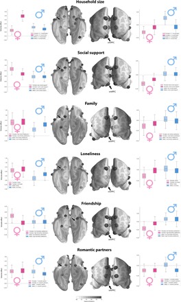
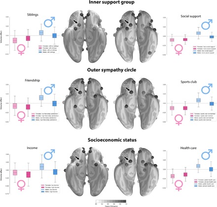
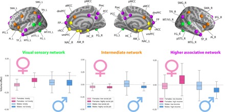
In human and nonhuman primates, sex differences typically explain much interindividual variability. Male and female behaviors may have played unique roles in the likely coevolution of increasing brain volume and more complex social dynamics. To explore possible divergence in social brain morphology between men and women living in different social environments, we applied probabilistic generative modeling to ~10,000 UK Biobank participants. We observed strong volume effects especially in the limbic system but also in regions of the sensory, intermediate, and higher association networks. Sex-specific brain volume effects in the limbic system were linked to the frequency and intensity of social contact, such as indexed by loneliness, household size, and social support. Across the processing hierarchy of neural networks, different conditions for social interplay may resonate in and be influenced by brain anatomy in sex-dependent ways.


Figures

Fig. 1
Fig. 1. Sex disparity in the association between limbic system morphology and the richness of social interaction.
Marginal posterior population distributions uncover the degree of sex overlap in social brain anatomy for several social traits. Among all target regions, many strong volume effects were apparent in the AM and vmPFC of the limbic network (cf. the Supplementary Materials). For each index of less and more rich social life (light versus dark colors), boxplots show the estimated population volume parameter distribution of the AM and vmPFC for men and women (blue versus pink colors). Each set of four boxplots juxtaposes the probabilistic relevance of a region’s volume in explaining sex and social trait, taking into account positive and negative volume effects of the 35 remaining social brain regions. Vertical axis indicates the brain volume effect obtained from z-scored region averages measured by T1-MRI. Black arrows indicate the brain region corresponding to the boxplots. Transparency level of the brain regions reflects the degree of divergence in population volume dispersion between the four social/sex groups (less transparent = more divergence according to Kullback-Leibler distance). The collective results may speak to sex-specific sensitivity of limbic system structures to the quality and quantity of social exchange.
Fig. 2
Fig. 2. Similar volumetric divergences in the reward circuitry in men with fewer social interactions and low indicators of social hierarchy.
Marginal posterior population distributions reveal similar volume parameter divergences in the reward circuitry of the social brain. Across analyses of six social traits, the NAC showed a sex-specific volume parameter pattern. In the six social traits, the female groups showed largely congruent population volume parameter distributions in NAC anatomy. The collective results suggest sex idiosyncrasies in neural processing of social and nonsocial reward cues.
Fig. 3
Fig. 3. Sex-specific volume effects at the network level.
In addition to the within-network volume effects (Figs. 1 and 2), our analyses estimated marginal posterior population distributions for the overall network volume effects of the social brain: visual sensory (green), limbic (yellow; not shown since weakest effects as a whole network), intermediate (orange), and higher associative networks (purple). As such, sex-related anatomical divergences also become apparent for subsets of social brain regions, known to be functionally cohesive. AI_L, left anterior insula; AI_R, right anterior insula; AM_L, left amygdala; AM_R, right amygdala; aMCC, anterior mid-cingulate cortex; Cereb_L, left cerebellum; Cereb_R, right cerebellum; dmPFC, dorsomedial prefrontal cortex; FG_L, left fusiform gyrus; FG_R, right fusiform gyrus; FP, medial frontal pole; HC_L, left hippocampus; HC_R, right hippocampus; IFG_L, left inferior frontal gyrus; IFG_R, right inferior frontal gyrus; MTG_L, left middle temporal gyrus; MTG_R, right middle temporal gyrus; MT/V5_L, left middle temporal V5 area; MT/V5_R, right middle temporal V5 area; NAC_L, left nucleus accumbens; NAC_R, right nucleus accumbens; PCC, posterior cingulate cortex; pMCC, posterior mid-cingulate cortex; Prec, precuneus; pSTS_L, left posterior superior temporal sulcus; pSTS_R, right posterior superior temporal sulcus; rACC, rostral anterior cingulate cortex; SMA_L: left supplementary motor area; SMA_R, right supplementary motor area; SMG_L, left supramarginal gyrus; SMG_R, right supramarginal gyrus; TP_L, left temporal pole; TP_R, right temporal pole; TPJ_L, left temporo-parietal junction; TPJ_R, right temporo-parietal junction; vmPFC, ventromedial prefrontal cortex.

References

    1. Dunbar R. I. M., Shultz S., Evolution in the social brain. Science 317, 1344–1347 (2007). - PubMed
    1. Bickart K. C., Wright C. I., Dautoff R. J., Dickerson B. C., Barrett L. F., Amygdala volume and social network size in humans. Nat. Neurosci. 14, 163–164 (2010). - PMC - PubMed
    1. Lewis P. A., Rezaie R., Brown R., Roberts N., Dunbar R. I. M., Ventromedial prefrontal volume predicts understanding of others and social network size. Neuroimage 57, 1624–1629 (2011). - PMC - PubMed
    1. Silk J. B., Alberts S. C., Altmann J., Social bonds of female baboons enhance infant survival. Science 302, 1231–1234 (2003). - PubMed
    1. Key C., Ross C., Sex differences in energy expenditure in non–human primates. Proc. Biol. Sci. 266, 2479–2485 (1999). - PMC - PubMed

Publication types

LinkOut - more resources