

Identification of Superficial White Matter Abnormalities in Alzheimer's Disease and Mild Cognitive Impairment Using Diffusion Tensor Imaging
- PMID: 32206757
- PMCID: PMC7081087
- DOI: 10.3233/ADR-190149
Identification of Superficial White Matter Abnormalities in Alzheimer's Disease and Mild Cognitive Impairment Using Diffusion Tensor Imaging
Abstract
Background: Diffusion tensor imaging (DTI) estimates the microstructural alterations of the brain, as a magnetic resonance imaging (MRI)-based neuroimaging technique. Prior DTI studies reported decreased structural integrity of the superficial white matter (SWM) in the brain diseases.
Objective: This study aimed to determine the diffusion characteristics of SWM in Alzheimer's disease (AD) and mild cognitive impairment (MCI) using tractography and region of interest (ROI) approaches.
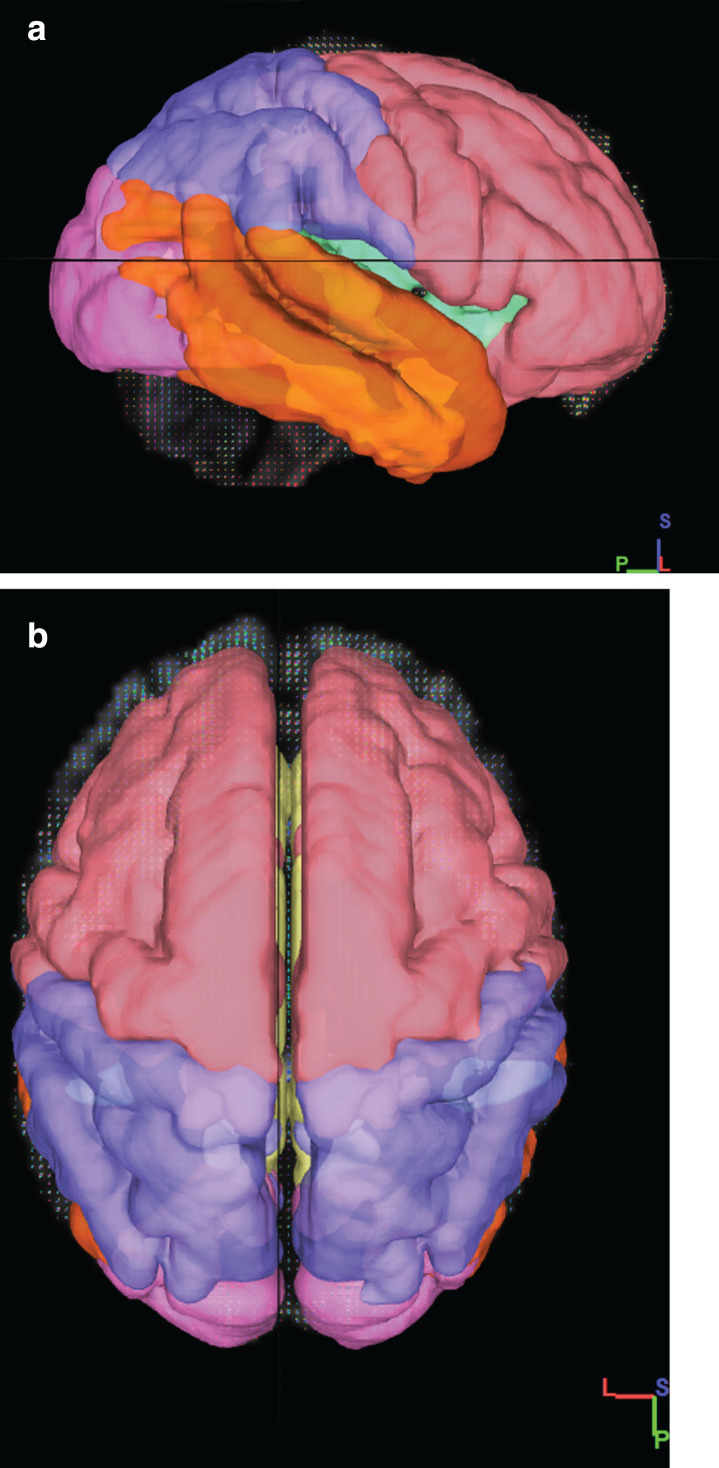
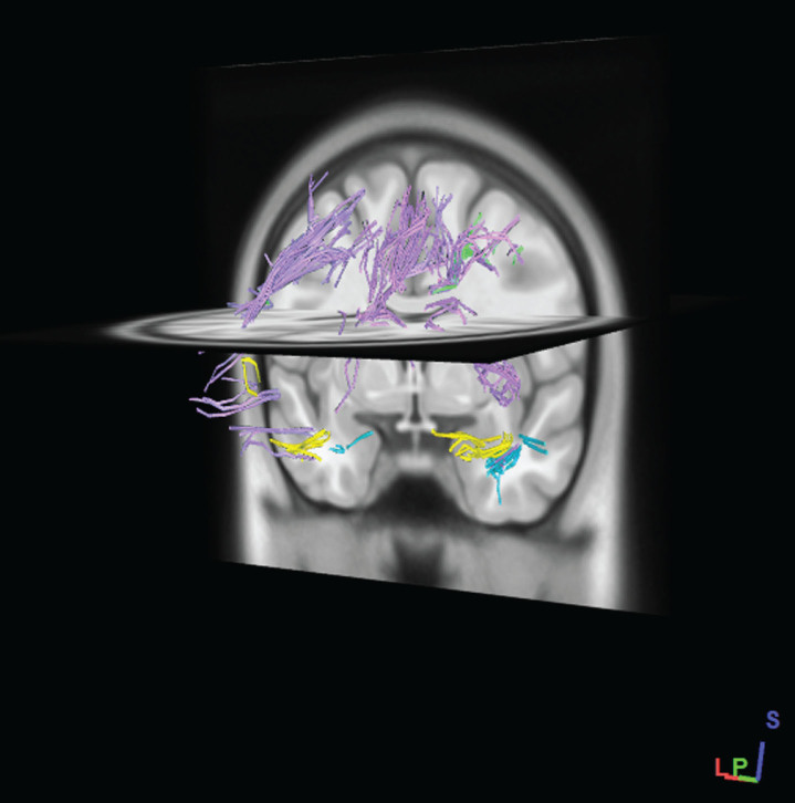
Methods: The diffusion MRI data were downloaded from the Alzheimer's Disease Neuroimaging Initiative (ADNI) database on 24 patients with AD, 24 with MCI, and 24 normal control (NC) subjects. DTI processing was performed using DSI Studio software. First, for ROI-based analysis, The superficial white matter was divided into right and left frontal, parietal, temporal, insula, limbic and occipital regions by the Talairach Atlas, Then, for tractography-based analysis, the tractography of each of these regions was performed with 100000 seeds. Finally, the average diffusion values were extracted from voxels within the ROIs and tracts.
Results: Both tractography and ROI analyses showed a significant difference in radial, axial and mean diffusivity values between the three groups (p < 0.05) across most of the SWM. Furthermore, The Mini-Mental State Examination was significantly correlated with radial, axial, and mean diffusivity values in parietal and temporal lobes SWM in the AD group (p < 0.05).
Conclusion: DTI provided information indicating microstructural changes in the SWM of patients with AD and MCI. Therefore, assessment of the SWM using DTI may be helpful for the clinical diagnosis of patients with AD and MCI.
Keywords: Alzheimer’s disease; diffusion tensor imaging; mild cognitive impairment; superficial white matter; tractography.
© 2020 – IOS Press and the authors. All rights reserved.
Conflict of interest statement
The authors disclose no conflicts of interest.
Figures

References
-
- Tang SX, Feng QL, Wang GH, Duan S, Shan BC, Dai JP (2017) Diffusion characteristics of the fornix in patients with Alzheimer’s disease. Psychiatry Res Neuroimaging 265, 72–76. - PubMed
-
- Schouten TM, Koini M, Vos F, Seiler S, Rooij M, Lechner A, Schmidt R, Heuvel MVD, Grond JV, Rombouts S (2017) Individual classification of Alzheimer’s disease with diffusion magnetic resonance imaging. Neuroimage 152, 476–481. - PubMed
-
- Hesseltine SM, Ge Y, Law M (2007) Applications of diffusion tensor imaging and fiber tractography. Appl Radiol 36, 8.
-
- Mori S, Crain BJ, Chacko VP, van Zijl PC (1999) Three-dimensional tracking of axonal projections in the brain by magnetic resonance imaging. Ann Neurol 45, 265–269. - PubMed
Grants and funding
LinkOut - more resources
Full Text Sources
