

Mediastinal Emphysema, Giant Bulla, and Pneumothorax Developed during the Course of COVID-19 Pneumonia
- PMID: 32207255
- PMCID: PMC7183834
- DOI: 10.3348/kjr.2020.0180
Mediastinal Emphysema, Giant Bulla, and Pneumothorax Developed during the Course of COVID-19 Pneumonia
Abstract
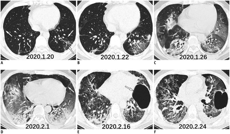
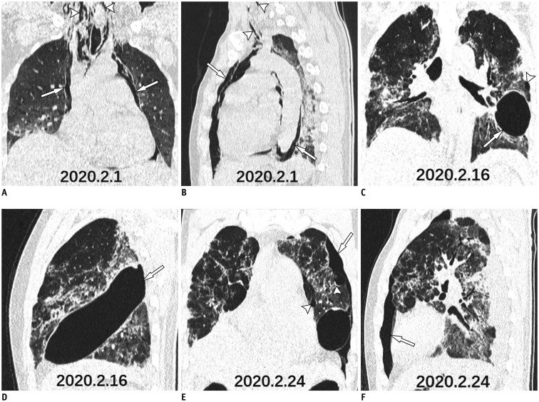
The coronavirus disease 2019 (COVID-19) pneumonia is a recent outbreak in mainland China and has rapidly spread to multiple countries worldwide. Pulmonary parenchymal opacities are often observed during chest radiography. Currently, few cases have reported the complications of severe COVID-19 pneumonia. We report a case where serial follow-up chest computed tomography revealed progression of pulmonary lesions into confluent bilateral consolidation with lower lung predominance, thereby confirming COVID-19 pneumonia. Furthermore, complications such as mediastinal emphysema, giant bulla, and pneumothorax were also observed during the course of the disease.
Keywords: COVID-19; Complication; Coronavirus; Pneumonia; Tomography, X-ray computed.
Copyright © 2020 The Korean Society of Radiology.
Conflict of interest statement
The authors have no potential conflicts of interest to disclose.
Figures
Comment in
-
Spontaneous Pneumomediastinum: A Probable Unusual Complication of Coronavirus Disease 2019 (COVID-19) Pneumonia.Korean J Radiol. 2020 May;21(5):627-628. doi: 10.3348/kjr.2020.0281. Korean J Radiol. 2020. PMID: 32323507 Free PMC article. No abstract available.
-
Spontaneous Pneumomediastinum in a Patient with Coronavirus Disease 2019 Pneumonia and the Possible Underlying Mechanism.Korean J Radiol. 2020 Jul;21(7):929-930. doi: 10.3348/kjr.2020.0426. Korean J Radiol. 2020. PMID: 32524794 Free PMC article. No abstract available.
References
-
- World Health Organization. Coronavirus disease 2019 (COVID-19) situation report–36. [Accessed February 25, 2020]. Available at: https://www.who.int/docs/default-source/coronaviruse/situation-reports/2....
Publication types
MeSH terms
LinkOut - more resources
Full Text Sources
Medical
