

Skin Infections Caused by Staphylococcus aureus
- PMID: 32207539
- PMCID: PMC9128951
- DOI: 10.2340/00015555-3466
Skin Infections Caused by Staphylococcus aureus
Abstract
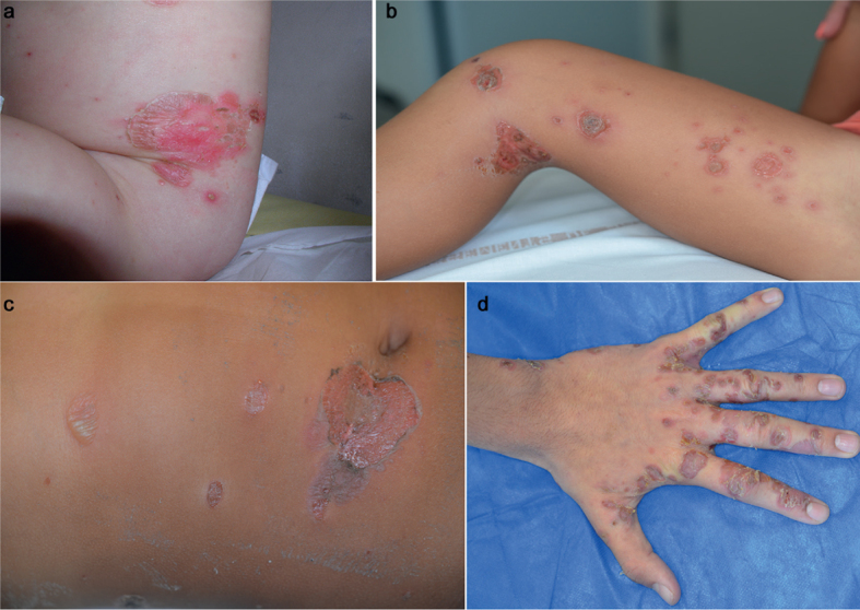
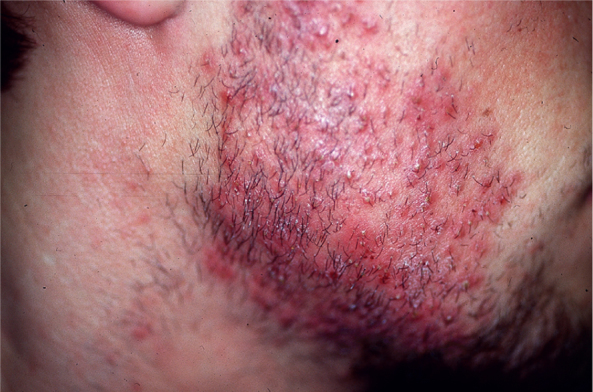
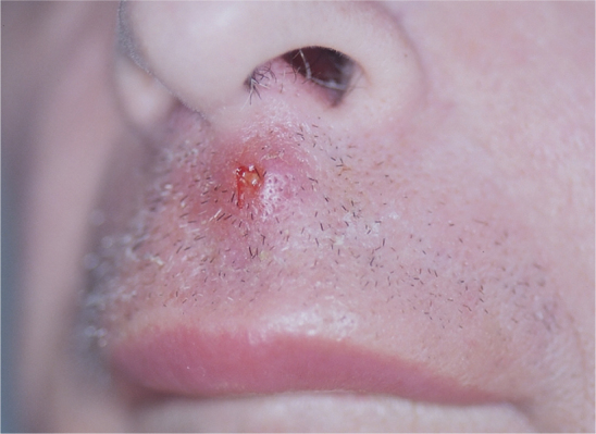
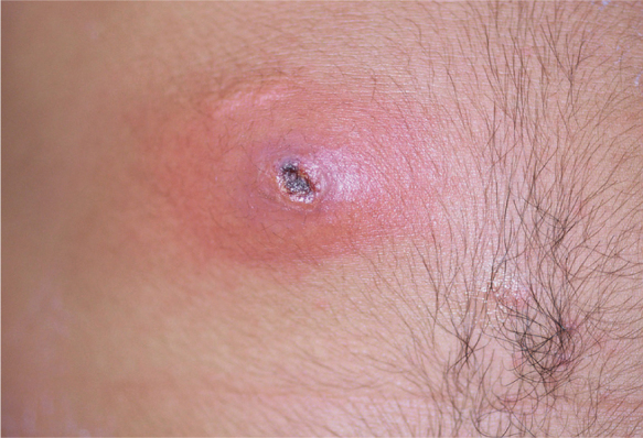
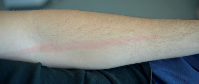
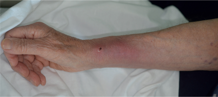
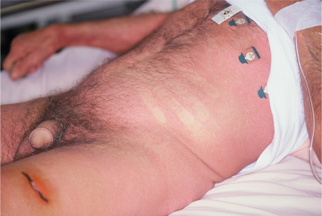
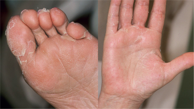
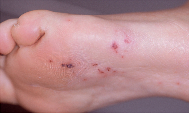
Staphylococcus aureus is the most common pathogen involved in skin infections worldwide, regardless of the patient's age, the climate or geographical area. The main skin clinical manifestations can be linked to a few toxins produced by the bacteria, which give rise to a rich and varied clinical spectrum. Panton Valentine leucocidin, exfoliatins, enterotoxins and toxin shock syndrome toxin 1 are the main toxins involved in most dermatological manifestations associated with S. aureus. Other less frequent cutaneous manifestations can occur in endocarditis, bacteraemia. Currently, the most important event is worldwide emergence of community-acquired S. aureus resistant to methicillin (CA-MRSA), mainly causing skin infections.
Keywords: abscess; bacterial skin infections; cellulitis; furuncle; staphylococcus aureus; skin infections.
Figures
References
-
- Stanley JR, Amagai M. Pemphigus, bullous impetigo and staphylococcal scald skin syndrome. N Engl J Med 2006; 355: 1800–1810. - PubMed
-
- Salah LA, Faergemann J. A retrospective analysis of skin bacterial colonisation, susceptibility and resistance in atopic dermatitis and impetigo patients. Acta Derm Venereol 2015; 95: 532–535. - PubMed
MeSH terms
Substances
LinkOut - more resources
Full Text Sources
Medical
