. 2020 Apr 17;477(7):1345-1362.
doi: 10.1042/BCJ20200146.

A bacteriophage mimic of the bacterial nucleoid-associated protein Fis

Affiliations

A bacteriophage mimic of the bacterial nucleoid-associated protein Fis

Soumyananda Chakraborti et al. Biochem J. .

Abstract

We report the identification and characterization of a bacteriophage λ-encoded protein, NinH. Sequence homology suggests similarity between NinH and Fis, a bacterial nucleoid-associated protein (NAP) involved in numerous DNA topology manipulations, including chromosome condensation, transcriptional regulation and phage site-specific recombination. We find that NinH functions as a homodimer and is able to bind and bend double-stranded DNA in vitro. Furthermore, NinH shows a preference for a 15 bp signature sequence related to the degenerate consensus favored by Fis. Structural studies reinforced the proposed similarity to Fis and supported the identification of residues involved in DNA binding which were demonstrated experimentally. Overexpression of NinH proved toxic and this correlated with its capacity to associate with DNA. NinH is the first example of a phage-encoded Fis-like NAP that likely influences phage excision-integration reactions or bacterial gene expression.

Keywords: DNA bending; DNA binding; Fis; NinH; bacteriophage λ; nucleoid-associated proteins.


Conflict of interest statement

The authors declare that there are no competing interests associated with the manuscript.

Figures
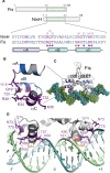
Figure 1.
Figure 1.. Sequence and structural similarities between NinH and Fis.
(A) Sequence homology between NinH and the Fis DNA binding domain. The position of homologous regions is depicted on the 98-residue Fis and 68-residue NinH proteins. The sequence alignment highlights identical residues (purple), functionally equivalent amino acids (blue) and others (gray). Residues in the Fis-DNA structure that participate in DNA binding are indicated by a filled circle. With the exception of Asn84 (orange circle), all of these are conserved in λ NinH and labeled above the primary sequence. Helices are labeled αA, αB and αC in NinH and correspond to helices αB, αC and αD in Fis, the latter two helices comprise the helix-turn-helix. (B) The NinH structure modeled by Phyre2 [42] on the apo Fis structure (PDB 1FIP). Residues are colored as in (A) and those matching the key residues in Fis that contact DNA are shown in stick format and labeled. (C) Crystal structure of the Fis–DNA complex [14]; PDB 3IV5) also showing residues conserved between NinH and Fis. (D) Detail of the Fis–DNA structure with residues involved in DNA contact depicted in stick format. Residues are colored as in (A), with N84 (orange) being replaced by serine in the NinH sequence. The orientation of the complex differs slightly from (C) to make it easier to visualize the contacts with DNA. Key guanosine and cytosine base pairs recognized by Fis are indicated at −7 and 7.
Figure 2.
Figure 2.. Modeling and structural analyses of NinH.
(A) Primary sequence of λ NinH protein highlighting the predicted secondary structure elements. (B) Modeled structure of the NinH dimer with secondary structure features indicated. (C) Top, front and side views of the NinH dimer model (green) with N-terminal His-tags (blue) fitted into the low molecular mass envelope generated from the SAXS profile. The high-resolution model fits the triangular prism shape well with some empty space suggesting flexibility of the N-terminal His-tags. (D) Overlay of SAXS scattering profile of His-NinH (black dots) and putative scattering profile generated from the high-resolution model using Crysol software (blue). The overall quality of the fit is high (χ2- test value of 1.03). (E) Front and side views of the structural alignment of the NinH modeled complex with Fis (PDB: 3IV5).
Figure 3.
Figure 3.. DNA binding by NinH protein.
(A) Gel mobility shift assays contained 50 or 500 nM NinH protein and 0.15 nM of 32P-labeled 60 bp (DS60) dsDNA (lanes 1–3) and bent DNA (BT1–3) containing 1 (lanes 4–6), 2 (lanes 7–9) or 3 (lanes 10–12) additional adenine residues in one of the DNA strands. (B) NinH and His-NinH binding to 10 nM fluorescein-labeled 20 bp dsDNA (DS20) as determined by fluorescence anisotropy. Data are the mean and standard deviation of two (His-NinH) and three (NinH) independent experiments. (C,D) NinH binding to linear dsDNA and bent DNA. Binding reactions contained 0.15 nM DNA with 0.781, 1.562, 3.125, 6.25, 12.5, 25, 50 and 100 nM protein. Data are the mean and standard deviation of three independent experiments. The relative proportion of the two protein–DNA complexes was determined by ImageJ [68] analysis of phosphorimaged gels.
Figure 4.
Figure 4.. Bending of dsDNA by NinH.
(A,B) Energy transfer was determined in steady state and lifetime FRET assays. Reactions contained 1 μM NinH protein or controls without protein or addition of 3 µM BSA with 50 nM DNA and were incubated for 10 min at room temperature before analysis. Experiments were performed in triplicate; error bars indicate the standard error of the mean. Negative controls have been used previously [69].
Figure 5.
Figure 5.. Identification of the NinH DNA binding motif.
(A) The GST-NinH binding site obtained by PBM using HK and ME microarrays. (B) The GST-NinH binding motif determined by HT-SELEX. (C) Published Fis DNA binding site based on SELEX, ChIP and EMSA data [3,13–15]. Residues below the motif correspond to those that exert a negative effect on Fis binding. Motif residues in (A) and (B) are numbered and aligned according to the Fis recognition sequence shown in (C) and are also highlighted in Figure 1D. Motif alignments were performed manually.
Figure 6.
Figure 6.. Oligomeric state and DNA binding properties of NinH site-directed mutants.
(A) Structure of the modeled NinH dimer highlighting the position of residues mutated to alanine. (B) Analytical centrifugation analysis assessing the monomeric or dimeric state of NinH proteins in solution. Peaks are labeled with the calculated molecular mass. (C) DNA binding by His-NinH wt and T32A proteins. Gel shift assays were performed with 10 nM Cy5-labeled 20 bp dsDNA (Cy520) matching the NinH consensus sequence (Supplementary Table S1). DNA–protein interactions were assessed by 7% neutral PAGE. (D) NinH mutant binding assessed by thermophoresis using 10 nM Cy520 dsDNA.
Figure 7.
Figure 7.. Effect of NinH wt and mutant overexpression on E. coli growth.
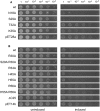
BL21-AI carrying pHis-NinH, pHis-NinH mutants and appropriate vector controls, pET28a(+) and pET14b, were grown to an A650nm of 0.4 in LB at 37°C. Serial dilutions (10 µl) were spotted onto agar plates containing either (A) kanamycin or (B) ampicillin without (uninduced) or with the addition of 0.2% arabinose and 1 mM IPTG (induced).

References

    1. Schneider R.,Lurz R.,Luder G.,Tolksdorf C.,Travers A. and Muskhelishvili G. (2001) An architectural role of the Escherichia coli chromatin protein Fis in organising DNA. Nucl. Acids Res. 29, 5107–5114 10.1093/nar/29.24.5107 - DOI - PMC - PubMed
    1. Dillon S.C. and Dorman C.J. (2010) Bacterial nucleoid-associated proteins, nucleoid structure and gene expression. Nat. Rev. Microbiol. 8, 185–195 10.1038/nrmicro2261 - DOI - PubMed
    1. Cho B.K.,Knight E.M.,Barrett C.L. and Palsson B.O. (2008) Genome-wide analysis of Fis binding in Escherichia coli indicates a causative role for A-/AT-tracts. Genome Res. 18, 900–910 10.1101/gr.070276.107 - DOI - PMC - PubMed
    1. Duprey A.,Reverchon S. and Nasser W. (2014) Bacterial virulence and Fis: adapting regulatory networks to the host environment. Trends Microbiol. 22, 92–99 10.1016/j.tim.2013.11.008 - DOI - PubMed
    1. Johnson R.C. (2015) Site-specific DNA inversion by serine recombinases. Microbiol. Spectr. 3, 1–36 10.1128/microbiolspec.MDNA3-0047-2014 - DOI - PMC - PubMed

Publication types

MeSH terms