. 2020 Apr 7;59(13):1309-1313.
doi: 10.1021/acs.biochem.0c00067. Epub 2020 Mar 27.

Fluorescent Probes for Monitoring Serine Ubiquitination

Affiliations

Fluorescent Probes for Monitoring Serine Ubiquitination

Kedar Puvar et al. Biochemistry. .

Abstract

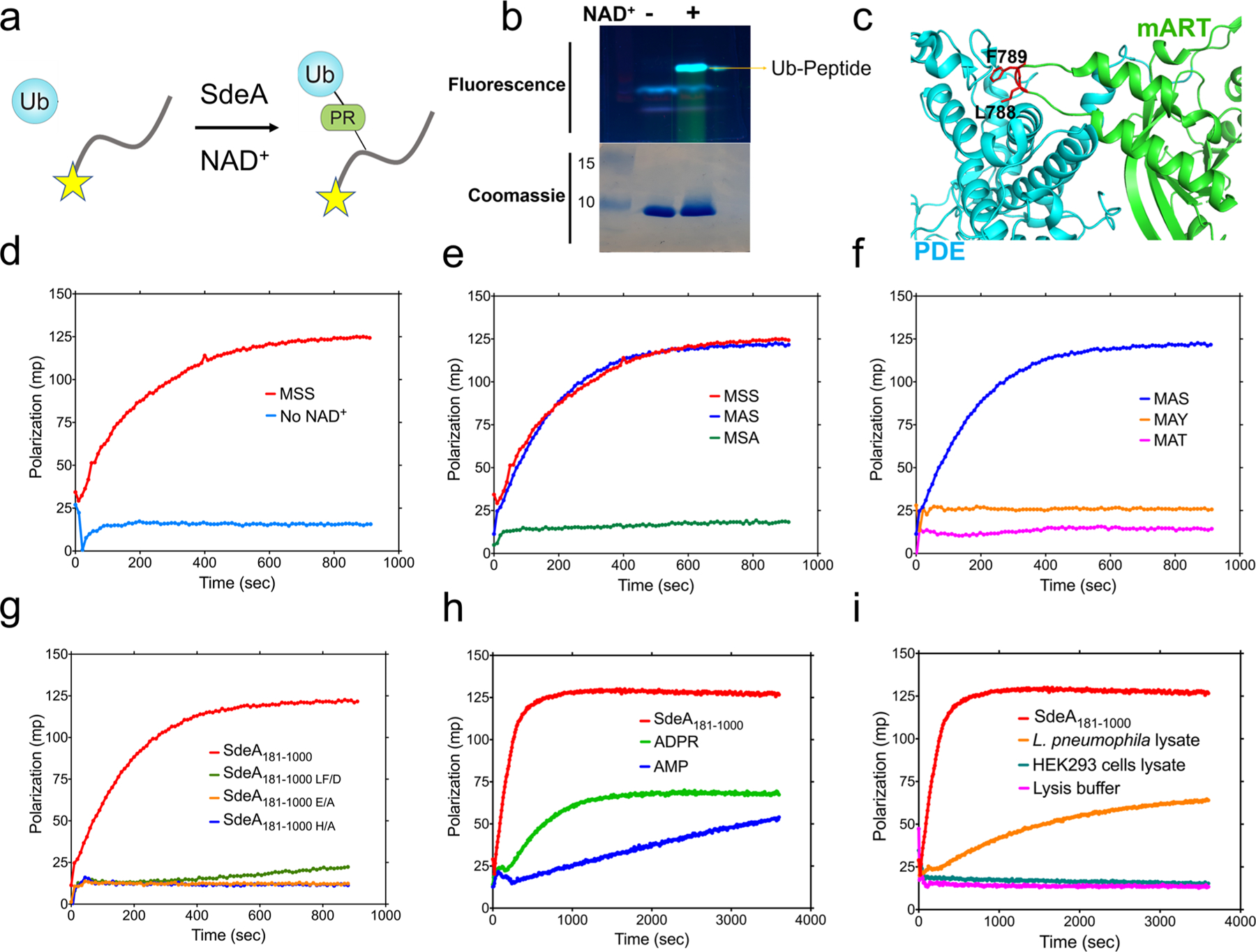
In a radical departure from the classical E1-E2-E3 three-enzyme mediated ubiquitination of eukaryotes, the recently described bacterial enzymes of the SidE family of Legionella pneumophila effectors utilize NAD+ to ligate ubiquitin onto target substrate proteins. This outcome is achieved via a two-step mechanism involving (1) ADP ribosylation of ubiquitin followed by (2) phosphotransfer to a target serine residue. Here, using fluorescent NAD+ analogues as well as synthetic substrate mimics, we have developed continuous assays enabling real-time monitoring of both steps of this mechanism. These assays are amenable to biochemical studies and high-throughput screening of inhibitors of these effectors, and the discovery and characterization of putative enzymes similar to members of the SidE family in other organisms. We also show their utility in studying enzymes that can reverse and inhibit this post-translational modification.


Conflict of interest statement

The authors declare no competing financial interest.

Figures

Figure 1.
Figure 1.
General features of SdeA. (a) Three key constructs were utilized in this study: full length enzyme, a construct containing both PDE and mART domains, and a construct containing only the mART domain. (b) Overall SidE mechanism of action that involves first mono-ADP ribosylation of ubiquitin at Arg42, catalyzed by the mART domain. Next, this ADP-ribosylated intermediate reacts with the PDE domain to be transferred to a Ser residue of the substrate protein.
Figure 2.
Figure 2.
Fluorescent NAD+ analogues used for monitoring the first step of SdeA-catalyzed ubiquitination. (a) Structures of NAD+, εNAD+, and NtzAD+. (b) When SdeA and Ub are incubated with εNAD+, a marked increase in fluorescence is observed in a time-dependent manner, consistent with the liberation of the nicotinamide group and loss of quenching. The activities of three distinct constructs of SdeA are compared.
Figure 3.
Figure 3.
Structure of the synthetic peptide substrate for SdeA ubiquitination assays. A nine-residue peptide with an N-terminal fluorescein group was designed, derived from the N-terminus of the known ubiquitination substrate protein Rab1.
Figure 4.
Figure 4.
(a) Overall scheme of a ubiquitination assay. (b) Fluorescence imaging of SDS–PAGE analysis of SdeA reaction in the presence of the peptide substrate reveals a fluorescent band when NAD+ is included, indicative of successful reaction. (c) Crystal structure of SdeA mART and PDE domains showing interdomain interactions, the importance of which can be tested via this fluorometric assay (Protein Data Bank entry 5ZQ2). (d) Including the fluorescent Rab1 peptide in the SdeA reaction caused a significant increase in FP. (e) Selective mutation of the Ser residues in the peptide substrate shows that the third Ser is likely targeted. (f) Mutation of the Ser to other hydroxyl-containing residues, such as Thr or Tyr, causes activity to be lost, indicating that SdeA specifically targets Ser residues. (g) Analysis of SdeA mutants using the fluorometric assay allows us to compare their activity in real time. The catalytic mutants (E/A and H/A) as well as the intradomain binding mutant (SdeA LF/D) were utilized. (h) Analysis of inhibition of SdeA-catalyzed ubiquitination. The nucleotides ADPR and AMP were included in the reaction mixture, resulting in inhibition of peptide ubiquitination. (i) Substituting cell lysates for purified SdeA protein allows this assay to be used to detect ubiquitination of the synthetic peptide. The L. pneumophila lysate caused an increase in the level of polarization. Within the parameters of this assay, the HEK cell lysate did not show a detectable increase.
Figure 5.
Figure 5.
(a) Analysis of SidE regulators. SidJ was preincubated with SdeA for the indicated time points, and ubiquitination was measured. (b) Purified DupB (SdeD) was added to the preubiquitinated peptide, demonstrating removal of PR-linked Ub. (c) The L. pneumophila lysate without SidEs was also utilized in the two-step assay described above to show Dup activity.

References

    1. Komander D, and Rape M (2012) The Ubiquitin Code. Annu. Rev. Biochem 81 (1), 203–229. - PubMed
    1. Ashida H, Kim M, and Sasakawa C (2014) Exploitation of the Host Ubiquitin System by Human Bacterial Pathogens. Nat. Rev. Microbiol 12 (6), 399–413. - PubMed
    1. Qiu J, and Luo Z-Q (2017) Legionella and Coxiella Effectors: Strength in Diversity and Activity. Nat. Rev. Microbiol 15 (10), 591–605. - PubMed
    1. Qiu J, Sheedlo MJ, Yu K, Tan Y, Nakayasu ES, Das C, Liu X, and Luo Z-Q (2016) Ubiquitination Independent of E1 and E2 Enzymes by Bacterial Effectors. Nature 533 (7601), 120–124. - PMC - PubMed
    1. Bhogaraju S, Kalayil S, Liu Y, Bonn F, Colby T, Matic I, and Dikic I (2016) Phosphoribosylation of Ubiquitin Promotes Serine Ubiquitination and Impairs Conventional Ubiquitination. Cell 167 (6), 1636–1649.e13. - PubMed

Publication types