. 2020 Apr 9;5(7):e133125.
doi: 10.1172/jci.insight.133125.

Aberrant cell migration contributes to defective airway epithelial repair in childhood wheeze

Affiliations

Aberrant cell migration contributes to defective airway epithelial repair in childhood wheeze

Thomas Iosifidis et al. JCI Insight. .

Abstract

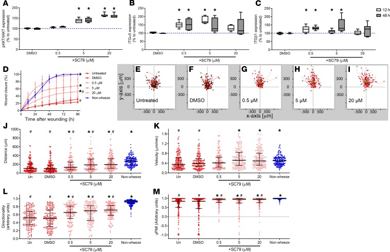
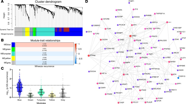
Abnormal wound repair has been observed in the airway epithelium of patients with chronic respiratory diseases, including asthma. Therapies focusing on repairing vulnerable airways, particularly in early life, present a potentially novel treatment strategy. We report defective lower airway epithelial cell repair to strongly associate with common pre-school-aged and school-aged wheezing phenotypes, characterized by aberrant migration patterns and reduced integrin α5β1 expression. Next generation sequencing identified the PI3K/Akt pathway as the top upstream transcriptional regulator of integrin α5β1, where Akt activation enhanced repair and integrin α5β1 expression in primary cultures from children with wheeze. Conversely, inhibition of PI3K/Akt signaling in primary cultures from children without wheeze reduced α5β1 expression and attenuated repair. Importantly, the FDA-approved drug celecoxib - and its non-COX2-inhibiting analogue, dimethyl-celecoxib - stimulated the PI3K/Akt-integrin α5β1 axis and restored airway epithelial repair in cells from children with wheeze. When compared with published clinical data sets, the identified transcriptomic signature was also associated with viral-induced wheeze exacerbations highlighting the clinical potential of such therapy. Collectively, these results identify airway epithelial restitution via targeting the PI3K-integrin α5β1 axis as a potentially novel therapeutic avenue for childhood wheeze and asthma. We propose that the next step in the therapeutic development process should be a proof-of-concept clinical trial, since relevant animal models to test the crucial underlying premise are unavailable.

Keywords: Asthma; Cell Biology; Cell migration/adhesion; Integrins; Pulmonology.


Conflict of interest statement

Conflict of interest: TI, DAK, SMS, and AK have registered an international patent related to findings presented in this manuscript (21 May 2019, PCT/AU2019/050493).

Figures

Figure 1
Figure 1. Defective cell migration of leading edge cells in pAEC of children with wheeze.
(A) Cultures from children without wheeze had the capacity to repair by 72 hours after wounding. (B) In contrast, cultures from children with wheeze failed to close the wound by 96 hours after wounding. (C) Leading edge pAEC of children without wheeze responded to the scratch wounding stimulus by migrating directionally, toward the center of the wound site. (D) Leading edge pAEC of children with wheeze showed a dysregulated response to wounding, where some cells migrated into the wound site in an uncoordinated manner and other cells did not migrate very far into the wound and even migrated backward into the leading edge. The green dot represents the mean center of mass of the endpoints of all tracked cells. (E and F) Leading edge pAEC from children without wheeze migrated far (E) and fast (F) into the wound site by 10 hours after wounding, although response to wounding was varied. However, leading edge cells of children with wheeze migrated shorter average distances (E) and at slower velocity (F) than their nonwheezing counterparts (P < 0.050). (G and H) Notably, leading edge cells of children without wheeze migrated directionally (G) and collectively into the center of the wound, as shown with high y axis forward migration index (yFMI) values (H). Conversely, leading edge pAEC of children with wheeze demonstrated migration trajectories with significantly less directionality (G) and yFMI (H), indicating a loss of coordination in their response to wounding. Cell migration trajectory data were generated from 296 and 228 leading edge cell tracks of children with wheeze (n = 14) and without wheeze (n = 9), respectively. All experiments were completed in 2 technical replicates. The data were represented as median ± IQR, *P < 0.050, Mann-Whitney U test.
Figure 2
Figure 2. Role of integrins in pAEC from children with and without wheeze.
(A and B) Protein expression of integrin subunits α5 (A) and β1 (B) were found to be significantly lower in pAEC from children with wheeze compared with their nonwheezing counterparts. (C–E) Strong immunofluorescence staining of both integrin α5 (C) and β1 (D) on leading edge pAEC from children without wheeze was demonstrated that was almost undetectable in sites distal to the wound (E). (F–H) In contrast, pAEC cultures of children with wheeze exhibited weak integrin α5 (F) and β1 (G) staining along the leading edge cells (H). The slides were counterstained with Hoechst nuclear stain (blue). Representative images of samples from 5 children with wheeze and 5 children without wheeze (CH). Oil immersion objective, optical magnification of 600×, imaged using 60× objective and 10× ocular lens; NA 1.4. Scale bar: 50 μm. (I) Blocking β1 integrin (1:40 dilution, IgG1κ, P5D2) function significantly reduced pAEC wound closure rates to similar rates observed in their wheezing counterparts (red dashed line). Matching dilution of isotype control antibody (IgG1κ, MOPC-21) or untreated pAEC from children without wheeze reached full closure by 72 hours after wounding. n = 4 children. Median ± IQR, *P < 0.050, Mann-Whitney U test. (J) Cultures treated with isotype antibody (1:40 dilution) displayed comparable migration patterns with untreated pAEC from children without wheeze. (K) However, cultures treated with 1:40 dilution of anti–β1 integrin antibody in culture media migrated less far into the wound, lacking cell directionality and specificity toward the wound center. Individual cell tracks were transposed so that each track had its start at the origin. n = 60 tracks from 4 children without wheeze (IK).
Figure 3
Figure 3. Transcriptional response to in vitro pAEC wounding.
(A) Two-dimensional principal component analysis (PCA) plot displays samples from children without wheeze (blue; n = 4 children) and samples from children with wheeze (red; n = 6 children) to cluster separately, indicating large transcriptional differences. (B) Volcano plot. Scattered points represent genes: the x axis is the log2-transformed fold-change values per gene for leading edge samples at 24 hours after wounding from children with wheeze relative to matching nonasthmatic controls. Log10-transformed P values above 1.3 were considered statistically significant (Benjamini-Hochberg–adjusted P < 0.050). Orange dots are genes significantly upregulated (log2 fold-change > 0.58, adjusted P < 0.050), and purple dots are significantly downregulated genes (log2 fold-change < –0.58) in samples from children with wheeze relative to matching nonwheezing controls. Genes colored in black are either not differentially expressed (–0.58 < log2 fold-change < 0.58) or did not reach statistical significance (adjusted P > 0.050). (C) Top 5 downregulated canonical pathways associated with differentially expressed genes in leading edge pAEC at 24 hours after wounding from children with wheeze compared with corresponding samples of nonwheezing children. (D) Network map of genes associated with PI3K/Akt signaling pathway. Genes are highlighted as upregulated (orange) or downregulated (purple). (E) Top 5 upstream transcriptional regulators of integrin α5 and β1 ranked with predicted Z scores. All upstream regulators were determined to be statistically significant (adjusted P < 0.050). (F) Lower levels of activated Akt (phosphorylated Ser473) in pAEC from children with wheeze (n = 6 children) compared with their nonasthmatic counterparts (*P < 0.050; Mann-Whitney U test; n = 6 children). Levels of pAKT were normalized to total Akt levels, and data were represented as median ± IQR.
Figure 4
Figure 4. Inhibition of Akt signaling abrogates pAEC repair and integrin expression.
pAEC from children without wheeze were treated with different concentrations (0.01, 0.1, 1 μM) of the specific Akt inhibitor MK2206. (A) MK2206 inhibited phosphorylation of Akt (serine residue 473) at 12 and 48 hours after treatment. (B and C) Inhibition of Akt in pAEC from children without wheeze resulted in significant reduction of integrin subunit α5 (B) cell membrane expression in a concentration-dependent manner at 12 and 48 hours after MK2206 treatment. Although no differences were observed in cell membrane integrin subunit β1 at 12 hours following Akt inhibition an increase was seen at 48 hours following treatment compared to vehicle control (C). (D) Treatment of pAEC cultures from nonwheezing children with MK2206 at the time of scratch wounding resulted in a concentration-dependent reduction in closure rates, although DMSO vehicle (0.05% v/v) control was not significantly altered compared with untreated cultures. (E–M) Although treatment of pAEC from nonwheezing children with 0.05% (v/v) DMSO vehicle control had no effect on cell migration (E and F), MK2206 treatment attenuated cell migration in a concentration-dependent manner (G–I) by inhibiting distance migrated (J), velocity (K), directionality (L), and centrality (yFMI, M). All experiments were completed with pAEC cultures from 6 children without wheeze, and data were represented as either box and whisker (min/max) or dot plots with median ± IQR. Statistical differences between treatment and untreated control (*P < 0.050) or wheezing group (#P < 0.050) were determined using 2-way Kruskal-Wallis ANOVA with Dunn’s post hoc test for multiple comparisons.
Figure 5
Figure 5. Activation of Akt signaling enhances repair of pAEC from children with wheeze and increased integrin α5β1 expression.
pAEC from children with wheeze were treated with different concentrations (0.5, 5, 20 μM) of the specific Akt activator SC79. (A) SC79 treatment resulted in phosphorylation of Akt (serine residue 473), at 12 and 48 hours after treatment. (B) Significant increase of integrin subunit α5 cell membrane expression was observed in pAEC from children with wheeze for all concentrations at 12 hours and 0.5, 5, and 20 μM SC79 at 48 hours. (C) Also, integrin subunit β1 cell membrane expression was increased in pAEC from children with wheeze treated with 0.5, 2, and 5 μM SC79 at 12 and 48 hours. (D) Treatment of pAEC from wheezers with SC79 at the time of scratch wounding resulted in a concentration-dependent increase in closure rates, although DMSO vehicle (0.08% v/v) control was not significantly altered compared with untreated cultures. (E–M) Although treatment of pAEC from wheezers with 0.08% (v/v) DMSO vehicle control had no effect on cell migration (E and F), SC79 treatment enhanced cell migration in a concentration-dependent manner (G–I) by stimulating distance migrated (J), velocity (K), directionality (L), and centrality (yFMI, M). All experiments were completed with pAEC cultures from 6 children with wheeze, and data were represented as either box and whisker (min/max) or dot plots with median ± IQR. Statistical differences between treatment and untreated control (*P < 0.050) or nonwheezing group (#P < 0.050) were determined using 2-way Kruskal-Wallis ANOVA with Dunn’s post hoc test for multiple comparisons. The wound closure (D) and cell migration parameters (J–M) for the untreated wheeze and nonwheeze groups were also presented in Figure 4 and were utilized for baseline response purposes.
Figure 6
Figure 6. Repair of pAEC from children with wheeze is restored by celecoxib and dimethyl-celecoxib treatment via the PI3K/Akt–integrin α5β1 axis.
pAEC from children with wheeze were treated with 10 μM of COX2 inhibitor celecoxib or its analogue, dimethyl-celecoxib. (A) Celecoxib and its analogue treatment resulted in phosphorylation of Akt (serine residue 473) at 12 and 48 hours after treatment. (B and C) Significant increase of integrin subunits α5 (B) and β1 (C) cell membrane expression was observed in treated cultures from children with wheeze at both 12 and 48 hours. (D) Treatment of pAEC cultures from wheezers with celecoxib or dimethyl-celecoxib at the time of scratch wounding resulted in complete repair comparable with their nonwheezing counterparts. (E–K) Although treatment of pAEC from wheezers with 0.13% (v/v) DMSO vehicle control had no effect on cell migration (E), celecoxib or dimethyl-celecoxib treatments enhanced cell migration (F and G) by stimulating distance migrated (H), velocity (I), directionality (J), and centrality (yFMI, K). All experiments were completed with pAEC cultures from 6 children with wheeze, and data were represented as either box and whisker (min/max) or dot plots with median ± IQR. Statistical differences between treatment and untreated control (*P < 0.050) or nonwheezing group (#P < 0.050) were determined using 2-way Kruskal-Wallis ANOVA with Dunn’s post hoc test for multiple comparisons. The wound closure (D) and cell migration parameters (J–M) for the untreated nonwheeze groups were also presented in Figure 4 and were utilized for baseline response purposes.
Figure 7
Figure 7. Evaluation of the defective pAEC repair gene signature in published transcriptomic data sets.
(A) Weighted gene coexpression network analysis (WGCNA) cluster dendrogram. Hierarchical cluster analysis was conducted to detect gene coexpression clusters with corresponding color assignments using data from 64 children with or without recurrent wheeze. Each color represents a module in the constructed gene coexpression network by WGCNA. (B) WGCNA modules and sample trait heatmap. Using the default parameter settings and genes filtered based on probeset concordance (n = 1737), 5 gene modules were identified to correlate with recurrence of respiratory wheeze. Data are presented with the correlation coefficient (P value), where positive correlations are red, and negative correlations are blue. (C) Weighted gene coexpression network analysis calculation of gene significance (GS) to sample trait of interest, recurrence of wheeze, in each gene module. The blue module had an overrepresented number of genes associating with wheeze recurrence. (D) Minimum network map of blue module genes that strongly associate with wheeze recurrence in the pediatric acute wheeze data set. Genes are highlighted as highly interconnected (pink) or weakly interconnected (purple) according to known protein/protein interactions from published studies (prior knowledge). Size of nodes indicate a larger number of connections.

References

    1. Masoli M, Fabian D, Holt S, Beasley R, Global Initiative for Asthma (GINA) Program The global burden of asthma: executive summary of the GINA Dissemination Committee report. Allergy. 2004;59(5):469–478. - PubMed
    1. Childhood Asthma Management Program Research Group. et al. Long-term effects of budesonide or nedocromil in children with asthma. N Engl J Med. 2000;343(15):1054–1063. doi: 10.1056/NEJM200010123431501. - DOI - PubMed
    1. Bisgaard H, Hermansen MN, Loland L, Halkjaer LB, Buchvald F. Intermittent inhaled corticosteroids in infants with episodic wheezing. N Engl J Med. 2006;354(19):1998–2005. doi: 10.1056/NEJMoa054692. - DOI - PubMed
    1. Martinez FD. Inhaled corticosteroids and asthma prevention. Lancet. 2006;368(9537):708–710. doi: 10.1016/S0140-6736(06)69261-1. - DOI - PubMed
    1. O’Byrne PM, Pedersen S, Lamm CJ, Tan WC, Busse WW, START Investigators Group Severe exacerbations and decline in lung function in asthma. Am J Respir Crit Care Med. 2009;179(1):19–24. - PubMed

Publication types

Substances