. 2020 Mar 24;15(3):e0230668.
doi: 10.1371/journal.pone.0230668. eCollection 2020.

Histopathologic assessment of cultured human thymus

Affiliations

Histopathologic assessment of cultured human thymus

Laura P Hale et al. PLoS One. .

Abstract

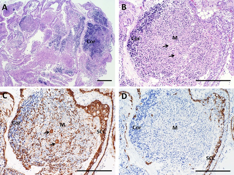
The maintenance and propagation of complex mixtures of cells in vitro in the form of native organs or engineered organoids has contributed to understanding mechanisms of cell and organ development and function which can be translated into therapeutic benefits. For example, allogeneic cultured postnatal human thymus tissue has been shown to support production of naïve recipient T cells when transplanted into patients with complete DiGeorge anomaly and other genetic defects that result in congenital lack of a thymus. Patients receiving such transplants typically exhibit reversal of their immunodeficiency and normalization of their peripheral blood T cell receptor V-beta repertoire, with long-term survival. This study was designed to assess the histopathologic changes that occur in postnatal human thymus slices when cultured according to protocols used for transplanted tissues. Results showed that as thymic organ cultures progressed from days 0 through 21, slices developed increasing amounts of necrosis, increasing condensation of thymic epithelium, and decreasing numbers of residual T cells. The architecture of the thymic epithelial network remained generally well-preserved throughout the 21 days of culture, with focal expression of cytokeratin 14, a putative biomarker of thymic epithelial cells with long-term organ-repopulating potential. All organ slices derived from the same donor thymus closely resembled one another, with minor differences in size, shape, and relative content of cortex versus medulla. Similarly, slices derived from different donors showed similar histopathologic characteristics when examined at the same culture time point. Taken together, these results demonstrate that diagnostic criteria based on structural features of the tissue identifiable via hematoxylin and eosin staining and cytokeratin immunohistochemistry can be used to evaluate the quality of slices transplanted into patients with congenital athymia.


Conflict of interest statement

Cultured postnatal human thymus (RVT-802) is an investigational product implanted into patients under an Investigational New Drug (IND) application with the United States Food and Drug Administration (FDA) that is sponsored by Dr. Markert. The technology for RVT-802 was developed by Dr. Markert and has been licensed to Enzyvant Therapeutics GmbH (Enzyvant) by Duke University. Dr. Markert and Duke University have received royalties from Enzyvant. If the technology is commercially successful in the future, Dr. Markert and Duke University may benefit financially. Portions of the salaries of Drs. Markert, Kurtzberg, and Hale and consultation fees for Dr. Neff were paid by funding to Duke University from Enzyvant. This does not alter our adherence to PLOS ONE policies on sharing data and materials.

Figures

Fig 1
Fig 1. Architecture of cultured thymus, day 0.
A, B. Hematoxylin and eosin (H&E) staining shows well-defined cortical and lighter-staining medullary areas, as expected for normal pediatric thymus. C. Immunohistochemistry with a cocktail of antibodies (AE1/AE3) that together detect all types of epithelial cells demonstrates that thymic epithelial cells are present beneath the capsule and in a light lacy network in both cortex and medulla. Arrows in B and C point to a Hassall body. D. Cytokeratin 14 (CK14) antibody reacts with thymic epithelial cells in the sub-capsular cortex and in the medulla, as well as with scattered thymic epithelial cells in the cortex. The dotted line highlights an area of medulla that is surrounded by cortex. Brown color indicates a positive reaction with antibody. SCC denotes sub-capsular cortex, Cor denotes cortex, and M denotes medulla. Bar represents 1 mm in A and 500 μm in B–D.
Fig 2
Fig 2. Examples of Hassall bodies.
The histologic appearance of Hassall bodies is shown on day 0 (A, B) and day 9 (C, D) of culture. Hematoxylin and eosin (H&E) stain is shown in panels A and C. Reactivity with pan-cytokeratin (AE1/AE3) antibodies is shown in panels B and D; brown color indicates a positive reaction. Arrowheads point out representative Hassall bodies, which appear less prominent on H&E-stained sections of cultured thymus due to the depletion and necrosis of the surrounding thymocytes. However, Hassall bodies can still be readily identified by careful examination or by using immunohistochemistry. Bar represents 100 μm.
Fig 3
Fig 3. Architecture of cultured thymus, day 7.
A, B. Hematoxylin and eosin (H&E) stain shows marked depletion of thymocytes, although some cortical areas (Cor) still contain large numbers of thymocytes with retained nuclei. Pan-cytokeratin (AE1/AE3) (C) and cytokeratin 14 (CK14) immunohistochemistry (D) show condensation of the thymic epithelium in the subcapsular cortex (SCC) and in the medulla (M). Brown color indicates a positive reaction with antibody. Bar represents 1 mm in A and 500 μm in B–D.
Fig 4
Fig 4. Architecture of cultured thymus, day 9.
A, B. Hematoxylin and eosin (H&E) stain. Few if any live T cells or thymic epithelial cells are present in the pale-staining area in A that is enclosed by the dotted line, which is almost completely necrotic (Necr). Most nuclei formerly present in this region have been degraded, a process called karyolysis. Other areas where the nuclei from residual thymocytes have not been completely degraded continue to stain dark blue with hematoxylin. Arrow in B points to a Hassall body. C. Pan-cytokeratin (AE1/AE3) immunoreactivity. D. Cytokeratin 14 (CK14) immunoreactivity. Brown color indicates a positive reaction with antibody. Bar represents 1 mm in A and 500 μm in B–D.
Fig 5
Fig 5. Architecture of cultured thymus, day 12.
A, B. Hematoxylin and eosin (H&E) stain. At this time point, many thymocytes have either been lost from the tissue or they have died and their nuclei have been dissolved, making the tissue more eosinophilic (pink). Some areas retain architecture characteristic of normal uncultured thymus with cortical-like areas (Cor) that stain more basophilic (blue) and medullary-like areas (M), although with greatly decreased thymocyte cellularity. C. Pan-cytokeratin (AE1/AE3) immunoreactivity. D. Cytokeratin 14 (CK14) immunoreactivity. Brown color indicates a positive reaction with antibody. At this time point, the sub-capsular cortex (SCC) has thickened and epithelial cells appear more prominent due to the decreased numbers of thymocytes present. Arrows point to representative Hassall bodies. Bar represents 1 mm in A and 500 μm in B–D.
Fig 6
Fig 6. Architecture of cultured thymus, day 20.
A, B. Hematoxylin and eosin (H&E) stain. At this time point, most thymocytes have either been lost from the tissue or they have died and their nuclei have been dissolved, making the tissue more eosinophilic (pink). Large groups of residual thymocytes are rare, although scattered cells with nuclear characteristics of thymocytes are evident. C. Pan-cytokeratin (AE1/AE3) immunoreactivity. Much of the epithelium present in formerly medullary areas (M) is condensed due to loss of medullary thymocytes, but scattered epithelial cells indicative of a residual light, lacy, three-dimensional network of thymic epithelial cells remain. D. Cytokeratin 14 (CK14) immunohistochemistry highlights former medullary areas and the subcapsular cortex. Brown color indicates a positive reaction with antibody. Arrows point to representative Hassall bodies. Bar represents 1 mm in A and 500 μm in B–D.
Fig 7
Fig 7. Assessing intact nuclei in thymus slices.
Examples of intact thymic epithelial cell nuclei (arrows) are shown in the subcapsular cortex on day 9 (panel A) and in the medulla on day 21 (panel B). Hematoxylin and eosin stain; bar represents 50 μm.
Fig 8
Fig 8. Comparison of thymic epithelial network of cultured thymus tissue at different time points.
A. Day 0. B. Day 5. C. Day 9. D. Day 12. E. Day 21. Although there are time point-related differences in thymocyte depletion and the amount of necrosis such that the tissue become less basophilic (blue color) with time, the structure of the thymic epithelial network (brown color) remains intact as the culture progresses. Both cortical and medullary epithelium may condense as intervening thymocytes are depleted. Brown color indicates a positive reaction with a cocktail of anti-cytokeratin antibodies (AE1 + AE3); Hematoxylin counterstain. Bar represents 400 μm.
Fig 9
Fig 9. CD3 immunohistochemistry in thymus slices as a function of time in culture.
A, B. On day 0, essentially all immature T cells in the cortex and more mature cells in the medulla react strongly with CD3 antibody. Higher magnification (B) shows pale blue nuclei surrounded by a ring of brown immunoreactivity, consistent with membrane expression of CD3. C—E. On day 7, the tissue still shows extensive reactivity with CD3 antibody. However, higher magnification (D, E) shows that the majority of the immunoreaction (brown color) is associated with debris from dead thymocytes, as most brown foci lack evidence of nuclei (D). Small foci of cells that demonstrate intact nuclei and membrane staining (arrows) can still be identified in areas away from the debris (E). As cultures progress through day 9 (F–G), day 12 (H–I), and day 21 (J–K), reactivity with thymocyte cellular debris remains strong, making it difficult to reliably detect potentially intact cells amidst the debris. The slices shown are all from a single lot that is representative of multiple lots examined at these time points. Bar represents 500 μm in panels A, C, F, H, and J and 50 μm in B, D, E, G, I, and K.
Fig 10
Fig 10. CD45RO immunoreactivity in thymus slices as a function of time in culture.
A, B. On day 0, essentially all immature T cells in the cortex (Cor) and medulla (M) react strongly with antibody specific for CD45RO. Higher magnification (B) shows the strong brown positive membrane reactivity. C, D. By day 7, reactivity with CD45RO antibody is strongest in the medulla. Some positive cells exhibit the expected specific membrane staining, but there is also considerable reactivity with cellular debris. Loss of membrane immunostaining and intense reactivity primarily with residual thymocyte debris is seen on days 9 (E, F), 12 (G, H) and 21 (I, J), although some cells with intact membrane staining can occasionally still be detected (arrows in H). Bar represents 500 μm in panels A, C, E, G, and I and 50 μm in B, D, F, H, and J.
Fig 11
Fig 11. TdT immunoreactivity in thymus slices as a function of time in culture.
A, B. On day 0, essentially all immature T cells in the cortex (Cor) react strongly with antibody specific for TdT. Higher magnification (B) shows strong brown positive reactivity with the nuclei of cortical thymocytes with rare positive cells in the medulla (M). C, D. By day 7, the nuclei of most thymocytes that remain in cortical areas are small and strongly basophilic (blue) consistent with apoptosis and they fail to react with TdT antibody. The antibody reactivity shown in the center of D is with cellular debris. Similar lack of TdT immunostaining of residual thymocyte nuclei is seen on days 9 (E, F), 12 (G, H) and 21 (I, J). Bar represents 500 μm in panels A, C, E, G, and I and 50 μm in B, D, F, H, and J.
Fig 12
Fig 12. CD43 immunoreactivity in thymus slices as a function of time in culture.
A, B. On day 0, immature thymocytes in the cortex (Cor) react weakly and those in the medulla (M) react more strongly with antibody specific for CD43. The dark bands in A are sectioning artifacts (wrinkles and folds in the tissue). Higher magnification (B) shows the brown positive membrane reactivity with both cortical and medullary thymocytes. C, D. By day 7, reactivity with CD43 antibody is generally limited to the medullary thymocytes. Some positive cells exhibit the expected specific membrane staining, but there is also considerable reactivity with cellular debris in cortical areas. Loss of membrane immunostaining and intense reactivity primarily with residual thymocyte debris is seen on days 9 (E, F), 12 (G, H) and 21 (I, J), although some cells with intact membrane staining can be detected at these later time points. Bar represents 500 μm in panels A, C, E, G, and I and 50 μm in B, D, F, H, and J.
Fig 13
Fig 13. βF1 immunostaining in thymus slices as a function of time in culture.
A, B. On day 0, a subset of immature T cells in the cortex (Cor) and essentially all of the medullary thymocytes react strongly with βF1 antibody that is specific for the β chain of the αβ T cell receptor. Higher magnification (B) shows strong brown positive membrane reactivity; M denotes medulla. C, D. Reactivity with antibody remains strong on day 7, however the specific membrane staining is lost and reactivity is primarily with cellular debris in medullary areas. Similar loss of membrane immunostaining and reactivity primarily with residual thymocyte debris is seen on days 9 (E, F), 12 (G, H) and 21 (I, J), but cells with intact membranes can be detected (see arrows in J). Bar represents 500 um in panels A, C, E, G, and I and 50 um in B, D, F, H, and J.
Fig 14
Fig 14. Ki-67 immunohistochemistry in thymus slices as a function of time in culture.
A, B. On day 0, the nuclei of the majority of immature T cells in the cortex (Cor) react strongly with antibody specific for Ki-67. Higher magnification (B) shows strong brown positive reactivity with the nuclei of cortical thymocytes. Only rare lymphocytes in the medulla (M) react with antibody. C- E. By day 7, the nuclei of most thymocytes that remain in cortical areas are small with indistinct nuclear borders consistent with apoptosis, and they fail to react with Ki-67-specific antibody (D). The cells that react with antibody (E, arrows) have larger nuclei that suggest they are thymic epithelial cells. Similar lack of Ki-67 labeling of residual thymocyte nuclei is seen on days 9 (F, G), 12 (H, I) and 21 (J, K). The slices shown are all from a single lot that is representative of multiple lots examined at similar time points. Bar represents 500 μm in panels A, C, E, G, and I and 50 μm in B, D, F, H, and J.

References

    1. Markert ML, Boeck A, Hale LP, Kloster AL, McLaughlin TM, Batchvarova MN, et al. Transplantation of thymus tissue in complete DiGeorge syndrome. New Engl J Med. 1999;341: 1180–1189. 10.1056/NEJM199910143411603 - DOI - PubMed
    1. Markert ML, Sarzotti M, Ozaki DA, Sempowski GD, Rhein ME, Hale LP, et al. Thymic transplantation in complete DiGeorge syndrome: Immunologic and safety evaluation in twelve patients. Blood. 2003;102: 1121–1130. 10.1182/blood-2002-08-2545 - DOI - PubMed
    1. Markert ML, Alexieff MJ, Li J, Sarzotti M, Ozaki DA, Devlin BH, et al. Postnatal thymus transplantation with immunosuppression as treatment for DiGeorge syndrome. Blood. 2004;104: 2574–2581. 10.1182/blood-2003-08-2984 - DOI - PubMed
    1. Markert ML, McLaughlin TM, Davis CM, Watson TJ, Ward FE, Kostyu D, et al. Successful formation of a chimeric human thymus allograft following transplantation of partially HLA-matched postnatal thymus. J Immunol. 1997;158: 998–1005. - PubMed
    1. Markert ML, Devlin BH, Alexieff MJ, Li J, McCarthy EA, Gupton SE, et al. Review of 54 patients with complete DiGeorge anomaly enrolled in protocols for thymus transplantation: Outcome of 44 consecutive transplants. Blood. 2007;109: 4539–4547. 10.1182/blood-2006-10-048652 - DOI - PMC - PubMed

Publication types