. 2020 Mar 24;4(6):1145-1158.
doi: 10.1182/bloodadvances.2019000785.

Runx1 negatively regulates inflammatory cytokine production by neutrophils in response to Toll-like receptor signaling

Affiliations

Runx1 negatively regulates inflammatory cytokine production by neutrophils in response to Toll-like receptor signaling

Dana C Bellissimo et al. Blood Adv. .

Abstract

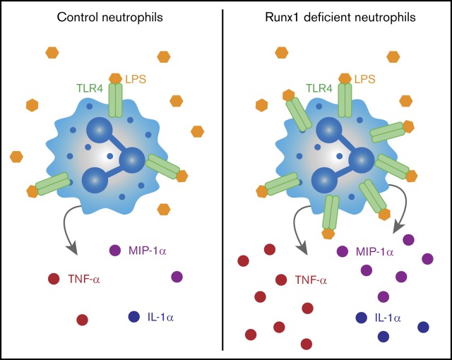
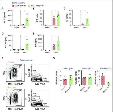
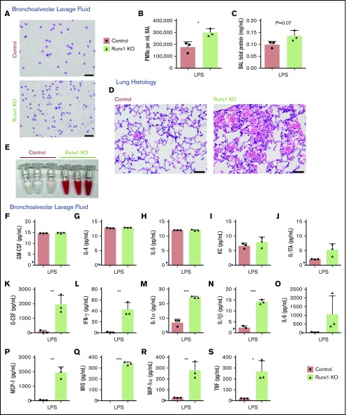
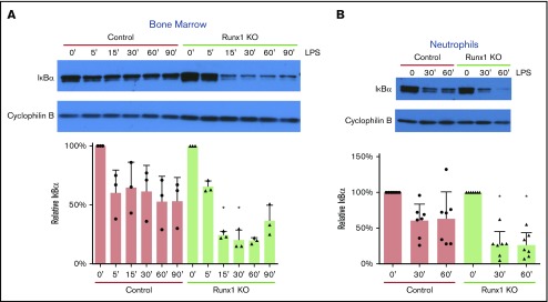
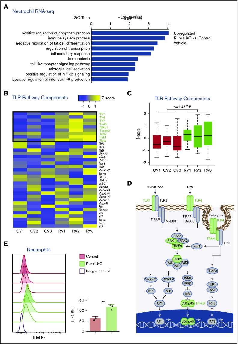
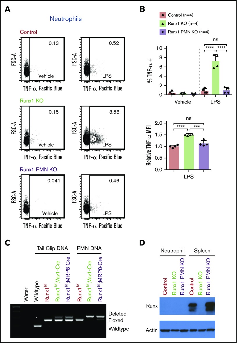
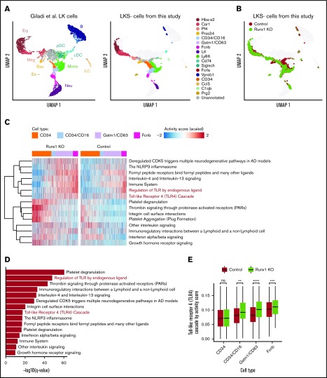
RUNX1 is frequently mutated in myeloid and lymphoid malignancies. It has been shown to negatively regulate Toll-like receptor 4 (TLR4) signaling through nuclear factor κB (NF-κB) in lung epithelial cells. Here we show that RUNX1 regulates TLR1/2 and TLR4 signaling and inflammatory cytokine production by neutrophils. Hematopoietic-specific RUNX1 loss increased the production of proinflammatory mediators, including tumor necrosis factor-α (TNF-α), by bone marrow neutrophils in response to TLR1/2 and TLR4 agonists. Hematopoietic RUNX1 loss also resulted in profound damage to the lung parenchyma following inhalation of the TLR4 ligand lipopolysaccharide (LPS). However, neutrophils with neutrophil-specific RUNX1 loss lacked the inflammatory phenotype caused by pan-hematopoietic RUNX1 loss, indicating that dysregulated TLR4 signaling is not due to loss of RUNX1 in neutrophils per se. Rather, single-cell RNA sequencing indicates the dysregulation originates in a neutrophil precursor. Enhanced inflammatory cytokine production by neutrophils following pan-hematopoietic RUNX1 loss correlated with increased degradation of the inhibitor of NF-κB signaling, and RUNX1-deficient neutrophils displayed broad transcriptional upregulation of many of the core components of the TLR4 signaling pathway. Hence, early, pan-hematopoietic RUNX1 loss de-represses an innate immune signaling transcriptional program that is maintained in terminally differentiated neutrophils, resulting in their hyperinflammatory state. We hypothesize that inflammatory cytokine production by neutrophils may contribute to leukemia associated with inherited RUNX1 mutations.


Conflict of interest statement

Conflict-of-interest disclosure: The authors declare no competing financial interests.

Figures

None
Graphical abstract
Figure 1.
Figure 1.
Increased secretion of inflammatory cytokines, chemokines, and growth factors by Runx1 KO BM. (A-E) Absolute quantification by CBA of inflammatory factor levels in the supernatant of whole BM cells stimulated for 8 hours with vehicle or 100 ng/mL LPS. Bar graphs include independent data points. Error bars represent mean ± standard deviation (SD). Five replicates from 4 experiments were performed for each condition with all results above the limit of detection (blue arrowhead) plotted. For all factors except KC (C), a 2-tailed unpaired Student t test was performed comparing the factor concentration between the control and Runx1 KO LPS-treated samples. Because of limited detection of KC in the control LPS-treated sample (C), a 1-sample Student t test was performed comparing the mean of the Runx1 KO LPS-treated sample to a hypothetical mean = 0. (F-G) Representative FACS plots gated on live singlets and corresponding quantification of the frequencies of monocytes, neutrophils, and eosinophils in the BM (n = 3 from 3 experiments, mean ± SD, 2-tailed unpaired Student t test). Eos, eosinophils; FSC-A, forward scatter area; Mo, monocytes; PE, phycoerythrin. *P ≤ .05; **P ≤ .01.
Figure 2.
Figure 2.
Increased TNF-α production by Runx1 KO neutrophils in response to TLR4 stimulation. (A) Representative FACS plots of intracellular TNF-α production by monocytes (CD11b+Ly6G) after stimulation of whole BM with vehicle or 100 ng/mL LPS for 4 hours. (B) Quantification of the frequency of TNF-α+ monocytes and relative MFI of TNF-α in the TNF-α+ monocytes normalized to control monocytes run in the same experiment (n = 6 from 4 experiments). (C) Representative FACS plots of intracellular TNF-α production by neutrophils (CD11b+Ly6G+) after stimulation of whole BM with vehicle or 100 ng/mL LPS for 4 hours. (D) Quantification of the frequency of TNF-α+ neutrophils and relative MFI of TNF-α in the TNF-α+ neutrophils normalized to control neutrophils run in the same experiment (n = 6 from 4 experiments). (B,D) Bar graphs depict independent data points with the mean ± SD; 2-tailed unpaired Student t tests. (E) Quantification of the frequency of TNF-α+ neutrophils (CD11b+Ly6G+) after stimulation of whole BM for 4 hours with TLR agonists (n = 4 to 6, as indicated from 6 experiments). Bar graphs depict independent data points with the mean ± SD. Statistics represent the results of a 1-way analysis of variance followed by Sidak’s multiple comparison test to compare the means of the control and Runx1 KO samples for each TLR agonist. (F-H) Absolute quantification by CBA of inflammatory factor levels in the supernatant of 200 000 FACS-purified neutrophils (CD11b+SiglecFF4/80Ly6G+) stimulated for 8 hours with vehicle or 100 ng/mL LPS. (I) Quantification by CBA of TNF in the supernatant of 200 000 purified monocytes stimulated for 8 hours with vehicle or 100 ng/mL LPS. (F-I) Bar graphs depict independent data points with the mean ± SD. Five replicates from 3 experiments were performed for each condition with all results above the limit of detection (blue arrowhead) plotted. Statistics represent 2-tailed unpaired Student t tests. **P ≤ .01; ***P ≤ .001; ****P ≤ .0001.
Figure 3.
Figure 3.
Increased activation of Runx1 KO neutrophils in vivo in response to TLR4 stimulation. (A-C) Data derived from mice in 1 experiment exposed to nebulized LPS simultaneously for 30 minutes and harvested 24 hours later (n = 5 for each genotype). Three mice were used for BAL analysis, and 2 mice were used for lung histology from each genotype. (A) Representative cytospins of BAL fluid used to quantify the differential inflammatory infiltrates (n = 3). Scale bars, 50 μm; Hema 3 stain. (B) Absolute neutrophil counts in BAL fluid determined by calculating white blood cell count and multiplying it by the percent PMNs determined from the cytospins (n = 3, mean ± SD, unpaired 2-tailed Student t tests). (C) Quantification of BAL fluid total protein levels (n = 3). (D-S) Data from a second experiment in which mice were exposed to LPS simultaneously and harvested 24 hours later (n = 5 for each genotype). Three mice were used for BAL analysis, and 2 mice were used for lung histology from each genotype. Four out of 5 Runx1 KO mice in this experiment had profound alveolar hemorrhage indicated by either BAL appearance (3/3) or lung histology (1/2). (D) Lung histology showing degree of inflammatory infiltrate, alveolar hemorrhage, and gross damage (n = 2). Second Runx1 KO replicate for this experiment is shown in supplemental Figure 5B. Scale bars, 50 μm; hematoxylin and eosin stain. (E) Gross appearance of BAL fluid (n = 3). (F-S) Absolute quantification by CBA of inflammatory factor levels in the BAL fluid (mean ± SD). Three replicates were performed for each condition with all results above the limit of detection (blue arrowhead) plotted. For all factors except MIG (Q), a 2-tailed unpaired Student t test was performed. Because of limited detection of MIG in the control BAL fluid (Q), a 1-sample Student t test was performed comparing the mean of the Runx1 KO BAL fluid to a hypothetical mean = 0. *P ≤ .05; **P ≤ .01; ***P ≤ .001. GM-CSF, granulocyte-macrophage colony-stimulating factor.
Figure 4.
Figure 4.
Increased TLR4 pathway activation in Runx1 KO neutrophils. (A) Representative western blot of IκBα degradation in whole BM over a time course of 0 to 90 minutes (′) of stimulation with 100 ng/mL LPS (n = 3). Bar graphs depict IκBα levels relative to the 0-minute time point for each genotype after normalization to the Cyclophilin B loading control for each sample (mean ± SD, 2-tailed unpaired Student t tests comparing Runx1 KO and control samples at each time point). Representative of 7 experiments. (B) Representative western blot of IκBα degradation in FACS-purified neutrophils (CD11b+SiglecFF4/80Ly6G+) over a time course of 0 to 60 minutes of stimulation with 100 ng/mL LPS (n = 7 from 5 experiments). *P ≤ .05.
Figure 5.
Figure 5.
Dysregulated expression of inflammatory pathways in Runx1 KO neutrophils. (A-D) RNA-seq data collected from FACS-purified neutrophils (CD11b+SiglecFF4/80Ly6G+) stimulated with vehicle or 100 ng/mL LPS for 2 hours (n = 3 from 3 experiments). Expression values are normalized to total cell number with a spike in control. (A) GO analysis of differentially expressed genes upregulated in Runx1 KO neutrophils as compared with control neutrophils with vehicle treatment. GO analysis of differentially expressed genes downregulated in vehicle-treated Runx1 KO neutrophils as compared with controls and up- or downregulated in LPS-treated Runx1 KO neutrophils as compared with controls is shown in supplemental Figure 6A. (B) Heat maps of TLR4 pathway genes in vehicle-treated Runx1 KO (RV1, RV2, RV3) compared with control neutrophils (CV1, CV2, CV3) ordered by fold-change in expression (high to low). Statistically significantly upregulated genes are marked with an asterisk and green text (false discovery rate <0.05). (C) Normalized expression of TLR4 pathway genes in vehicle-treated neutrophils (P = 1.45e-5, Student t test) (D) Schematic of TLR4 pathway genes with all statistically significantly upregulated genes in vehicle-treated Runx1 KO neutrophils denoted in green. Schematic adapted from O’Neill et al. (E) Cell surface TLR4 on control and Runx1 KO neutrophils (CD11b+Ly6G+) as compared with a representative isotype control (normalized to mode). Bar graphs depict absolute TLR4 MFI of individual samples (n = 3, mean ± SD, 2-tailed unpaired Student t test). Data are representative of 3 experiments. **P ≤ .001.
Figure 6.
Figure 6.
Dysregulation of TLR4 signaling pathway in Runx1 KO neutrophils occurs prior to the committed neutrophil stage. (A) Representative FACS plots of intracellular TNF-α production by neutrophils (CD11b+Ly6G+) after stimulation of whole BM with vehicle or 100 ng/mL LPS for 4 hours. Runx1 PMN KO = Runx1f/f; MRP8-Cre. (B) Quantification of the frequency of TNF-α+ neutrophils and relative MFI of the TNF-α+ neutrophils normalized to control neutrophils run in the same experiment (n = 4 from 4 experiments, mean ± SD, 1-way analysis of variance and Tukey’s multiple comparisons test). (C) Presence of the expected wild-type, floxed (f), or deleted Runx1 polymerase chain reaction products from tail clips (with a small amount of contaminating blood) and FACS-purified neutrophils (CD11b+SiglecFF4/80Ly6G+). (D) Western blot showing total RUNX1 levels in FACS-purified neutrophils and splenocytes from the same mice. ***P ≤ .001; ****P ≤ .0001. ns, not significant.
Figure 7.
Figure 7.
scRNA-seq suggests that dysregulation of TLR signaling pathways begins in CD34+/CD16+GMPs. (A) Left panel shows UMAP of 11 956 LK cells (Lin, c-Kit+) from Giladi et al, with refined cell type labeling. Right panel shows projection of 14 795 LKS cells from this study onto the same UMAP, with cell type labels transferred from Giladi et al by k-nearest-neighbor algorithm (see supplemental Methods). The control sample includes LKS cells of pooled BM from 1 Runx1f/f and 1 Runx1f/ mouse (ie, with a monoallelic germline Runx1 mutation). (B) Same UMAP from panel A, colored by genotype. (C) Top upregulated pathways in Runx1 KO neutrophil HPs compared with control cells. Color indicates pathway activity score computed using the AUCell package. Cells were down-sampled so that Runx1 KO and control have equal cell numbers across stages. (D) Significance level computed by 2-tailed unpaired Student t test of pathways shown in panel C. (E) Activity score of “Toll Like Receptor 4 Cascade” between Runx1 KO and control cells across stages. Two-tailed unpaired Student t tests were performed between genotypes for each stage. **q value ≤ 0.01; ***q value ≤ 0.001; ****q value ≤ 0.0001. AD, Alzheimer disease.

References

    1. Bellissimo DC, Speck NA. RUNX1 mutations in inherited and sporadic leukemia. Front Cell Dev Biol. 2017;5:111. - PMC - PubMed
    1. Mangan JK, Speck NA. RUNX1 mutations in clonal myeloid disorders: from conventional cytogenetics to next generation sequencing, a story 40 years in the making. Crit Rev Oncog. 2011;16(1-2):77-91. - PMC - PubMed
    1. Mevel R, Draper JE, Lie-A-Ling M, Kouskoff V, Lacaud G. RUNX transcription factors: orchestrators of development. Development. 2019;146(17):dev148296. - PubMed
    1. Songdej N, Rao AK. Inherited platelet dysfunction and hematopoietic transcription factor mutations. Platelets. 2017;28(1):20-26. - PMC - PubMed
    1. Kitoh A, Ono M, Naoe Y, et al. Indispensable role of the Runx1-Cbfbeta transcription complex for in vivo-suppressive function of FoxP3+ regulatory T cells. Immunity. 2009;31(4):609-620. - PubMed

Publication types

MeSH terms