. 2021 Feb;41(2):342-358.
doi: 10.1177/0271678X20908355. Epub 2020 Mar 24.

Neuroprotective role of lactate in rat neonatal hypoxia-ischemia

Affiliations

Neuroprotective role of lactate in rat neonatal hypoxia-ischemia

Hélène Roumes et al. J Cereb Blood Flow Metab. 2021 Feb.

Abstract

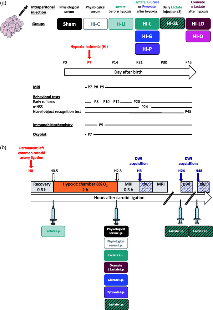
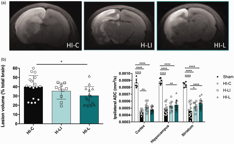
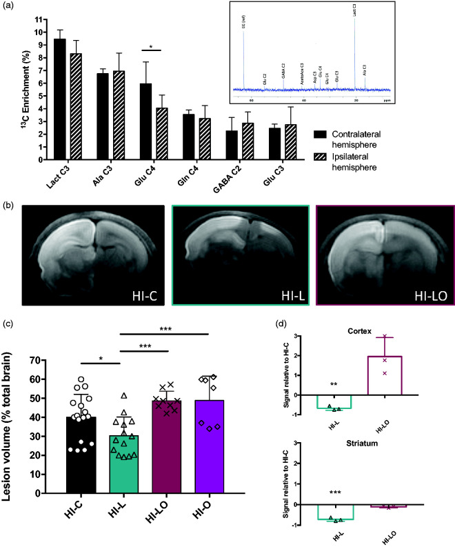
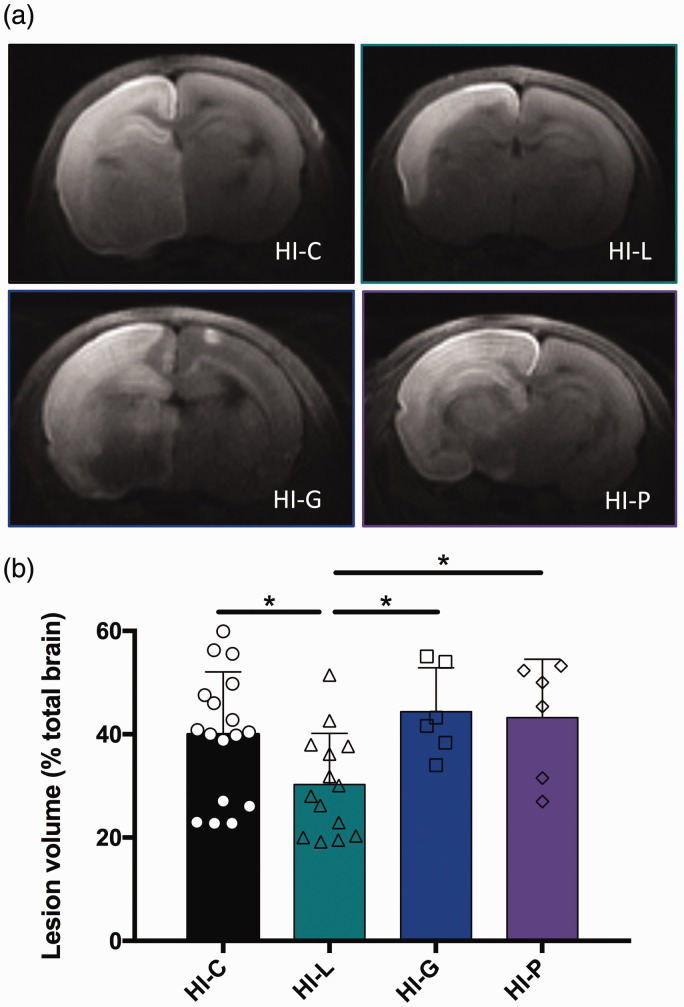
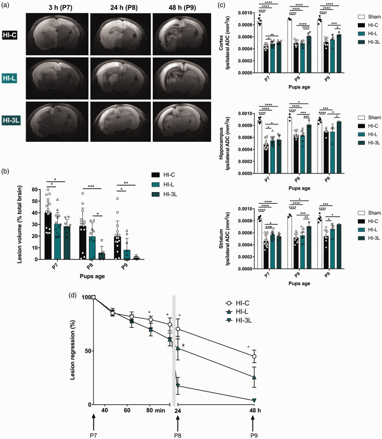
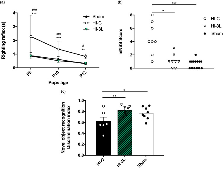
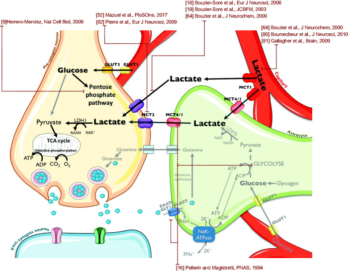
Hypoxic-ischemic (HI) encephalopathy remains a major cause of perinatal mortality and chronic disability in newborns worldwide (1-6 for 1000 births). The only current clinical treatment is hypothermia, which is efficient for less than 60% of babies. Mainly considered as a waste product in the past, lactate, in addition to glucose, is increasingly admitted as a supplementary fuel for neurons and, more recently, as a signaling molecule in the brain. Our aim was to investigate the neuroprotective effect of lactate in a neonatal (seven day old) rat model of hypoxia-ischemia. Pups received intra-peritoneal injection(s) of lactate (40 μmol). Size and apparent diffusion coefficients of brain lesions were assessed by magnetic resonance diffusion-weighted imaging. Oxiblot analyses and long-term behavioral studies were also conducted. A single lactate injection induced a 30% reduction in brain lesion volume, indicating a rapid and efficient neuroprotective effect. When oxamate, a lactate dehydrogenase inhibitor, was co-injected with lactate, the neuroprotection was completely abolished, highlighting the role of lactate metabolism in this protection. After three lactate injections (one per day), pups presented the smallest brain lesion volume and a complete recovery of neurological reflexes, sensorimotor capacities and long-term memory, demonstrating that lactate administration is a promising therapy for neonatal HI insult.

Keywords: MRI; Neonatal hypoxia-ischemia; astrocyte to neuron lactate shuttle; lactate; neuroprotection.


Conflict of interest statement

Declaration of conflicting interests: The author(s) declared no potential conflicts of interest with respect to the research, authorship, and/or publication of this article.

Figures

Figure 1.
Figure 1.
Experimental design and groups. (a) Groups studied and chronology. (b) Time course between carotid artery ligation, hypoxia exposure and DWI acquisition. Nine different groups were studied; the sham group in which pups underwent only the exposition of the left common carotid artery (no HI insult), the HI-C group in which pups were exposed to the HI procedure (left common carotid artery ligation + 2 h hypoxia) without any treatment (intraperitoneal injection of physiological saline solution), the H-LI group in which pups received a single intraperitoneal lactate injection after the carotid artery ligation and before entering the hypoxic chamber, the HI-L group in which pups received a single intraperitoneal lactate injection when leaving the hypoxic chamber (corresponding to 150 min after the carotid artery ligation and 30 min before the DWI acquisition), the HI-G group in which pups received a single intraperitoneal glucose injection when leaving the hypoxic chamber,), the HI-P group in which pups received a single intraperitoneal pyruvate injection when leaving the hypoxic chamber, the HI-3L group in which pups received a daily post-HI injection during three days (when leaving the hypoxic chamber, then 24 h and 48 h after the HI insult) of lactate and the HI-LO group in which both lactate and oxamate (LDH inhibitor) were injected to the pups. HI-O group: HI pups, which received only oxamate.
Figure 2.
Figure 2.
Evaluation of a preventive or curative effect of lactate injection on neonate brain lesions and edema volumes 3 h after carotid artery ligation. Lactate was intraperitoneally injected before (H-LI group) or after (HI-L group) the brain insult. In the control group (HI-C), physiological serum was injected when leaving the hypoxic chamber. (a) Trace DWI of P7 brains (from HI-C, H-LI, and HI-L groups) performed at 4.7 T. Damages appeared as a hypersignal (decrease of water movement, which reflects cytotoxic edema). (b) Quantification of lesion volume (%, relative to total brain volume) for the different groups. (c) Quantification of the ipsilateral ADC in cortex, hippocampus and striatum for sham, HI-C, H-LI and HI-L groups. * P <0.05, ** P <0.01, **** P <0.0001 by one-way ANOVA, followed by Fisher’s LSD test.
Figure 3.
Figure 3.
Metabolic role of lactate in the neuroprotection. (a) 13C-NMR spectrum and 13C-specific enrichments (% of 13C-enriched molecules) of brain metabolites 30 min after [3-13C]lactate intraperitoneal injection. Results are mean values ± SD. * P <0.05 by one-way ANOVA, followed by Fisher’s LSD test. Insert: Typical 13C-NMR spectrum obtained from brain extract 30 min after i.p. injection of [3-13C]lactate. EG: ethylene glycol, internal reference; AcetoAce C3: acetaoacetate carbon 3; Glu C4, C3 or C2: glutamate carbon 4, 3 or 2; Gln C4, glutamine carbon 4; GABA C2: γ-aminobutyrate carbon 2; Asp C3: aspartate carbon 3; Lact C3: lactate carbon 3; Ala C3: alanine carbon 3. (b, c and d) Effect of oxamate, a lactate dehydrogenase (LDH) inhibitor on neonates’ brain lesions and edema volumes 3 h after the carotid artery ligation. In the HI-L group, lactate was intraperitoneally injected after the brain insult, while in the HI-LO group, both lactate and oxamate (a LDH inhibitor) were co-injected (in the HI-O group, only oxamate was injected). In the control group (HI-C), lactate was replaced by physiological serum. (b) Trace DWI of P7 brains (HI-C, HI-L, and HI-LO groups) obtained at 4.7 T. (c) Quantification of lesion volume for the different groups (%, relative to total brain volume). * P <0.05, *** P <0.001, by one-way ANOVA, followed by Fisher’s LSD test. (d) Quantification of oxiblots in cortex and striatum for HI-C, HI-L, and HI-LO groups (n = 3 per group). Results are mean values ± SEM. ** P <0.01, *** P <0.001, paired t-test.
Figure 4.
Figure 4.
Comparison of glucose, pyruvate and lactate injections on neonate brain lesions and edema volumes 3 h after carotid artery ligation. Lactate (HI-L), pyruvate (HI-P), glucose (HI-G) or physiological serum (HI-C) were intraperitoneally injected after the brain insult. (a) Trace DWI of P7 brains. Damages appeared as a hypersignal (decrease of water movement, which reflects edema). (b) Quantification of lesion volume (%, relative to total brain volume) for the different groups. * P <0.05, by one-way ANOVA, followed by Fisher’s LSD test.
Figure 5.
Figure 5.
Longitudinal study of the neuroprotective effect of lactate. (a) Brain trace DWI of P7, P8 and P9 pups of HI-C, HI-L and HI-3L groups. Images were obtained at 4.7 T, 3 h, 24 h and 48 h after the insult. (b) Quantification of lesion volumes (%, relative to total brain volume) and comparison between the control group, one injection of lactate and three injections of lactate (daily). (c) Quantification of ADC in cortical, hippocampal and striatal lesions at P7, P8 and P9. (d) Evaluation of lesion regression as a function of time (%, relative to original lesion size). Results are mean values ± SD. (b)(c) * P <0.05, ** P <0.01, *** P <0.001, by one-way ANOVA, followed by Fisher’s LSD test. (c) *statisticaly significant between HI-C and HI-L, °statisticaly significant between HI-C and HI-3L, #statisticaly significant between HI-L and HI-3L.
Figure 6.
Figure 6.
Effect of lactate therapy on neurological reflexes as well as juvenile somatosensory and memory capacities. (a) Performances on righting reflexes at P8, P10 and P12. Significantly different from sham group: ** P <0.01, *** P <0.001, significantly different from HI-3L: # P <0.05, ### P <0.001. (b) Modified neurological severity scores (mNSS) performed at P24. * P <0.05, *** P <0.001. (c) Novel object recognition test performed at P45. Results are mean values +/_ SEM. * P <0.05, ** P <0.01.
Figure 7.
Figure 7.
Mechanistic hypothesis of lactate neuroprotection on HI damages. Injected lactate could be taken from the bloodstream by astrocytes. Then, through the astrocyte-neuron lactate shuttle, lactate could be metabolized by neurons as an energy source to overcome the lack of glucose induced by HI insult. The small amount of available neuronal glucose could thus be saved to integrate the pentose phosphate pathway to maintain the Redox balance together with the NADH provided by lactate metabolism.

References

    1. Kurinczuk JJ, White-Koning M, Badawi N.Epidemiology of neonatal encephalopathy and hypoxic-ischaemic encephalopathy. Early Hum Develop 2010; 86: 329–338. - PubMed
    1. Davidson JO, Wassink G, van den Heuij LG, et al.. Therapeutic hypothermia for neonatal hypoxic-ischemic encephalopathy – where to from here? Front Neurol 2015; 6: 198. - PMC - PubMed
    1. Shalak L, Perlman JM.Hypoxic-ischemic brain injury in the term infant-current concepts. Early Hum Develop 2004; 80: 125–141. - PubMed
    1. Cotten CM, Shankaran S.Hypothermia for hypoxic-ischemic encephalopathy. Exp Rev Obstetr Gynecol 2010; 5: 227–239. - PMC - PubMed
    1. Allen KA, Brandon DH.Hypoxic ischemic encephalopathy: pathophysiology and experimental treatments. Newborn Infant Nursing Rev 2011; 11: 125–133. - PMC - PubMed

Publication types

LinkOut - more resources