. 2020 Mar 24;18(1):136.
doi: 10.1186/s12967-020-02309-9.

Pathogenesis of chronic rhinosinusitis with nasal polyps: role of IL-6 in airway epithelial cell dysfunction

Affiliations

Pathogenesis of chronic rhinosinusitis with nasal polyps: role of IL-6 in airway epithelial cell dysfunction

Emilie Bequignon et al. J Transl Med. .

Abstract

Background: Chronic rhinosinusitis with nasal polyps (CRSwNP) is characterized by an alteration in airway epithelial cell functions including barrier function, wound repair mechanisms, mucociliary clearance. The mechanisms leading to epithelial cell dysfunction in nasal polyps (NPs) remain poorly understood. Our hypothesis was that among the inflammatory cytokines involved in NPs, IL-6 could alter epithelial repair mechanisms and mucociliary clearance. The aim of this study was to evaluate the in vitro effects of IL-6 on epithelial repair mechanisms in a wound repair model and on ciliary beating in primary cultures of Human Nasal Epithelial Cells (HNEC).

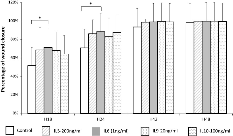
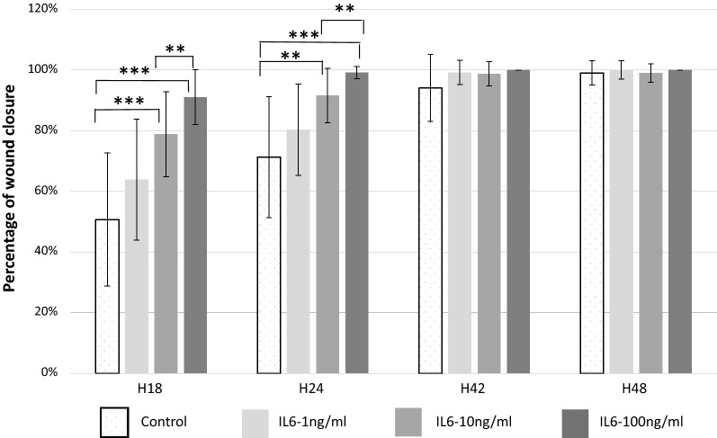
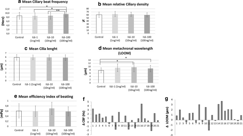
Methods: Primary cultures of HNEC taken from 38 patients during surgical procedures for CRSwNP were used in an in vitro model of wound healing. Effects of increasing concentrations of IL-6 (1 ng/mL, 10 ng/mL, and 100 ng/mL) and other ILs (IL-5, IL-9, IL-10) on wound closure kinetics were compared to cultures without IL-modulation. After wound closure, the differentiation process was characterized under basal conditions and after IL supplementation using cytokeratin-14, MUC5AC, and βIV tubulin as immunomarkers of basal, mucus, and ciliated cells, respectively. The ciliated edges of primary cultures were analyzed on IL-6 modulation by digital high-speed video-microscopy to measure: ciliary beating frequency (CBF), ciliary length, relative ciliary density, metachronal wavelength and the ciliary beating efficiency index.

Results: Our results showed that: (i) IL-6 accelerated airway wound repair in vitro, with a dose-response effect whereas no effect was observed after other ILs-stimulation. After 24 h, 79% of wounded wells with IL6-100 were fully repaired, vs 46% in the IL6-10 group, 28% in the IL6-1 group and 15% in the control group; (ii) specific migration analyses of closed wound at late repair stage (Day 12) showed IL-6 had the highest migration compared with other ILs (iii) The study of the IL-6 effect on ciliary function showed that CBF and metachronal wave increased but without significant modifications of ciliary density, length of cilia and efficiency index.

Conclusion: The up-regulated epithelial cell proliferation observed in polyps could be induced by IL-6 in the case of prior epithelial damage. IL-6 could be a major cytokine in NP physiopathology.

Keywords: Chronic rhinosinusitis with nasal polyps (CRSwNP); Epithelial cell; IL-6; IL-9; Inflammation; Interleukin 6; Mucociliary clearance; Nasal Polyps; Repair mechanisms; Wound healing.


Conflict of interest statement

The authors declare that they have no competing interests.

Figures

Fig. 1
Fig. 1
Study of the effect of ILs on wound closure. Comparison of the mean percentage of wound repair after wound healing of HNEC cultures (n = 11 patients) at each regular interval between control wells and each IL tested (IL-5, IL-6, IL-9, IL-10). *p value < 0.05
Fig. 2
Fig. 2
Study of IL6 dose–response effect on wound closure. Comparison of the mean percentage of wound repair after wound healing of HNEC cultures (n = 6 patients) at each regular interval between controls wells and each concentration of IL-6 tested: IL6-1, IL6-10; IL6-100 (1 ng/ml, 10 ng/ml, 100 ng/ml, respectively). *p value < 0.05; **p < 0.005; ***p < 0.0005
Fig. 3
Fig. 3
Study of the effect of ILs on the differentiation process after wound closure. a Diagram of well cutting and immunolabeling made in the three different thirds of wells (CK14 in the upper third, MUC5AC in the intermediate third and ATA in the lower third) and method of analysis: Effects of ILs on differentiation in HNEC cultures after wound closure (2 (F2), 7 (F7) and 12 (F12) days after wound closure) was characterized by immunolabeling: each well was separated in three areas for each immunolabeling marker before IL-staining in order to avoid mixed antibodies contaminations (by cutting each well in three areas): either with mouse monoclonal anti-cytokeratin 14 (CellMarque 314 M-14, 1/100) (picture a-1) as a basal cell marker, mouse monoclonal anti-mucin-5AC (Abcam ab3649, 1/200) (picture a-2) as a goblet cell marker, or mouse monoclonal anti-acetylated α-tubulin (Abcam ab24610, 1/500) (picture a-3) as a ciliated cell marker, before being revealed by a secondary goat anti-mouse Alexa Fluor- 488 antibody (Molecular Probes 1/500). Nuclei were stained by DAPI. In order to evaluate the “wound” effect and overcome the IL effect on healthy epithelium, we measured the ratio of the number of nuclei stained by DAPI or the percentage of cells positive for each immunolabeling (ATA, MUC5AC, and CK14) in two wounded areas on the number of nuclei for the DAPI in the corresponding two non-wounded control areas of the same well. be Comparison of ratios (percentage of positive cells in wounded area/Percentage of positive cells in non-wounded control areas) after 2, 7 and 12 days after wound closure between culture wells modulated by each IL (IL-5, IL-6, IL-9, IL-10) and not (controls wells). b Comparison of nuclei ratios (wounded areas/non-wounded areas) between culture wells with IL and without *p < 0.05. c Comparison of ciliated cell ratios (wounded areas/non-wounded areas) between culture wells with IL and without *p < 0.05. d Comparison of goblet cell ratios (wounded areas/non-wounded areas) between culture wells with IL and without *p < 0.05. e Comparison of basal cell ratios (wounded areas/non-wounded areas) between culture wells with IL and without *p < 0.05
Fig. 4
Fig. 4
Study of IL-6 effect on ciliary beating. Comparison of ciliary beating parameters between IL-6 groups at increased concentrations (IL6-1, IL6-10, IL6-100) (1 ng/ml, 10 ng/ml, 100 ng/ml respectively)) and control group after 21 days of stimulation by IL-6 in culture medium of HNEC (n = 21 controls wells and n = 21 wells with IL6-100 and n = 21 wells with IL6-10 and and n = 21 wells with IL6-1) (paired analysis): comparison of mean ciliary beat frequency (CBF) (in Herz) (a), mean relative ciliary density (b), mean cilia length (c), mean metachronal wavelength (LOOM) (in μm) (d), mean efficiency index of beating (in mPa) (e). f, g Focus of significant results of paired analysis: variation of ciliary beat frequency (CBF) between each well with IL-6 stimulation at 100 ng/ml and its respective control well (n = 21) (f) and focus on variation of metachronal wavelength (LOOM) between each well with IL-6 stimulation at 100 ng/ml and its respective control well (n = 21) (g)

Similar articles

Cited by

References

    1. Sharma R, Lakhani R, Rimmer J, Hopkins C. Surgical interventions for chronic rhinosinusitis with nasal polyps. Cochrane Database Syst Rev. 2014;11:006990. - PMC - PubMed
    1. Fokkens WJ, Lund VJ, Mullol J, Bachert C, Alobid I, Baroody F, et al. European position paper on rhinosinusitis and nasal Polyps 2012. Rhinol Suppl. 2012;23:3. - PubMed
    1. Wladislavosky-Waserman P, Kern EB, Holley KE, Eisenbrey AB, Gleich GJ. Epithelial damage in nasal polyps. Clin Allergy. 1984;14(3):241–247. doi: 10.1111/j.1365-2222.1984.tb02203.x. - DOI - PubMed
    1. Coste A, Rateau JG, Bernaudin JF, Peynegre R, Escudier E. Nasal polyposis pathogenesis: a flow cytometric and immunohistochemical study of epithelial cell proliferation. Acta Otolaryngol. 1996;116(5):755–761. doi: 10.3109/00016489609137920. - DOI - PubMed
    1. Coste A, Rateau JG, Roudot-Thoraval F, Chapelin C, Gilain L, Poron F, et al. Increased epithelial cell proliferation in nasal polyps. Arch Otolaryngol Head Neck Surg. 1996;122(4):432–436. doi: 10.1001/archotol.1996.01890160072013. - DOI - PubMed

Publication types