. 2020 May 15;390(2):111961.
doi: 10.1016/j.yexcr.2020.111961. Epub 2020 Mar 21.

N-terminal acetylation of actin by NAA80 is essential for structural integrity of the Golgi apparatus

Affiliations

N-terminal acetylation of actin by NAA80 is essential for structural integrity of the Golgi apparatus

Tobias B Beigl et al. Exp Cell Res. .

Abstract

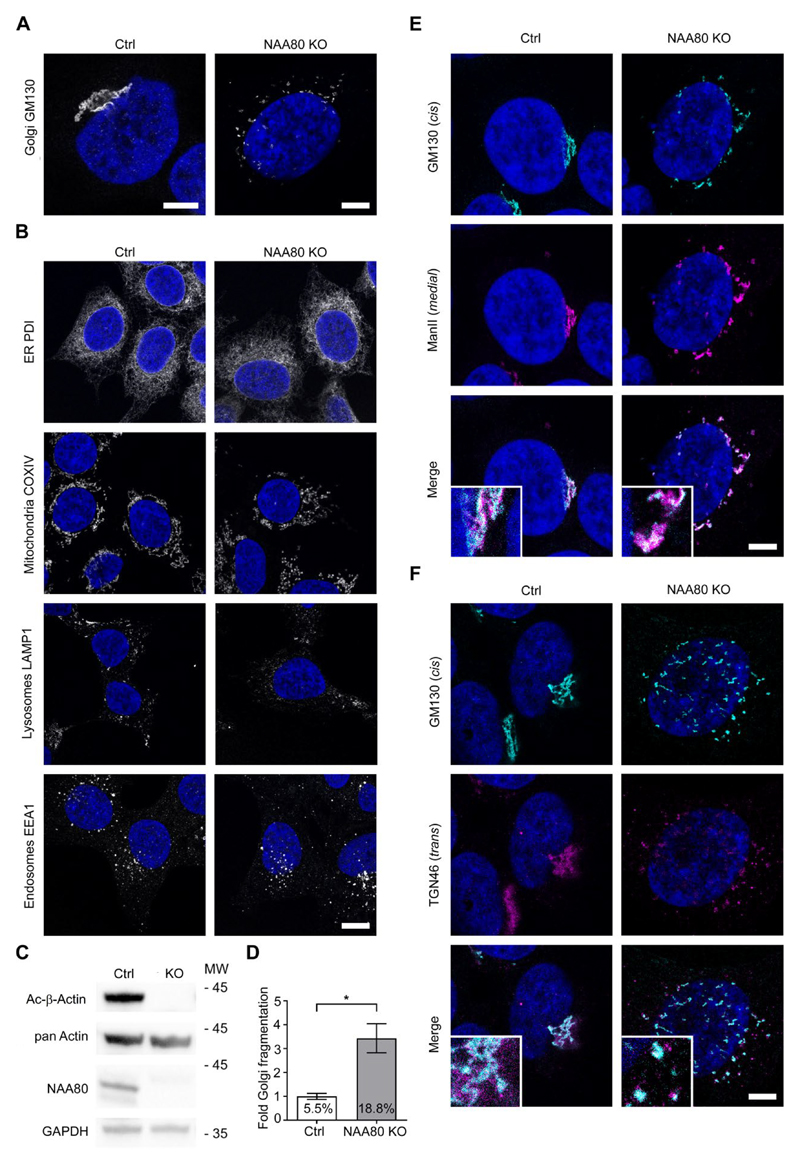
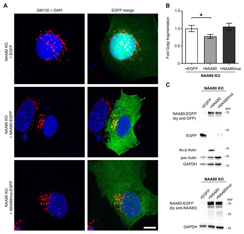
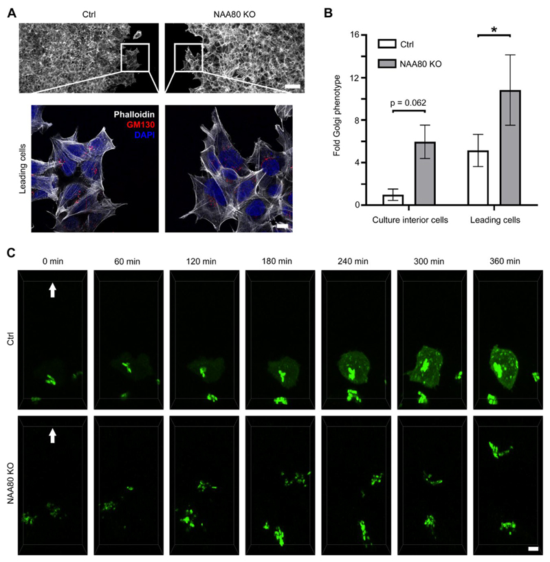
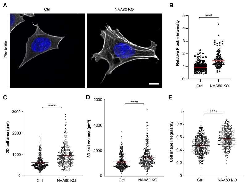
N-alpha-acetyltransferase 80 (NAA80) was recently demonstrated to acetylate the N-terminus of actin, with NAA80 knockout cells showing actin cytoskeleton-related phenotypes, such as increased formation of membrane protrusions and accelerated migration. Here we report that NAA80 knockout cells additionally display fragmentation of the Golgi apparatus. We further employed rescue assays to demonstrate that this phenotype is connected to the ability of NAA80 to modify actin. Thus, re-expression of NAA80, which leads to re-establishment of actin's N-terminal acetyl group, rescued the Golgi fragmentation, whereas a catalytic dead NAA80 mutant could neither restore actin Nt-acetylation nor Golgi structure. The Golgi phenotype of NAA80 KO cells was shared by both migrating and non-migrating cells and live-cell imaging indicated increased Golgi dynamics in migrating NAA80 KO cells. Finally, we detected a drastic increase in the amount of F-actin in cells lacking NAA80, suggesting a causal relationship between this effect and the observed re-organization of Golgi structure. The findings further underscore the importance of actin Nt-acetylation and provide novel insight into its cellular roles, suggesting a mechanistic link between actin modification state and Golgi organization.

Keywords: Actin cytoskeleton; Cell migration; Golgi structure; N-alpha-acetyltransferase 80 (NAA80); N-terminal acetylation; Posttranslational modification.


Conflict of interest statement

Declaration of competing interests The authors declare no competing or financial interests.

Figures

Fig. 1
Fig. 1. NAA80 knockout results in Golgi fragmentation.
A Confocal STED images showing the Golgi morphology in fixed HAP1 Ctrl and NAA80 KO cells labelled with GM130, a cis-Golgi protein. Nuclei were stained with DAPI. Representative images from three independent experiments, scale bars = 5 μm. B. Fixed HAP1 Ctrl and NAA80 KO cells were IF labelled with different subcellular markers as indicated in addition to DAPI nuclear stain. Representative confocal STED images from two independent experiments, scale bar = 10 μm. C. Western blot analysis of HAP1 Ctrl and NAA80 KO whole cell lysates showing the Nt-acetylation status of actin (Ac-β-actin) and detection of pan actin and NAA80. GAPDH was used as a loading control. One of three independent experiments is shown. D. Quantification of Golgi fragmentation in HAP1 NAA80 KO cells, expressed relative to Ctrl and with absolute % of fragmented Golgi complexes in the culture indicated. The mean ± sd from three independent experiments (each with n ≥ 500 cells) is shown. *p ≤ 0.05, unpaired t-test with Welch’s correction. E. HAP1 Ctrl and NAA80 KO cells co-stained for GM130 (cis-Golgi) and Mannosidase II (medial-Golgi). Confocal STED images out of two independent experiments are shown. Zoomed-in inset images are in 3x magnification, scale bar = 5 μm. F. As in E, but showing co-staining of GM130 (cis-Golgi) and TGN46 (trans-Golgi).
Fig. 2
Fig. 2. Restoring actin Nt-acetylation decreases Golgi fragmentation in NAA80 KO cells.
A. HAP1 NAA80 KO cells were transfected with either EGFP-control vector, NAA80-EGFP or catalytically dead NAA80mut-EGFP, fixed 24 h post transfection and IF stained for GM130. Representative confocal STED images from three independent experiments are shown; scale bar = 20 μm. B. Quantification of Golgi fragmentation in the transfected NAA80 KO cells shown in A. The mean ± sd of three independent experiments (each with n ≥ 200 cells) is shown; *p ≤ 0.05, one-way ANOVA with Tukey’s multiple comparison test. C. Western blot analysis of EGFP, actin Nt-acetylation (Ac-β-actin), pan actin and NAA80 in whole cell lysates of transfected NAA80 KO cells. Cells were transfected as in A and harvested 24 h post transfection. Representative blots from two independent experiments are shown.
Fig. 3
Fig. 3. Increased Golgi fragmentation in NAA80 KO cells is independent of cell motility and involves enhanced Golgi dynamics.
A. HAP1 Ctrl and NAA80 KO cells in a wound-healing assay, fixed 18 h post wounding, labelled with GM130 antibodies and stained with phalloidin. Confocal STED images show an overview of the wound edge (top, only phalloidin, 25x objective) and zoomed-in images showing the Golgi morphology in cells at the leading edge (100x objective). Representative images from three independent experiments are shown; scale bars correspond to 50 or 10 μm (overview or zoomed-in images, respectively). B. Quantification of Golgi fragmentation in the motile (leading edge) and relatively stationary (culture interior) cells from A. The mean ± sd from three independent experiments is shown, including n ≥ 200 cells per experiment. * p ≤ 0.05, one-way ANOVA with Tukey’s multiple comparison test. C. Selected images from live-cell recordings of HAP1 Ctrl or NAA80 KO cells transiently transfected with mannosidase II–mNeonGreen and migrating into the wound area during the total 6 h period of imaging. Cells were seeded 24 h and transfected 22 h prior to wound infliction and subsequent initiation of imaging. Shown are maximum projections of selected z-stacks (5 optical sections) acquired with spinning disk microscopy. Arrows point in the direction of cell movement. Representative cells at the very wound edge were chosen from a minimum of three independent experiments. Scale bar = 5 μm.
Fig. 4
Fig. 4. Increased level of F-actin and morphological differences in NAA80 KO cells.
A. HAP1 NAA80 KO and Ctrl cells were subjected to intra-well simultaneous fluorescent phalloidin staining and imaged with equal settings using a high-speed confocal spinning disk. Shown are representative cells for Ctrl and NAA80 KO imaged as z-stacks. DAPI was used to stain nuclei. Scale bar = 10 µm. B. The sum of F-actin intensity (based on the phalloidin staining) was measured in single HAP1 Ctrl and NAA80 KO cells from A. Shown are single values for the relative F-actin intensity normalised to the Ctrl. The mean is marked as a red line. A total of n ≥ 100 cells pooled out of three independent experiments are shown. **** p ≤ 0.0001, unpaired t-test with Welch’s correction. C-E. Morphological analysis measuring 2D cell area (C, 49 % increase), 3D cell volume (D, 32 % increase) and cell shape irregularity (E, 24 % increase) by live-cell holographic imaging using the HoloMonitor M4 system (see Methods). Shown is the mean (black line) with ± SEM (red lines) of n = 364 (NAA80 KO) and n = 383 (Ctrl) cells pooled together out of three independent experiments. **** p ≤ 0,0001, unpaired t-test with Welch’s correction.

Similar articles

Cited by

References

    1. Drazic A, et al. NAA80 is actin's N-terminal acetyltransferase and regulates cytoskeleton assembly and cell motility. Proc Natl Acad Sci U S A. 2018;115(17):4399–4404. - PMC - PubMed
    1. Goris M, et al. Structural determinants and cellular environment define processed actin as the sole substrate of the N-terminal acetyltransferase NAA80. Proc Natl Acad Sci U S A. 2018;115(17):4405–4410. - PMC - PubMed
    1. Wiame E, et al. NAT6 acetylates the N-terminus of different forms of actin. FEBS J. 2018;285(17):3299–3316. - PubMed
    1. Aksnes H, Ree R, Arnesen T. Co-translational, Post-translational, and Non-catalytic Roles of N-Terminal Acetyltransferases. Mol Cell. 2019;73(6):1097–1114. - PMC - PubMed
    1. Aksnes H, et al. First Things First: Vital Protein Marks by N-Terminal Acetyltransferases. Trends Biochem Sci. 2016;41(9):746–760. - PubMed

Publication types