. 2020 Mar 24;30(12):3981-3988.e3.
doi: 10.1016/j.celrep.2020.02.113.

Vitamin B12 Regulates Glial Migration and Synapse Formation through Isoform-Specific Control of PTP-3/LAR PRTP Expression

Affiliations

Vitamin B12 Regulates Glial Migration and Synapse Formation through Isoform-Specific Control of PTP-3/LAR PRTP Expression

Albert Zhang et al. Cell Rep. .

Abstract

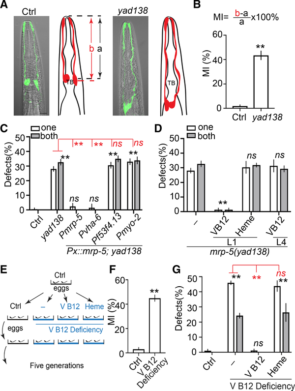
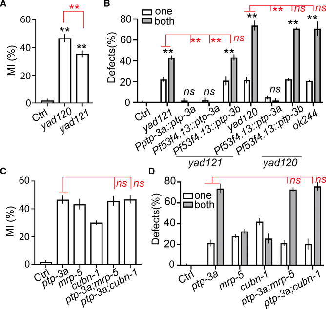
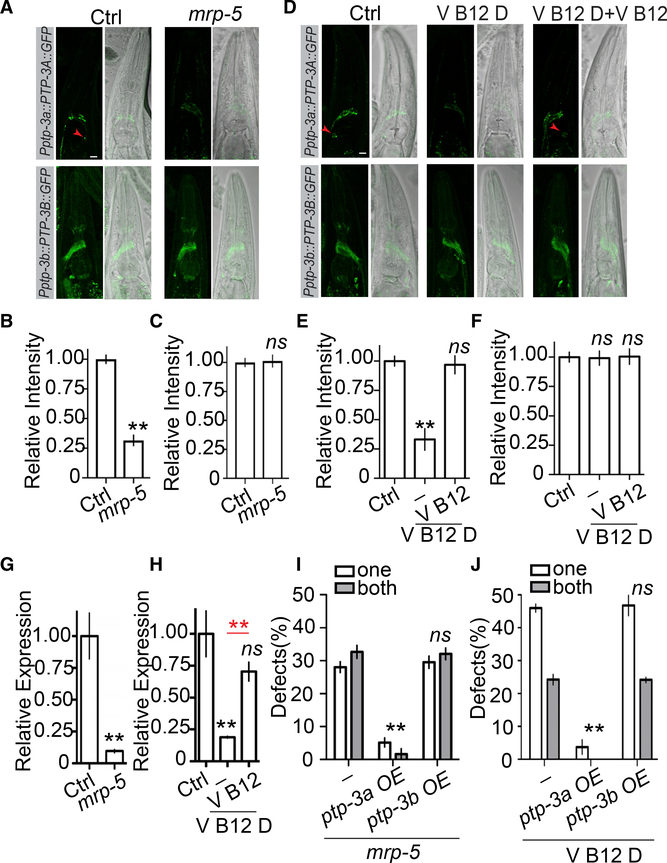
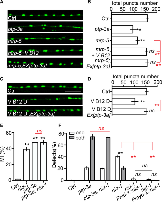
Vitamin B12 is known to play critical roles during the development and aging of the brain, and vitamin B12 deficiency has been linked to neurodevelopmental and degenerative disorders. However, the underlying molecular mechanisms of how vitamin B12 affects the development and maintenance of the nervous system are still unclear. Here, we report that vitamin B12 can regulate glial migration and synapse formation through control of isoform-specific expression of PTP-3/LAR PRTP (leukocyte-common antigen-related receptor-type tyrosine-protein phosphatase). We found the uptake of diet-supplied vitamin B12 in the intestine to be critical for the expression of a long isoform of PTP-3 (PTP-3A) in neuronal and glial cells. The expression of PTP-3A cell autonomously regulates glial migration and synapse formation through interaction with an extracellular matrix protein NID-1/nidogen 1. Together, our findings demonstrate that isoform-specific regulation of PTP-3/ LAR PRTP expression is a key molecular mechanism that mediates vitamin-B12-dependent neuronal and glial development.

Keywords: C. elegans; glial development; glial migration; neuronal development; synapse formation; vitamin B12.


Conflict of interest statement

Declaration of Interests The authors declare no competing interests.

Figures

Figure 1.
Figure 1.. Vitamin B12 Is Required for Terminating Glial Migration
(A) yad138 causes over-migration of AMsh glia. Confocal images and schematic representation of AMsh glia in control and mrp-5(yad138) animals expressing Pf53f4.13::GFP (yadIs48) are shown. TB, pharyngeal terminal bulb. Scale bar, 10 μm. (B) Quantification of Migration Index (MI) in control and mrp-5(yad138) animals. MI is calculated as (b − a)/a × 100%. As illustrated in (A), “a” represents the distance between the tip of nose and the center of the pharyngeal terminal bulb. “b” shows the distance between the tip of nose and the center of the AMsh cell bodies. (C) mrp-5 functions in the intestine to regulate Amsh migration. White and gray bars show the percentage of animals with over-migration defects in one AMsh or both AMsh glia, respectively. Pvha-6 is an intestine-specific promoter, Pf53f4.13 drives expression only in AMsh glia, and Pmyo-2 is a pharyngeal-muscle-specific promoter. (D) Injection of vitamin B12, but not heme, rescues mrp-5(lf) phenotypes. (E) A schematic showing how vitamin B12 deficiency was induced. (F) Quantification of MI of AMsh glia in animals cultured in control and vitamin-B12-deficient conditions. (G) Dietary supplementation of vitamin B12, but not heme, rescued AMsh migration defects in vitamin-B12-deficient conditions. Data show the percentage of animals with AMsh migration defects. In (B) and (F), data are represented as mean ± SEM. One-way ANOVA test; **p < 0.01; each point represents at least 30 worms. In (C), (D), and (G), data are represented as mean ± SEM. Two-way ANOVA test; **p < 0.01; ns, no significant difference. Each point represents three experiments of at least 50 worms each.
Figure 2.
Figure 2.. ptp-3a Functions in the Same Genetic Pathway as mrp-5 and Vitamin B12 in Regulating Glial Migration
(A) Quantification of MI in control, ptp-3a(yad120), and ptp-3a,b(yad121) animals. (B) ptp-3a, but not ptp-3b, regulates AMsh migration. White and gray bars show the percentage of animals with over-migration defects in one AMsh or both AMsh glia, respectively. Pf53f4.13 drives expression only in AMsh glia. (C and D) Double mutants of ptp-3a;mrp-5 and ptp-3a;cubn-1 display similar defects as in ptp-3a single mutants. Quantification of MI of AMsh glia (C) and the percentage of animals with over-migration defects (D) are shown. In (A) and (C), data are represented as mean ± SEM. One-way ANOVA test; **p < 0.01; each point represents at least 30 worms. In (B) and (D), data are represented as mean ± SEM. Two-way ANOVA test; **p < 0.01; each point represents three experiments of at least 50 worms each.
Figure 3.
Figure 3.. mrp-5 and Vitamin B12 Regulates AMsh Migration through Isoform-Specific Control of ptp-3a, but Not ptp-3b, Expression
(A–C) Loss offunction in mrp-5 decreases PTP-3A, but not PTP-3B, protein levels. Confocal images (A, left panel, GFP; right panel, differential interference contrast [DIC] merged with GFP; scale bar, 10 μm) and quantification (B and C) show the expression level of PTP-3A and PTP-3B in control and mrp-5(lf) animals. The red arrowheads point to AMsh cell bodies. (D–F) Vitamin B12 deficiency suppresses PTP-3A, but not PTP-3B, expression. Confocal images (D, left panel, GFP; right panel, DIC merged with GFP; scale bar, 10 μm) and quantification (E and F) show the expression level of PTP-3A and PTP-3B in control vitamin-B12-deficient and vitamin-B12- supplemented conditions. (G and H) Real-time PCR results show that loss of function in mrp-5 (G) or culturing animals in vitamin-B12-deficient conditions (H) decrease ptp-3a mRNA levels. A housekeeping gene, cdc-42, was used as an internal control. (I) Overexpression of ptp-3a, but not ptp-3b, in AMsh cells suppresses migration defects in mrp-5(lf) mutants. (J) Overexpression of ptp-3a, but not ptp-3b, in AMsh cells suppresses migration defects caused by vitamin B12 deficiency. In (B), (C), (E), and (F), data are represented as mean ± SEM. One-way ANOVA test; **p < 0.01; each point represents at least 20 worms. In (G)–(H), data are represented as mean ± SEM. One-way ANOVA test; **p < 0.01; each point represents three experimental replicates. In (I)–(J), data are represented as mean ± SEM. Two-way ANOVA test; **p < 0.01; each point represents three experiments of at least 50 worms each.
Figure 4.
Figure 4.. Vitamin B12 and mrp-5 Regulate Synapse Formation through ptp-3a, and the Interactions of nid-1 with ptp-3a Play an Important Role in Both Synapse Formation and Glial Migration
(A and B) mrp-5 regulates synapse formation through vitamin B12 and ptp-3a. Confocal images (A) show GABA neuron synapses visualized by SNB-1::GFP (juIs1) at the dorsal cord (scale bar, 10 μm), and quantification data (B) show the total SNB-1::GFP puncta number in the dorsal cord. (C and D) Vitamin B12 regulates synapse formation through ptp-3a. Confocal images (C) show GABA neuron synapses visualized by SNB-1::GFP (juIs1) at the dorsal cord (scale bar, 10 μm), and quantification data (D) show the total SNB-1::GFP puncta number in the dorsal cord. (E and F) nid-1 functions together with ptp-3a to regulate AMsh migration. (E) Quantification of MI in control, nid-1, ptp-3a(yad120), and nid-1;ptp-3a(yad120) animals. (F) The percentage of animals with over-migration defects. Pmyo-2 is a pharyngeal-muscle-specific promoter. In (B), (D), and (E), data are represented as mean ± SEM. One-way ANOVA test; **p < 0.01; each point represents at least 20 worms. In (F), data are represented as mean ± SEM. Two-way ANOVA test; **p < 0.01; each point represents three experiments of at least 50 worms each.

Similar articles

Cited by

References

    1. Ackley BD, Harrington RJ, Hudson ML, Williams L, Kenyon CJ, Chisholm AD, and Jin Y (2005). The two isoforms of the Caenorhabditis elegans leukocyte-common antigen related receptor tyrosine phosphatase PTP-3 function independently in axon guidance and synapse formation. J. Neurosci 25, 7517–7528. - PMC - PubMed
    1. Ahuja R, Yammani R, Bauer JA, Kalra S, Seetharam S, and Seetharam B (2008). Interactions of cubilin with megalin and the product of the amnion-less gene (AMN): effect on its stability. Biochem. J 410, 301–308. - PubMed
    1. Bacaj T, Tevlin M, Lu Y, and Shaham S (2008). Glia are essential for sensory organ function in C. elegans. Science 322, 744–747. - PMC - PubMed
    1. Barres BA (2008). The mystery and magic of glia: a perspective on their roles in health and disease. Neuron 60, 430–440. - PubMed
    1. Bito T, Matsunaga Y, Yabuta Y, Kawano T, and Watanabe F (2013). Vitamin B12 deficiency in Caenorhabditis elegans results in loss of fertility, extended life cycle, and reduced lifespan. FEBS Open Bio 3, 112–117. - PMC - PubMed

Publication types

MeSH terms

Substances