Review
. 2020 May;26(3):160-167.
doi: 10.5152/dir.2019.19391.

Abdominal and pelvic radiographs of medical devices and materials- part 2: neurologic and genitourinary devices and materials

Affiliations
Review

Abdominal and pelvic radiographs of medical devices and materials- part 2: neurologic and genitourinary devices and materials

Rishi Philip Mathew et al. Diagn Interv Radiol. 2020 May.

Abstract

Radiographs of the abdomen and pelvis are routinely obtained as a standard part of clinical care for the abdomen and pelvis. Brisk advances in technology over the last few decades have resulted in a multitude of medical devices and materials. Recognizing and evaluating these devices on abdominal and pelvic radiographs are critical, yet increasingly a difficult endeavor. In addition, multiple devices serving different purposes may have a similar radiographic appearance and position causing confusion for the interpreting radiologist. The role of the radiologist is to not only identify accurately these medical objects, but also to confirm for their accurate placement and to recognize any complications that could affect patient care, management or even be potentially life threatening. An extensive online search of literature showed our review article to be the most comprehensive work on medical devices and materials of the abdomen and pelvis, and in this second part of our two-part series, we discuss in depth about the neurologic and genitourinary devices seen on abdominal and pelvic radiographs.


Conflict of interest statement

Conflict of interest disclosure

The authors declared no conflicts of interest.

Figures
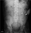
Figure 1
Figure 1
AXR shows a spinal cord stimulator placed for chronic back pain. Note its various parts: the pulse generator (thick arrow), its wire lead (arrowhead) and metallic contact points (thin arrows).
Figure 2
Figure 2
AXR shows a sacral nerve stimulator placed in a patient with a neurogenic bladder. Note its various parts: the pulse generator (arrowhead), its wire lead (thick arrow), and quadripolar electrodes (thin arrow).
Figure 3. a–c
Figure 3. a–c
Frontal radiograph (a) of the head and neck of a basic VP shunt series shows the proximal catheter (thick arrow) exiting the right lateral ventricle. Chest radiograph (b) of a basic VP shunt series shows the traversing shunt catheter (arrowheads). Abdominal radiograph (c) of a basic VP shunt series shows the distal catheter (arrow) in the right lower pelvis.
Figure 4
Figure 4
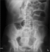
AXR shows a lumboperitoneal (LP) shunt. Note the lumbar catheter (thin arrow) enters the CSF space at L4 level, while the peritoneal catheter (thick arrow) is in the left lower quadrant.
Figure 5
Figure 5
AXR shows an epidural catheter with its tip in the epidural space (arrow).
Figure 6. a, b
Figure 6. a, b
AXR (a) shows an IsoMed baclofen pump (arrowhead) with its infusion port (thin arrow) accurately positioned for infusion of the drug (baclofen). AXR (b) of the same patient taken a few years later shows that the IsoMed baclofen pump has been replaced by a programmable SynchroMed pump (thick arrow). Its infusion port is satisfactorily positioned for infusion of the drug (baclofen).
Figure 7
Figure 7
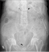
AXR shows a malpositioned baclofen pump that has flipped subcutaneously. Note its infusion port (thin arrow) is oriented medially preventing infusion of the drug. An intrauterine contraceptive device (IUCD) (thick arrow) is noted within the pelvis.
Figure 8
Figure 8
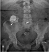
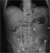
AXR shows a left nephrostomy tube (arrowhead) and a right double J (DJ) stent with its proximal (thin arrow) and distal (thick arrow) ends accurately reconstituted in the right renal pelvis and bladder respectively.
Figure 9
Figure 9
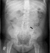
AXR shows bilateral ureteric stents placed into an ileal conduit. Note that the right ureteric stent (thin arrow) is accurately positioned in the right renal pelvis, while the left ureter stent (thick arrow) has migrated distally into the proximal ureter.
Figure 10
Figure 10
Pelvic radiograph shows a urinary bladder catheter (arrow) malpositioned in the urethra.
Figure 11
Figure 11
Pelvic radiograph shows a suprapubic catheter (arrow).
Figure 12. a–c
Figure 12. a–c
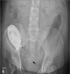
AXR (a) shows a continuous ambulatory peritoneal dialysis (CAPD) catheter with its tip (arrow) accurately positioned in the lower pelvis. AXR (b) shows a CAPD catheter with its tip (arrow) malpositioned in the right lower quadrant. AXR (c) shows a kinked (arrow) CAPD catheter making it nonfunctional.
Figure 13. a–c
Figure 13. a–c
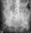
Pelvic radiograph shows a Mirena IUCD. Pelvic radiograph (b) shows a Multiload- 375 IUCD. Pelvic radiograph (c) shows a Lippes loop IUCD.
Figure 14. a, b
Figure 14. a, b
Pelvic radiograph (a) shows an IUCD after initial placement. Pelvic radiograph (b) of the same patient taken 2 years later shows that the IUCD has perforated and migrated and was confirmed on ultrasound to be extrauterine in location.
Figure 15. a, b
Figure 15. a, b
AXR (a) shows a Filshie clip in the left lower pelvis (arrow). Chest radiograph (b) of the same patient shows that the right Filshie clip (arrow) had migrated to the right upper quadrant and sits just under the right hemidiaphragm.
Figure 16
Figure 16
Pelvic radiograph shows a menstrual cup (arrow).
Figure 17. a–c
Figure 17. a–c
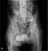
Pelvic radiograph (a) shows an accurately positioned vaginal Gellhorn pessary (arrow). Pelvic radiograph (b) shows a vaginal ring pessary. Pelvic radiograph (c) shows a malpositioned vaginal Gellhorn pessary (arrow).
Figure 18. a, b
Figure 18. a, b
Pelvic radiograph shows a vaginal tampon (arrow). Sagittal reformatted CT image (b) of the same patient shows the vaginal tampon (thick arrow) and its string (thin arrow).
Figure 19. a–c
Figure 19. a–c
Pelvic radiograph (a) shows the two metallic flexible rods (thin arrows) of a malleable penile prosthesis. Pelvic radiograph (b) shows an inflatable penile prosthesis (IPP). Note that only the metallic rear tip extender (thin arrows) of the IPP is visible on the radiograph. Coronal reformatted CT image (c) of the same patient shows the silicon tubes filled with saline (thick arrows) and the metallic rear tip extender (thin arrow) of the inflatable penile prosthesis.
Figure 20. a, b
Figure 20. a, b
Pelvic radiograph (a) shows prostate brachytherapy seeds. Chest radiograph (b) of the same patient taken 2 years later shows one of the brachy therapy seed has migrated into the left lung (arrow).

Similar articles

Cited by

References

    1. Kalia V, Bizzell C, Obray R, Obray J, Lamer T, Carrino JA. Spinal cord stimulation: the types of neurostimulation devices currently being used, and what radiologists need to know when evaluating their appearance on imaging. Curr Probl Diagn Radiol. 2010;39:227–233. doi: 10.1067/j.cpradiol.2009.07.010. - DOI - PubMed
    1. Gahzi AA, Banakhar MA, Elterman DS, Hassouna M. Radiographic position of the electrode as a predictor of the outcome of interstim therapy. Int Neurourol J. 2017;21:289–294. doi: 10.5213/inj.1734942.471. - DOI - PMC - PubMed
    1. Wallace AN, McConathy J, Menias CO, Bhalla S, Wippold FJ., 2nd Imaging evaluation of CSF shunts. AJR Am J Roentgenol. 2014;202:38–53. doi: 10.2214/AJR.12.10270. - DOI - PubMed
    1. Al-Rashed S, Kareem H, Kalra N, D’Antona L, Obeidat M, Patel B, Toma A. Lumboperitoneal shunt insertion without fluoroscopy guidance: Accuracy of placement in a series of 107 procedures. Version 2. F1000Res. 2017;6:565. doi: 10.12688/f1000research.11089.1. - DOI - PMC - PubMed
    1. Sbardelotto C, Yoshimi MM, da Pereira RR, de Castro RA. Breakage of a catheter in the epidural space. Rev Bras Anestesiol. 2008;58:643–650. doi: 10.1590/S0034-70942008000600009. - DOI - PubMed

MeSH terms

LinkOut - more resources