. 2020 Mar 24;11(2):e00349-20.
doi: 10.1128/mBio.00349-20.

Lipid Anchoring of Archaeosortase Substrates and Midcell Growth in Haloarchaea

Affiliations

Lipid Anchoring of Archaeosortase Substrates and Midcell Growth in Haloarchaea

Mohd Farid Abdul-Halim et al. mBio. .

Abstract

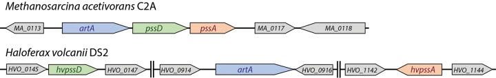
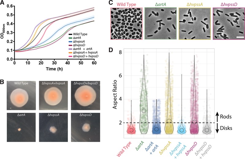
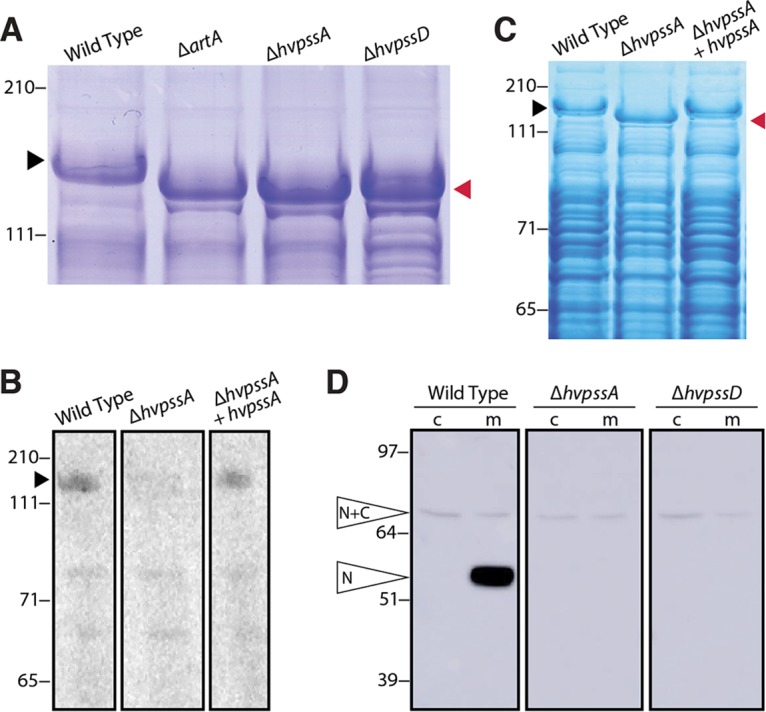
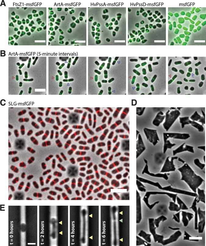
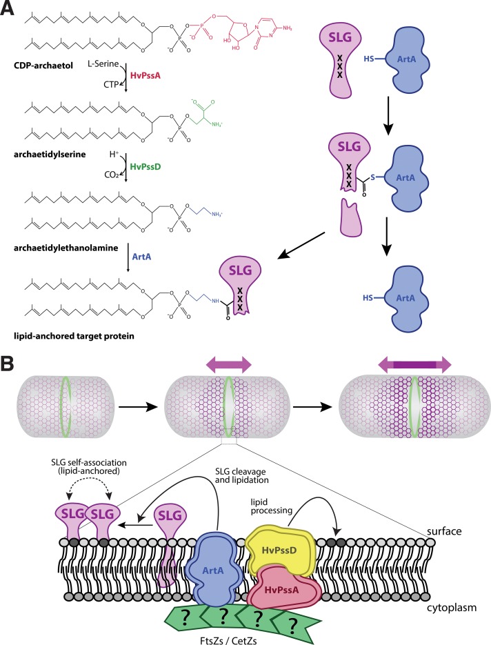
The archaeal cytoplasmic membrane provides an anchor for many surface proteins. Recently, a novel membrane anchoring mechanism involving a peptidase, archaeosortase A (ArtA), and C-terminal lipid attachment of surface proteins was identified in the model archaeon Haloferax volcanii ArtA is required for optimal cell growth and morphogenesis, and the S-layer glycoprotein (SLG), the sole component of the H. volcanii cell wall, is one of the targets for this anchoring mechanism. However, how exactly ArtA function and regulation control cell growth and morphogenesis is still elusive. Here, we report that archaeal homologs to the bacterial phosphatidylserine synthase (PssA) and phosphatidylserine decarboxylase (PssD) are involved in ArtA-dependent protein maturation. Haloferax volcanii strains lacking either HvPssA or HvPssD exhibited motility, growth, and morphological phenotypes similar to those of an ΔartA mutant. Moreover, we showed a loss of covalent lipid attachment to SLG in the ΔhvpssA mutant and that proteolytic cleavage of the ArtA substrate HVO_0405 was blocked in the ΔhvpssA and ΔhvpssD mutant strains. Strikingly, ArtA, HvPssA, and HvPssD green fluorescent protein (GFP) fusions colocalized to the midcell position of H. volcanii cells, strongly supporting that they are involved in the same pathway. Finally, we have shown that the SLG is also recruited to the midcell before being secreted and lipid anchored at the cell outer surface. Collectively, our data suggest that haloarchaea use the midcell as the main surface processing hot spot for cell elongation, division, and shape determination.IMPORTANCE The subcellular organization of biochemical processes in space and time is still one of the most mysterious topics in archaeal cell biology. Despite the fact that haloarchaea largely rely on covalent lipid anchoring to coat the cell envelope, little is known about how cells coordinate de novo synthesis and about the insertion of this proteinaceous layer throughout the cell cycle. Here, we report the identification of two novel contributors to ArtA-dependent lipid-mediated protein anchoring to the cell surface, HvPssA and HvPssD. ArtA, HvPssA, and HvPssD, as well as SLG, showed midcell localization during growth and cytokinesis, indicating that haloarchaeal cells confine phospholipid processing in order to promote midcell elongation. Our findings have important implications for the biogenesis of the cell surface.

Keywords: Haloferax volcanii; S-layer; archaea; archaeosortase; cell division; cell elongation; cell shape; cell surface; haloarchaea; lipid anchoring.


Figures

FIG 1
FIG 1
Schematic representation of artA, pssA, and pssD distribution across Euryarchaeota. Shown are M. acetivorans (top) and H. volcanii (bottom) genomic organization of artA and genes encoding homologs to PssA (HvPssA) and PssD (HvPssD). The M. acetivorans artA, pssA, and pssD gene lengths are 834 bp, 627 bp, and 744 bp, respectively. The H. volcanii artA, hvpssA, and hvpssD gene lengths are 912 bp, 672 bp, and 606 bp, respectively.
FIG 2
FIG 2
Absence of HvPssA or HvPssD leads to defects in growth, cell morphology, and motility. (A) Wild-type (strain H53) and ΔartA, ΔhvpssA, and ΔhvpssD mutant cells were grown with shaking in 96-well plates with a total volume of 200 μl of liquid semidefined CA medium, and the growth of six biological replicates was monitored at the OD600, with recordings taken every 30 min. For complementation analysis, artA, hvpssA, or hvpssD was expressed from pTA963 under the tryptophan-inducible p.tna promoter. The wild-type and deletion strains were transformed with an empty pTA963 plasmid as a control. (B) The wild-type (strain H53) and artA, hvpssA, or hvpssD deletion and complementation strains from individual colony on solid agar plates were individually stab inoculated with a toothpick into semisolid 0.35% agar in CA medium supplemented with tryptophan, followed by incubation at 45°C. (C) Phase-contrast images were taken from wild-type and mutant cells during mid-exponential-growth phase (OD600, 0.3) and immobilized under 0.5% agarose pads. (D) Violin distributions of aspect ratio measurements from single cells. Rodlike (aspect ratio, >2) cells were prevalent to disklike (aspect ratio, <2) cells in the mutant strains compared to the wild type. Biological replicates were collected on three different days, and data were analyzed from >1,000 cells under each condition by automated image segmentation. Scale bars = 5 μm.
FIG 3
FIG 3
HvPssA and HvPssD are critical for HVO_0405 C-terminal processing and SLG lipidation. (A) Coomassie-stained LDS-PAGE gel of cell extracts from H. volcanii H53 (wild-type [WT]) and ΔartA, ΔhvpssA, and ΔhvpssD mutant strains. The ΔartA, ΔhvpssA, and ΔhvpssD mutant SLG (red arrowhead) exhibited a mobility shift compared to the WT SLG (black arrowhead). (B) Fluorography of protein extracts isolated from H53 (WT), ΔhvpssA mutant, and hvpssA complementation (ΔhvpssA + hvpssA) cells grown in the presence of 1 μCi/ml [14C]mevalonic acid. Significant labeling of SLG (black arrowhead) is only detected in the WT and hvpssA complementation (ΔhvpssA + hvpssA) extracts. (C) Coomassie staining of the gel used for fluorography. The SLG mobility shift in the ΔhvpssA mutant (red arrowhead) is reverted upon hvpssA expression in trans. (D) Western blot analysis of cytoplasmic (c) and membrane (m) fractions of H53 (WT), ΔhvpssA mutant, and ΔhvpssD mutant strains expressing, in trans, HVO_0405-6×His. The N-terminal domain of HVO_0405 was detected using anti-HVO_0405-N-term antibodies. Hvo_0405 not processed by ArtA and the N-terminal HVO_0405 processed by ArtA are labeled “N+C” and “N,” respectively. The C-terminal domain, which carries a His tag, has not been analyzed in this experiment. Numbers indicate molecular mass in kilodaltons.
FIG 4
FIG 4
Midcell localization of the lipid-anchoring and processing machinery in H. volcanii. (A) Snapshots of merged phase-contrast (gray) and fluorescein isothiocyanate (FITC; green) channels of cells expressing FtsZ1-msfGFP, ArtA-msfGFP, HvPssA-msfGFP, HvPssD-msfGFP, and soluble msfGFP. Cells were immobilized under 0.5% agarose pads prepared with CA medium. (B) Time-lapse images of cells growing inside a CellASIC microfluidic device. Images of merged phase-contrast (gray) and FITC (green) channels were taken every 5 min for 12 h. Blue arrowheads indicate cell division events, while red arrowheads label one example of a cell elongating only after the arrival of ArtA-msfGFP to the midcell. (C) Snapshot of SLG-msfGFP (red) midcell localization. (D) Phase-contrast images of H. volcanii cells under prolonged overexpression (24 h) of the SLG-msfGFP fusion. (E) H. volcanii cells reshape and elongate preferentially at the midcell during protoplast recovery. Cells were loaded into the microfluidic chamber, and the S-layer was chemically removed by the addition of 1 mg/ml proteinase K and recovered with fresh medium (t = 0 h). Yellow arrowheads indicate the cell area extended until cell division (green arrowhead). Scale bars = 5 μm.
FIG 5
FIG 5
A model for lipid attachment and cell growth involving HvPssA, HvPssD, and ArtA. (A) In our speculative model, CDP-archaeol is converted to archaetidylethanolamine in two steps involving HvPssA and HvPssD. ArtA acts as a peptidase and covalently links its active-site cysteine to a newly generated C terminus of its target protein, simultaneously releasing the C-terminal peptide. Then, the free amino group of ethanolamine attacks the thiocarboxylate, which marks the covalent attachment of the target protein to the ArtA active-site cysteine. This results in covalent attachment of the lipid to the C terminus of the target protein as a carboxamide, simultaneously releasing ArtA. The process of cleavage and lipidation is dependent on HvPssA and HvPssD, either by binding of archaetidylethanolamine to ArtA or by protein-protein interaction between ArtA and HvPssA or HvPssD. (B) Recruitment of ArtA, HvPssA, or HvPssD to the midcell promotes anchoring of surface proteins and insertion of new SLG into the S-layer at midcell, contributing to cell elongation and division.

References

    1. Bisson-Filho AW, Zheng J, Garner E. 2018. Archaeal imaging: leading the hunt for new discoveries. Mol Biol Cell 29:1675–1681. doi: 10.1091/mbc.E17-10-0603. - DOI - PMC - PubMed
    1. de Pedro MA, Quintela JC, Höltje JV, Schwarz H. 1997. Murein segregation in Escherichia coli. J Bacteriol 179:2823–2834. doi: 10.1128/jb.179.9.2823-2834.1997. - DOI - PMC - PubMed
    1. May JW, Mitchison JM. 1986. Length growth in fission yeast cells measured by two novel techniques. Nature 322:752–754. doi: 10.1038/322752a0. - DOI
    1. Brown PJB, de Pedro MA, Kysela DT, Van der Henst C, Kim J, De Bolle X, Fuqua C, Brun YV. 2012. Polar growth in the alphaproteobacterial order Rhizobiales. Proc Natl Acad Sci U S A 109:1697–1701. doi: 10.1073/pnas.1114476109. - DOI - PMC - PubMed
    1. Sipiczki M, Takeo K, Grallert A. 1998. Growth polarity transitions in a dimorphic fission yeast. Microbiology 144:3475–3485. doi: 10.1099/00221287-144-12-3475. - DOI - PubMed

Publication types

MeSH terms

LinkOut - more resources