Review
. 2020 Mar 23;10(3):487.
doi: 10.3390/biom10030487.

TGF-β Signaling

Affiliations
Review

TGF-β Signaling

Kalliopi Tzavlaki et al. Biomolecules. .

Abstract

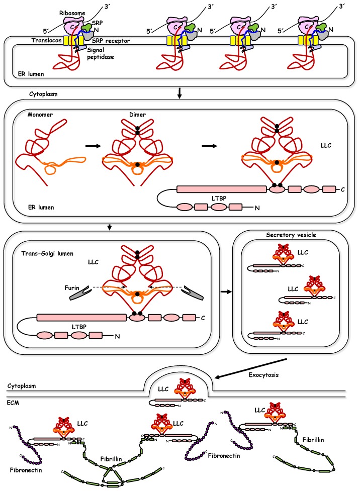
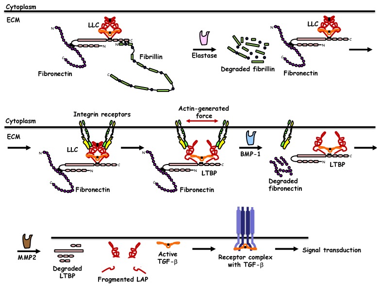
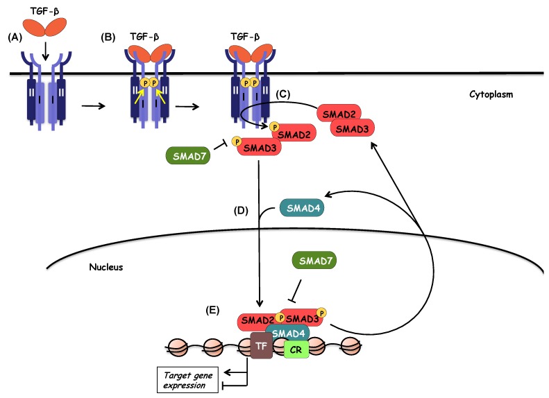
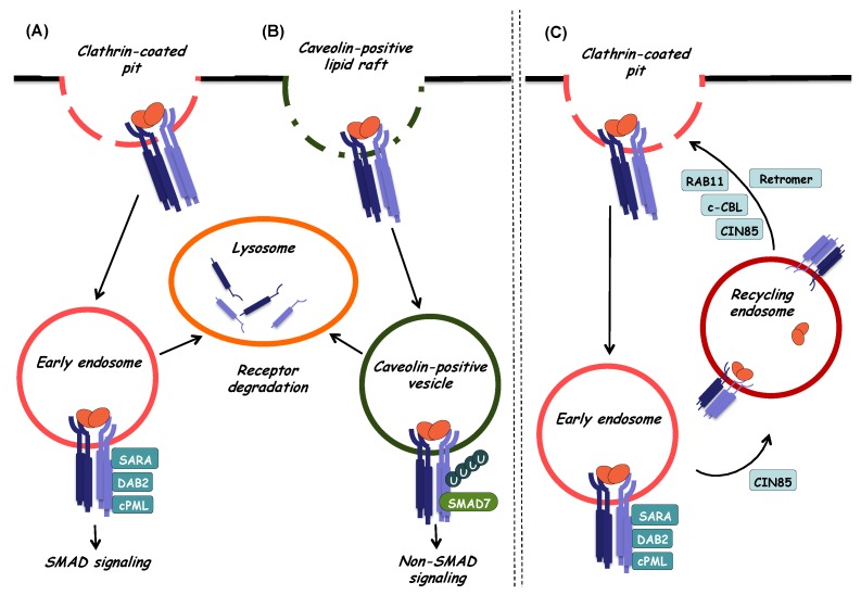
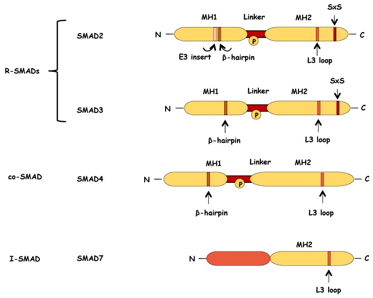
Transforming growth factor-β (TGF-β) represents an evolutionarily conserved family of secreted polypeptide factors that regulate many aspects of physiological embryogenesis and adult tissue homeostasis. The TGF-β family members are also involved in pathophysiological mechanisms that underlie many diseases. Although the family comprises many factors, which exhibit cell type-specific and developmental stage-dependent biological actions, they all signal via conserved signaling pathways. The signaling mechanisms of the TGF-β family are controlled at the extracellular level, where ligand secretion, deposition to the extracellular matrix and activation prior to signaling play important roles. At the plasma membrane level, TGF-βs associate with receptor kinases that mediate phosphorylation-dependent signaling to downstream mediators, mainly the SMAD proteins, and mediate oligomerization-dependent signaling to ubiquitin ligases and intracellular protein kinases. The interplay between SMADs and other signaling proteins mediate regulatory signals that control expression of target genes, RNA processing at multiple levels, mRNA translation and nuclear or cytoplasmic protein regulation. This article emphasizes signaling mechanisms and the importance of biochemical control in executing biological functions by the prototype member of the family, TGF-β.

Keywords: SMAD; extracellular matrix; phosphorylation; receptor serine/threonine kinase; signal transduction; transcription; transforming growth factor-β; ubiquitylation.


Conflict of interest statement

The authors declare no conflict of interest

Figures

Figure 1
Figure 1
Biosynthesis and extracellular deposition of transforming growth factor-β (TGF-β). A sequence of biochemical events is shown from the top left to the bottom, guided by black arrows. Ribosomes attached to the endoplasmic reticulum (ER) translate the TGF-β mRNA (black line 5′-3’) into TGF-β protein (red line with blue signal peptide). The signal peptide associates with the signal recognition protein (SRP), which associates with the SRP receptor and the translated polypeptide is transported through the translocon channel into the lumen of the ER where the signal peptidase cleaves the signal peptide, generating a pro-TGF-β monomer that folds in the lumen of the ER (red polypeptide corresponds to the N-terminal long polypeptide known as latency associated peptide (LAP) and orange polypeptide correspond to the mature C-terminal polypeptide). The architecture of pro-TGF-β follows the crystallographic structure of the molecule. Dimerization of pro-TGF-β takes place in the ER lumen via three disulfide bonds (black dots), two in the prodomain and one in the mature domain. The dimeric pro-TGF-β crosslinks via disulfide bonds (black dots) to the latent TGF-β binding protein (LTBP) in the ER lumen, forming the large latent complex (LLC). The LLC translocates from the ER lumen to the cis- (not shown) and then to the trans-Golgi cisternae. For simplicity, the prodomain N-linked glycosylation is not shown. In the trans-Golgi, furin protease cleaves at the junction of the prodomain with the mature domain (dotted arrows). The cleaved LLC accumulates in secretory vesicles that undergo exocytosis and secrete the LLC to the extracellular environment where the LLC incorporates into the matrix (ECM). LLC crosslinking to fibrillin (three disulfide bonds, black dots) and to fibronectin (three more disulfide bonds, black dots) is shown. All relevant proteins are drawn emphasizing their domain architecture, without clarifying the identity of each domain.
Figure 2
Figure 2
Activation of latent TGF-β. A sequence of biochemical events is shown from the top left to the bottom right, guided by black arrows. The large latent complex of TGF-β (LLC) deposited to the ECM via crosslinking to fibronectin and fibrillin is shown. Elastase proteolytically cleaves fibrillin. Integrin receptors on the plasma membrane associate with the RGD peptides (not shown) of the TGF-β prodomain. Integrin heterodimers of α- and β- integrin chains are shown in different color. Integrins, via their association to the actin cytoskeleton (not shown) exert force and change the conformation of the LLC prodomain, initiating the mature TGF-β release process. BMP-1 proteolytically cleaves fibronectin and MMP2 cleaves LTBP and the prodomain of TGF-β, generating fragmented prodomain, i.e., latency associated peptide (LAP), and releasing mature TGF-β. Active TGF-β associates with the signaling type II and type I receptors and initiates signal transduction. The role of coreceptors in ligand presentation is not shown.
Figure 3
Figure 3
The TGF-β/SMAD signaling pathway. During the first steps of TGF-β signaling, TGF-β ligand binds to a heteromeric complex of type II, and type I receptors (A). Upon ligand binding, type II receptor phosphorylates and activates type I receptor (B). Activated type I receptor in turn phosphorylates and activates the receptor-activated SMADs (R-SMADs), SMAD2 and SMAD3 (C). SMAD7 competes with R-SMADs for interacting with type I receptor, thus preventing R-SMAD activation and proper propagation of the signaling. Activated R-SMADs dissociate from type I receptors in order to form a complex with the common mediator SMAD4 (D). The trimeric complex translocates to the nucleus where it associates with high-affinity DNA binding transcription factors (TF) and chromatin remodeling proteins (CR) in order to positively or negatively regulate the transcription of target genes (E). SMAD7 can also inhibit the transcriptional activity of the nuclear SMAD complex.
Figure 4
Figure 4
Internalization and intracellular sorting of TGF-β receptors. Endocytosis and intracellular sorting of TGF-β receptors play an important role in the regulation of TGF-β signaling outcome and can be mediated via the two major endocytic pathways. (A) SMAD-dependent TGF-β signaling can initiate at the cell surface in clathrin-coated pits. When receptors internalize via clathrin-coated vesicles, they are directed to early endosomes. In these early stages of endocytosis, association of TGF-β receptors with SARA, cPML, and Dab-2 adaptor proteins, leads to the enhancement of TGF-β-induced SMAD activation and subsequent propagation of SMAD-dependent signaling. Internalized TGF-β receptors found in early endosomes can be sorted for degradation. In this case, they enter late endosomes (not shown) and finally reach lysosomes where degradation takes place. (B) Internalization of TGF-β receptors can also take place at caveolin-positive lipid raft compartments on the cell membrane, and in this case, internalized receptors enter caveolin-positive vesicles. There, the TGF-β type I receptor preferentially associates with SMAD7, which can control receptor turnover via recruitment of ubiquitin ligases and deubiquitylating enzymes thus regulating ubiquitylation and subsequent lysosomal degradation of receptors. Internalization of TGF-β receptors by lipid raft/caveolar-mediated endocytosis can also promote non-SMAD TGF-β signaling as SMAD7 competes with SMAD2/3 for interaction with the TGF-β type I receptor. (C) Once internalized via clathrin-coated pits, TGF-β receptors enter early endosomes. From there, receptors can be sorted to recycling endosomes in order to return back to the cell surface where they can respond again to ligand stimulation. The small GTPase RAB11, the adaptor protein CIN85, the ubiquitin ligase c-CBL and the retromer complex can facilitate recycling in different cell types.
Figure 5
Figure 5
Schematic representation of structure of the SMAD proteins. R-SMAD and SMAD4 proteins consist of two highly conserved domains, the MH1 and MH2 domains, which are separated by a non-conserved linker region. The N-terminal MH1 domain of R-SMADs contains a β-hairpin structure that is critical for DNA binding. In the case of SMAD2, the MH1 domain contains also an extra amino-acid sequence (E3 insert) that negatively regulates the DNA binding capacity of SMAD2. The C-terminal MH2 domain of R-SMADs contains an L3 loop that mediates the interaction between R-SMADs and the activated type I receptor. This L3 loop is also part of the SMAD4 structure and it is important for the formation of SMAD trimeric complexes. At their very C-terminus, R-SMADs, have a short conserved motif of two serines separated by one amino acid (Ser-X-Ser (SXS)) that are phosphorylated by the activated type I receptor, thus leading to the R-SMAD activation. The linker region encompasses multiple phosphorylation sites (P in circle), and it is targeted by various kinases that modulate SMAD stability and function. SMAD7, the inhibitory SMAD, retains the conserved MH2 domain but lacks the SXS motif at the C-terminus and the N-terminal region presents small similarity to the MH1 domain.
Figure 6
Figure 6
The TGF-β non-SMAD signaling pathways. (A) The ERK-MAP kinase pathway. First step for the activation of the TGF-β-induced ERK pathway is the phosphorylation of ShcA by the activated type I receptor. TGF-β-induced tyrosine phosphorylation of ShcA promotes formation of ShcA/Grb2/Sos complex and Ras activation. This leads to the sequential activation of Raf, MEK1/2 and finally ERK1/2. Activated ERK1/2 phosphorylate transcription factors (TF), thus contributing to TGF-β-induced transcriptional responses. Activated ERK1/2 can also phosphorylate SMADs at the linker region to regulate their activity. (B) The p38/JNK and NF-κB pathway (via TAK1). Upon ligand binding, TGF-β receptor complexes interact with TRAF6 promoting its autoubiquitylation. SMAD6 can inhibit TRAF6 ubiquitylation and activation by recruiting the deubiquitylating enzyme A20. TRAF6 activates TAK1 via Lys63-linked polyubiquitylation and activated TAK1 in turn activates through phosphorylation MAP kinase kinases (MKKs) MKK4, MKK3 and MKK6. MKKs activate their downstream kinases JNK and p38, which can then phosphorylate their target transcription factors (TF) in order to regulate transcription. SMAD7 enhances the activation of the p38 pathway as it acts as a scaffolding protein for TAK1, MKK3 and p38. Activated JNK and p38 phosphorylate also SMADs at the linker region, thus regulating SMAD-dependent transcriptional responses as well. Finally, TAK1 activates also IKK, which eventually leads to the activation of NF-κB signaling. (C) The PI3K/AKT/mTOR pathway. TGF-β promotes PI3K/AKT activation via direct interaction of the p85 subunit of PI3K (not shown) with TGF-β receptors. TGF-β-induced autoubiquitylation of TRAF6 results in recruitment and phosphorylation of AKT. TGF-β via PI3K, promotes also activation of mTORC2, which in turn can also phosphorylate and activate AKT promoting cell survival. Moreover, activated AKT prevents phosphorylation of SMAD3, thus attenuating SMAD3-dependent signaling. (D) TGF-β signaling by type I receptor intracellular domain signaling. The transmembrane metalloprotease TACE, promotes ectodomain cleavage of type I receptor, which is then followed by TRAF6-mediated ubiquitylation of the cytoplasmic domain of type I receptor and recruitment of presenilin-1 (PS1), part of the γ-secretase complex. PS1 proteolytically cleaves the ubiquitylated intracellular cytoplasmic domain of the receptor (TGFβRI ICD), which is released into the cytoplasm. Then, TGFβRI ICD translocates to the nucleus where it associates with other co-factors (not shown) and induces the expression of target genes. (E) MAP kinase pathway activation via TRAF4. The MAP kinase pathway can also be activated via TRAF4, another E3 ligase that upon ligand binding is recruited to the receptor complex, gets autoubiquitylated and then activates TAK1 via polyubiquitylation, eventually leading to activation of the p38 pathway. At the same time, TRAF4 targets SMURF2 for polyubiquitylation and subsequent degradation, thus contributing to the stability of TGF-β type I receptor. (F) The JAK-STAT pathway. STAT3 gets phosphorylated and activated by JAK (which interacts with the type I receptor) in response to TGF-β in order to regulate the expression of subset of TGF-β target genes. (G) The Rho-(like) GTPase pathway activation. TGF-β induces activation of RhoA GTPase (via both SMAD-independent and SMAD-dependent mechanisms), which eventually results in actin cytoskeleton reorganization and formation of stress fibers. Additionally, Par6, a regulator of cell polarity, once phosphorylated by TGF-β type II receptor, recruits Smurf1 E3 ligase that targets RhoA for degradation, eventually leading to tight junction dissociation. Upon TGF-β stimulation, Rho-like proteins Cdc42 and Rac1 are also activated and promote actin reorganization via activation of PAK2.

References

    1. Heldin C.H., Lu B., Evans R., Gutkind J.S. Signals and Receptors. Cold Spring Harb. Perspect. Biol. 2016;8:a005900. doi: 10.1101/cshperspect.a005900. - DOI - PMC - PubMed
    1. Derynck R., Miyazono K. The TGF-β Family. Cold Spring Harbor Laboratory Press; Woodbury, NY, USA: 2008. pp. 1–1114.
    1. Huminiecki L., Goldovsky L., Freilich S., Moustakas A., Ouzounis C.A., Heldin C.H. Emergence, development and diversification of the TGF-β signalling pathway within the animal kingdom. BMC Evol. Biol. 2009;9:28. doi: 10.1186/1471-2148-9-28. - DOI - PMC - PubMed
    1. Moustakas A., Heldin C.H. The regulation of TGFβ signal transduction. Development. 2009;136:3699–3714. doi: 10.1242/dev.030338. - DOI - PubMed
    1. Massagué J. TGFb signalling in context. Nat. Rev. Mol. Cell Biol. 2012;13:616–630. doi: 10.1038/nrm3434. - DOI - PMC - PubMed

Publication types

MeSH terms

LinkOut - more resources