Case Reports
. 2020 Mar 23;10(3):175.
doi: 10.3390/diagnostics10030175.

Acute Tentorial Subdural Hematoma Caused by Rupture of the Posterior Cerebral Artery after Minor Trauma-A Case Report

Affiliations
Case Reports

Acute Tentorial Subdural Hematoma Caused by Rupture of the Posterior Cerebral Artery after Minor Trauma-A Case Report

Urszula Maria Ciochon et al. Diagnostics (Basel). .

Abstract

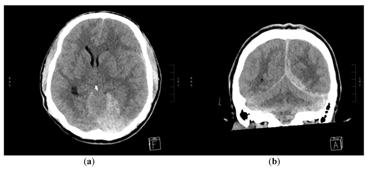
Acute subdural hematoma (aSDH) is a common pathology encountered after head trauma. Only a minority of aSDHs have an arterial source. In this article, we report a case of aSDH originating from a traumatic pseudoaneurysm of the distal segment of posterior cerebral artery (PCA), diagnosed several days after the initial minor trauma and successfully treated with endovascular coiling. This case emphasizes the importance of searching for vascular pathology when the localization, severity or relapsing course of the intracranial hemorrhage does not fully correspond to the severity of initial trauma and when the bleeding has a delayed onset. Characteristics, diagnostics and treatment possibilities of traumatic cerebral aneurysms, an important cause of arterial aSDH, are described in the article.

Keywords: acute subdural hematoma; endovascular coiling; pseudoaneurysm; traumatic brain aneurysm; traumatic brain injury.


Conflict of interest statement

The authors declare no conflict of interest.

Figures

Figure 1
Figure 1
Non-contrast CT scanning of the brain on the day of first admission. (a) Axial and (b) coronal multiplanar reconstruction (MPR) showing left-sided aSDH along tentorium cerebelli and posterior interhemispheric fissure. The right-sided epidural hematoma (EDH) is also seen on image (b).
Figure 2
Figure 2
Control non-contrast CT of the brain upon readmission. (a) Axial and (b) coronal MPR reconstruction showing increased mass effect from the growing left acute subdural hematoma (aSDH) with midline shift to the right side, obliteration of quadrigeminal and both ambient cisterns, compression of the left lateral ventricle and dilatation of the right ventricular trigonum. The right-sided EDH is not shown.
Figure 3
Figure 3
CT angiography of cerebral arteries. Axial maximum intensity projection (MIP) showing a spot sign close to the left tentorium cerebelli.
Figure 4
Figure 4
Digital subtraction angiography (DSA): (a) Injection in the right vertebral artery, anteroposterior (AP) view showing a pseudoaneurysm on a distal posterior cerebral artery (PCA) branch. (b) Zoomed post-embolization control AP image showing coils in the parent PCA segment and no filling of the pseudoaneurysm.

References

    1. Al-Mufti F., Mayer S. Neurocritical care of acute subdural hemorrhage. Neurosurg. Clin. N. Am. 2017;28:267–278. doi: 10.1016/j.nec.2016.11.009. - DOI - PubMed
    1. Maxeiner H., Wolff M. Pure subdural hematomas: A postmortem analysis of their form and bleeding points. Neurosurgery. 2002;50:503–508. doi: 10.1227/01.neu.0000279222.76375.d4. - DOI - PubMed
    1. Besenski N. Traumatic injuries: Imaging of head injuries. Eur. Radiol. 2002;12:1237–1252. doi: 10.1007/s00330-002-1355-9. - DOI - PubMed
    1. Murakami M., Kakita K., Hosokawa Y. Ruptured traumatic aneurysm after trivial injury mimicking acute spontaneous subdural hematoma - case report. Neurol. Med. Chir. (Tokyo) 2003;43:130–133. doi: 10.2176/nmc.43.130. - DOI - PubMed
    1. Holmes B., Harbaugh R.E. Traumatic intracranial aneurysms: A contemporary review. J. Trauma. 1993;35:855–860. doi: 10.1097/00005373-199312000-00009. - DOI - PubMed

Publication types

LinkOut - more resources