Review
. 2020 Mar 23;9(3):878.
doi: 10.3390/jcm9030878.

Natural History of Arrhythmogenic Cardiomyopathy

Affiliations
Review

Natural History of Arrhythmogenic Cardiomyopathy

Giulia Mattesi et al. J Clin Med. .

Abstract

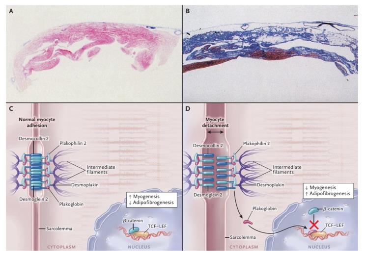
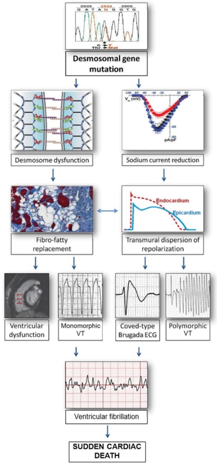
Arrhythmogenic cardiomyopathy (AC) is a heart muscle disease characterized by a scarred ventricular myocardium with a distinctive propensity to ventricular arrhythmias (VAs) and sudden cardiac death, especially in young athletes. Arrhythmogenic right ventricular cardiomyopathy (ARVC) represents the best characterized variant of AC, with a peculiar genetic background, established diagnostic criteria and management guidelines; however, the identification of nongenetic causes of the disease, combined with the common demonstration of biventricular and left-dominant forms, has led to coin the term of "arrhythmogenic cardiomyopathy", to better define the broad spectrum of the disease phenotypic expressions. The genetic basis of AC are pathogenic mutations in genes encoding the cardiac desmosomes, but also non-desmosomal and nongenetic variants were reported in patients with AC, some of which showing overlapping phenotypes with other non-ischemic diseases. The natural history of AC is characterized by VAs and progressive deterioration of cardiac performance. Different phases of the disease are recognized, each characterized by pathological and clinical features. Arrhythmic manifestations are age-related: Ventricular fibrillation and SCD are more frequent in young people, while sustained ventricular tachycardia is more common in the elderly, depending on the different nature of the myocardial lesions. This review aims to address the genetic basis, the clinical course and the phenotypic variants of AC.

Keywords: arrhythmogenic cardiomyopathy; sudden cardiac death.


Conflict of interest statement

The authors declared no potential conflicts of interest with respect to the research, authorship, and/or publication of this article

Figures

Figure 1
Figure 1
Histopathological Features and Pathogenesis of arrhythmogenic right ventricular cardiomyopathy (ARVC). With the azan trichrome stain, myocytes appear red, fibrous tissue appears blue, and fatty tissue appears white. Panel A shows a full-thickness histologic section (azan trichrome stain) of the anterior right ventricular wall in a normal heart; Panel B illustrates an analogous section from the heart of a patient with ARVC who died suddenly: Fibro-fatty tissue has replaced the muscular one. Desmosomes are not only structures supplying cell-cell adhesion, but they are also part of the Wnt–β-catenin signaling pathway, which suppresses the expression of adipogenic and fibrogenic genes (Panel C and D). Therefore, on one hand the impairment of desmosomal lead to detachment of cardiomyocytes (double-headed arrow), on the other in a gene transcriptional switch from myogenesis to adipogenesis and fibrogenesis (Panel C and D) [17]. Modified from Ref [4] with permission of the publisher.
Figure 2
Figure 2
Relationship between arrhythmogenic right ventricular cardiomyopathy (ARVC) and Brugada Syndrome. Mutant desmosomal proteins may induce potentially lethal ventricular arrhythmias by causing gap-junction remodeling and modifying the amplitude and kinetics of the sodium current, as a consequence of the cross-talk between these molecules at the intercalated discs. According to this view, Brugada syndrome and ARVC may share clinical features and arrhythmic mechanisms because of their common origin from the connexome, a coordinated network of proteins involving desmosomes, sodium channels, and gap-junction, aimed to control synergistically adhesion, excitability, and coupling of myocardial cells [18,19,20]. ECG = electrocardiogram; VT = ventricular tachycardia. Modified from Ref [20] with permission of the publisher.
Figure 3
Figure 3
Electrocardiogram of patient with desmoplakin (DSP)-related left dominant arrhythmogenic cardiomyopathy. Basal electrocardiogram showing T-wave inversion in lateral leads and low QRS voltages (<0.5 mV) in limb leads.
Figure 4
Figure 4
Cardiac magnetic resonance imaging of a patient with DSP-related left dominant arrhythmogenic cardiomyopathy. (A) End-diastolic frame of cine cardiac magnetic resonance sequence in long-axis four-chamber view showing fatty infiltration of the lateral wall of the left ventricle (red arrow). (B) Post-contrast image showing myocardial fibrosis in the form of extensive late gadolinium enhancement in the lateral wall and septum of the left ventricle (white arrows).

References

    1. Towbin J.A., McKenna W.J., Abrams D.J., Ackerman M.J., Calkins H., Darrieux F.C.C., Daubert J.P., De Chillou C., DePasquale E.C., Desai M.Y., et al. 2019 HRS Expert Consensus Statement on Evaluation, Risk Stratification, and Management of Arrhythmogenic Cardiomyopathy. Heart Rhythm. 2019;16:e301–e372. doi: 10.1016/j.hrthm.2019.05.007. - DOI - PubMed
    1. Thiene G., Nava A., Corrado D., Rossi L., Pennelli N. Right ventricular cardiomyopathy and sudden death in young people. N. Engl. J. Med. 1988;318:129–133. doi: 10.1056/NEJM198801213180301. - DOI - PubMed
    1. Marcus F.I., Fontaine G.H., Guiraudon G., Frank R., Laurenceau J.L., Malergue C., Grosgogeat Y. Right ventricular dysplasia: A report of 24 adult cases. Circulation. 1982;65:384–398. - PubMed
    1. Corrado D., Link M.S., Calkins H. Arrhythmogenic Right Ventricular Cardiomyopathy. N. Engl. J. Med. 2017;376:61–72. doi: 10.1056/NEJMra1509267. - DOI - PubMed
    1. Marcus F.I., McKenna W.J., Sherrill D., Basso C., Bauce B., Bluemke D.A., Calkins H., Corrado D., Cox M.G., Daubert J.P., et al. Diagnosis of arrhythmogenic right ventricular cardiomyopathy/dysplasia: Proposed modification of the task force criteria. Circulation. 2010;121:1533–1541. doi: 10.1161/CIRCULATIONAHA.108.840827. - DOI - PMC - PubMed

LinkOut - more resources