. 2020 Mar 24;10(1):5335.
doi: 10.1038/s41598-020-62091-y.

Sperm SPACA6 protein is required for mammalian Sperm-Egg Adhesion/Fusion

Affiliations

Sperm SPACA6 protein is required for mammalian Sperm-Egg Adhesion/Fusion

Sandrine Barbaux et al. Sci Rep. .

Abstract

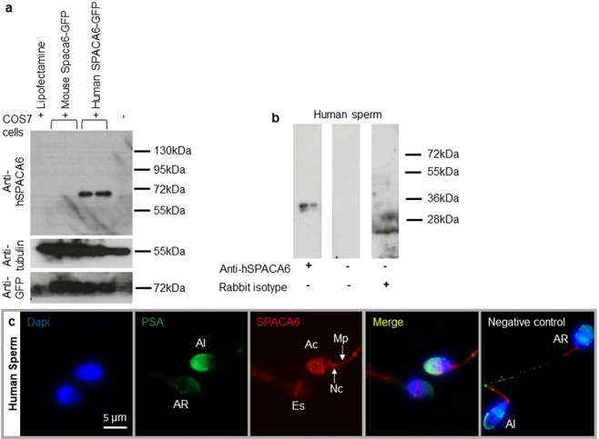
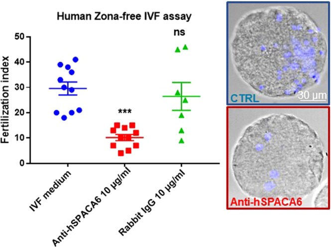
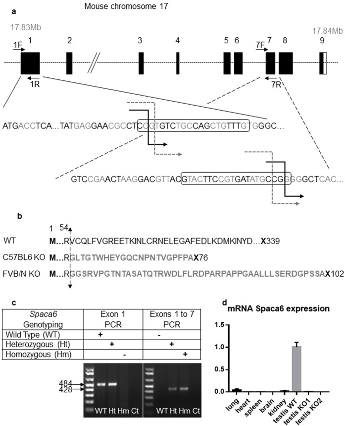
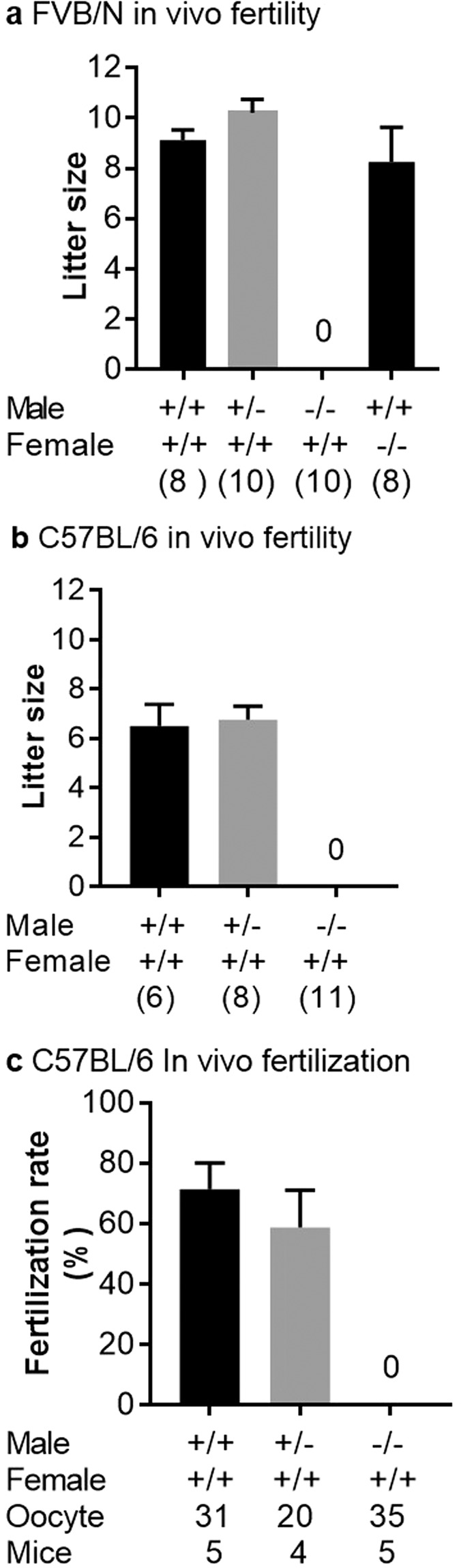
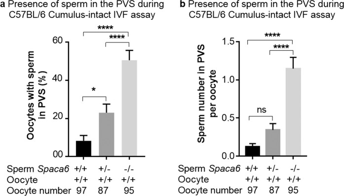
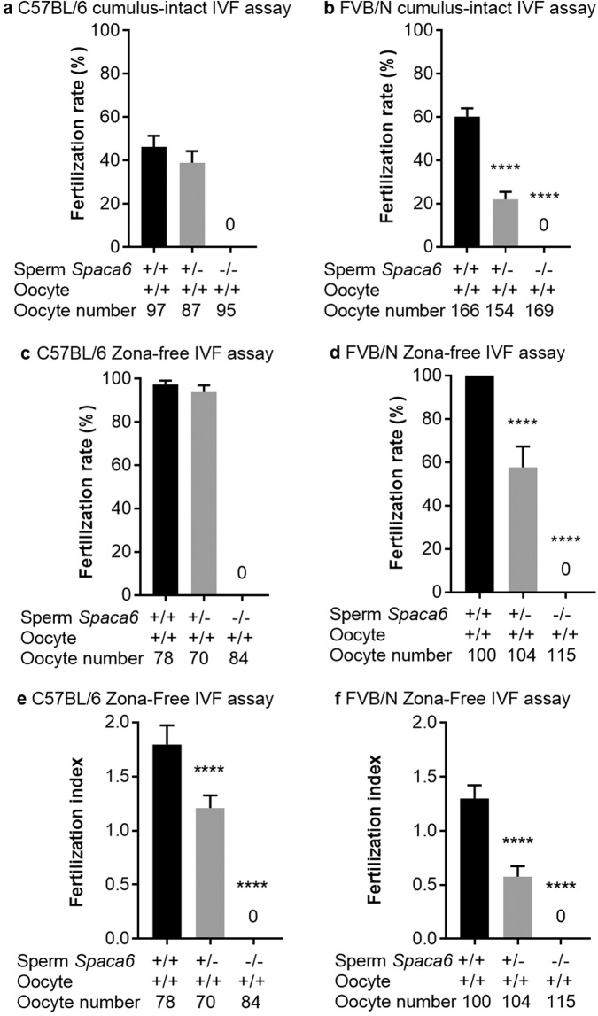
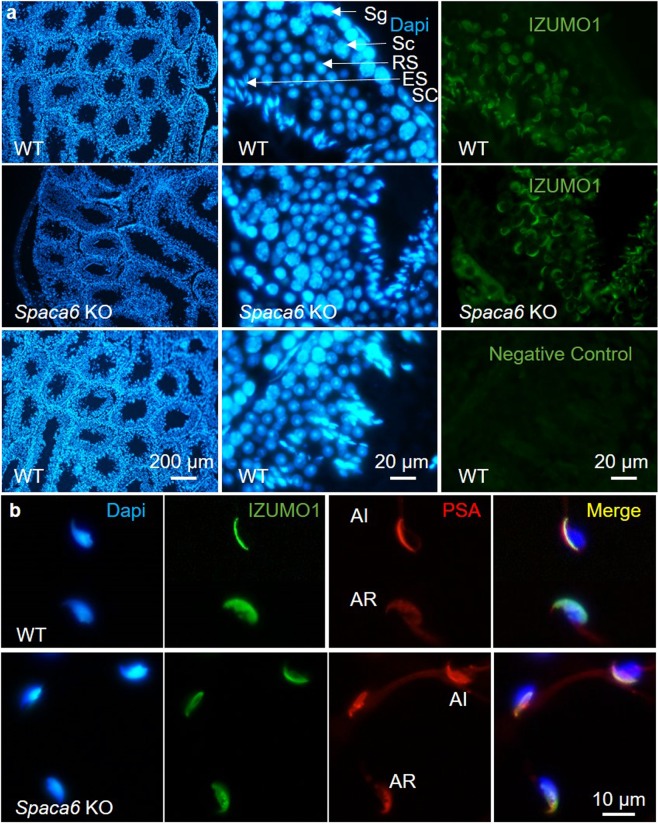
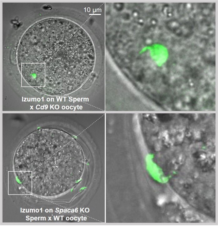
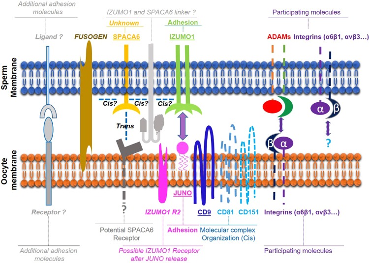
Three genes are known to be essential for gamete adhesion/fusion (Cd9, Izumo1 and Juno). Here, we confirmed that Spaca6 null males are infertile and showed that their sperm accumulate in the perivitelline space but are unable to fuse with oocyte. Like IZUMO1, SPACA6 which is expressed by human sperm, is remained on the equatorial segment after acrosomal reaction and is involved in human fertilization since an anti-SPACA6 antibody inhibited it. Despite the similarity of the phenotypes caused by Spaca6 and Izumo1 knockouts, these are not redundant and the essential relocation of IZUMO1 is not affected by the lack of SPACA6. We propose a model in which IZUMO1 and SPACA6 would be part of a molecular complex necessary for gamete fusion and that their concomitant presence would be required for the recruitment of another essential molecular actor, such as a fusogen, for the fusion to take place.


Conflict of interest statement

The authors declare no competing interests.

Figures

Figure 1
Figure 1
Human sperm SPACA6 expression. (a) Western blot using extracts of COS-7 cells transfected or not with mouse Spaca6-GFP and human SPACA6-GFP, revealed with rabbit polyclonal anti-human SPACA6, anti-Tubulin and anti-GFP antibodies. A band about 72 kDa (~36 kDa for SPACA6 + ~36 kDa for GFP) was visible only in COS-7 human SPACA6-GFP samples. In contrast, all samples were positive for Tubulin (55 kDa band) and those transfected with GFP were positive for it, demonstrating the quality of the deposited proteins and the efficiency of transfection respectively.(b) Western blot of human sperm protein extract using the anti-hSPACA6 antibody. The expected size band (~36 kDa) is only in the antibody lane. Controls with secondary antibody alone or with rabbit isotype (IgG) were negative. The corresponding full-length blots are presented in Supplementary Fig. S1. (c) Human SPACA6 distribution on sperm. Rabbit polyclonal anti-hSPACA6 revealed by a Donkey anti-rabbit Alexa 594 conjugated secondary antibody (red), localized SPACA6 on the acrosomal cap, equatorial and neck regions and midpiece of acrosomal intact sperm as attested by positive PSA-FITC conjugated (green) staining. After acrosome reaction (PSA-FITC negative), SPACA6 was remained essentially in the equatorial segment of sperm head. Nucleus was stained with Dapi (blue). AI: Acrosome Intact, AR: Acrosome Reacted, Es: Equatorial segment, Ac: Acrosomal cap, Nc: Neck, Mp: Midpiece.
Figure 2
Figure 2
Anti-hSPACA6 antibody inhibited Human fertilization. Overnight (18 h) insemination of zona-free human oocytes with human sperm in the presence of rabbit polyclonal anti-hSPACA6 (10 µg/ml), or rabbit IgG (10 µg/ml) or only IVF medium. The number of fused sperm heads into egg cytoplasm was recorded after insemination under fluorescent microscope (Nikon eclipse E600) and the fertilization index (FI: Number of decondensed sperm per oocyte, mean ± SEM) was calculated. FI decreased drastically from 29.5 ± 2.5 (n = 11) for the control group to 10.1 ± 1.1 (n = 11) for antibody treated group (10 µg/ml). No significant difference was observed between IVF medium control and IgG (at 10 µg/ml) control groups (FI: 26.4 ± 5.5 (n = 7)). Examples of illustrative images obtained after confocal analysis (Spinning Disk) are shown for the control and antibody groups. Transmission images were superimposed with Hoechst fluorescent signals (blue).
Figure 3
Figure 3
CRISPR-Cas9 Spaca6 gene deletion. (a) Genic organization of the mouse Spaca6 gene corresponding to cDNA NM_001162909. Black boxes are coding exons; the white box is the 3′ untranslated region. Black broken arrows: cuts in the C57BL/6 background. Grey dotted broken arrows: cuts in the FVB/N background. Sequences of the guides in exons 1 and 7 are in boxes. (b) Peptide sequences of the SPACA6 protein in the WT conditions (339 aa) and in both deleted versions: the deletion maintains the first 54 aa, then introduces 22 or 45 new different aa before a premature stop codon in the C57BL/6 and FVB/N background respectively. (c) Genotyping was performed by PCR on tail-tip DNA using Spaca6 Exon 1 F and 1 R primers and Spaca6 Exon1F and Exon 7 R. This last pair of primers gives a band (~428 bp) only when the deletion between exons 1 and 7 has taken place. (d) The absence of Spaca6 expression was verified by RT-qPCR. No transcripts were detected in the testis of two KO males, unlike the WT testis. Several WT tissues (lung, heart, spleen, brain and kidney) have also been shown to be negative for Spaca6 expression.
Figure 4
Figure 4
In vivo sterility of Spaca6 KO males. Litter size of crosses between FVB/N (a) or C57BL/6 (b) females and WT, heterozygotes, and KO males. Values indicate mean ± SEM. For both genetic backgrounds, no difference between WT and heterozygous males was found and no birth was obtained with KO males. FVB/N KO females were normally fertile (a). The numbers in parentheses indicate the numbers of mating pairs. (c) The same results were obtained when the fertilization rate of oocytes recovered in the oviduct the day after mating (as attested by the presence of a vaginal plug) was measured.
Figure 5
Figure 5
Abnormal frequency of Spaca6 KO sperm presence in the perivitelline space. After cumulus-intact IVF using sperm from Spaca6+/+, Spaca6+/− or Spaca6−/− C57BL/6 males, were reported: (a) the percentage of perivitelline space sperm containing-oocytes (mean ± SEM) which showed high and significant difference when comparing the KO to heterozygotes or WT groups (P < 0.0001) and less but significant difference when comparing heterozygotes and WT groups (P < 0.05), or (b) sperm number in perivitelline space per oocyte (mean ± SEM). In this case, only the difference between KO sperm and the two other groups was significant (P < 0.0001).
Figure 6
Figure 6
No fertilization was obtained with Spaca6 KO sperm in vitro. Fertilization rate (FR) (mean ± SEM) following cumulus-intact IVF assay at 106 sperm per ml or zona-free IVF assay at 105 sperm per ml for 3 hours. No fertilization was obtained with Spaca6 KO sperm in C57BL/6 (a) and FVB/N (b) lines. While no differences concerning the FR were observed when comparing the WT and heterozygous groups in C57BL/6 background (a,c), significant ones were obtained in FVB/N background both in cumulus-intact (b) and zona-free assays (d) (P < 0.0001). Regarding the fertilization index (FI; mean ± SEM), measurable only in zona-free IVF assay, and that gives the average number of fused sperm per oocyte observed after insemination, significant differences were observed in both backgrounds (e for C57BL/6 and f for FVB/N) when heterozygous groups were compared to WT ones (P < 0.0001). The sperm and oocyte Spaca6 genotypes are indicated below each figure.
Figure 7
Figure 7
Normal testis and epididymal sperm localization and relocation after acrosomal reaction of IZUMO1 in Spaca6 KO context. (a) Immunofluorescence staining of IZUMO1 and DAPI staining of permeabilized testicular sections from WT and Spaca6 KO C57BL/6 mice. The labeling of IZUMO1 (green) was carried out using a rat monoclonal anti-mouse IZUMO1 primary antibody, recognized by a secondary anti-rat goat antibody coupled to Alexa Fluor 488. The nuclei of the testicular cells were labeled with DAPI at 10 μg/mL. IZUMO1 was similarly detected in round and elongated spermatids (RS and ES) from WT and Spaca6 KO testes. A control condition with an incubation of the tissue with the secondary antibody alone was performed to check the specificity of the antibodies and to evaluate the background noise (negative control). Sg: Spermatogonia, Sc: Spermatocyte, RS, Round spermatid, ER: Elongated Spermatid, SC: Sertoli Cell. (b) Immunofluorescence staining of IZUMO1 and DAPI and PSA labeling of epididymal sperm from WT and Spaca6 KO males before or after acrosome reaction. While IZUMO1 is essentially localized in acrosomal cap in acrosome-intact sperm, it is more distributed on the sperm head after acrosome reaction. The absence of SPACA6 has no effect on IZUMO1 sperm localization and relocation after acrosome reaction.
Figure 8
Figure 8
Normal sperm IZUMO1 localization after physiological acrosomal reaction in Spaca6 KO context. Immunofluorescence staining of IZUMO1 of sperm obtained from WT and Spaca6 KO C57BL/6 mice after mating with Cd9 KO and WT females respectively and recovery of oocytes with physiologically acrosome-reacted sperm accumulated in the perivitelline space. The labeling of IZUMO1 (green) was carried out using a rat monoclonal anti-mouse IZUMO1 antibody, recognized by a secondary goat anti-rat antibody coupled to Alexa Fluor 488. Observation was performed under confocal Spinning Disk microscope (IMAG’IC facility). Zooms of the region with a spermatozoon indicate the IZUMO1 distribution. In the perivitelline space, IZUMO1 labeling is present along the entire head, of the WT and Spaca6 KO sperm heads meaning that even in the absence of SPACA6, the relocation of IZUMO1 after acrosomal reaction occurred normally.
Figure 9
Figure 9
Hypothetical model of mammalian gamete adhesion/fusion. The illustration represents the adhesion/fusion proteins involved in fertilization in mice and humans. The essential ones: SPACA6 and IZUMO1 on the sperm membrane, JUNO receptor of IZUMO1, and CD9 on the oocyte membrane. A non-exhaustive list of some molecules participating but not essential to gamete interaction: ADAMs and integrins on sperm, integrins and tetraspanins on the oocyte. Those that remain to be discovered: the fusogen actor and certainly other ligands and receptors on both sides. We propose a model in which a direct or indirect interaction between SPACA6 and IZUMO1 within a molecular complex would be required to recruit the fusogenic actor.

References

    1. Inoue N, Ikawa M, Isotani A, Okabe M. The immunoglobulin superfamily protein Izumo is required for sperm to fuse with eggs. Nature. 2005;434(7030):234–238. doi: 10.1038/nature03362. - DOI - PubMed
    1. Bianchi E, Doe B, Goulding D, Wright GJ. Juno is the egg Izumo receptor and is essential for mammalian fertilization. Nature. 2014;508(7497):483–487. doi: 10.1038/nature13203. - DOI - PMC - PubMed
    1. Kaji K, et al. The gamete fusion process is defective in eggs of Cd9-deficient mice. Nat. Genet. 2000;24(3):279–282. doi: 10.1038/73502. - DOI - PubMed
    1. Le Naour F, Rubinstein E, Jasmin C, Prenant M, Boucheix C. Severely reduced female fertility in CD9-deficient mice. Science. 2000;287(5451):319–321. doi: 10.1126/science.287.5451.319. - DOI - PubMed
    1. Miyado K, et al. Requirement of CD9 on the egg plasma membrane for fertilization. Science. 2000;287(5451):321–324. doi: 10.1126/science.287.5451.321. - DOI - PubMed

Publication types

MeSH terms