. 2020 Feb 10;6(2):1-13.
doi: 10.1212/NXG.0000000000000397. eCollection 2020 Apr.

Heterozygous STUB1 missense variants cause ataxia, cognitive decline, and STUB1 mislocalization

Affiliations

Heterozygous STUB1 missense variants cause ataxia, cognitive decline, and STUB1 mislocalization

Dong-Hui Chen et al. Neurol Genet. .

Abstract

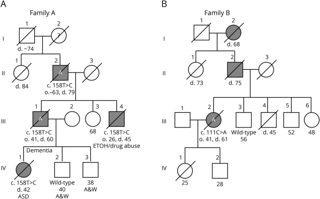
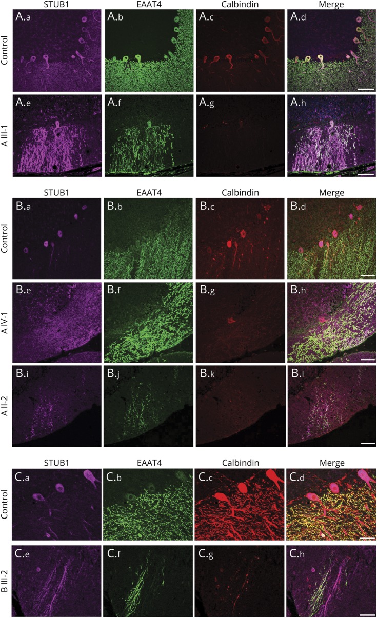
Objective: To identify the genetic cause of autosomal dominant ataxia complicated by behavioral abnormalities, cognitive decline, and autism in 2 families and to characterize brain neuropathologic signatures of dominant STUB1-related ataxia and investigate the effects of pathogenic variants on STUB1 localization.

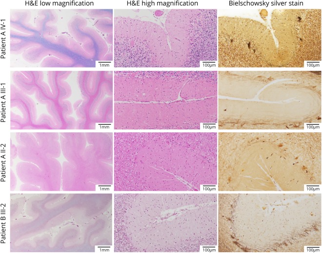
Methods: Clinical and research-based exome sequencing was used to identify the causative variants for autosomal dominant ataxia in 2 families. Gross and microscopic neuropathologic evaluations were performed on the brains of 4 affected individuals in these families.

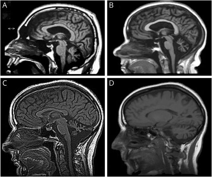
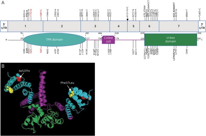
Results: Mutations in STUB1 have been primarily associated with childhood-onset autosomal recessive ataxia, but here we report heterozygous missense variants in STUB1 (p.Ile53Thr and p.The37Leu) confirming the recent reports of autosomal dominant inheritance. Cerebellar atrophy on imaging and cognitive deficits often preceded ataxia. Unique neuropathologic examination of the 4 brains showed the marked loss of Purkinje cells (PCs) without microscopic evidence of significant pathology outside the cerebellum. The normal pattern of polarized somatodendritic STUB1 protein expression in PCs was lost, resulting in aberrant STUB1 localization in the distal PC dendritic arbors.

Conclusions: This study confirms a dominant inheritance pattern in STUB1-ataxia in addition to a recessive one and documents its association with cognitive and behavioral disability, including autism. In the most extensive analysis of cerebellar pathology in this disease, we demonstrate disruption of STUB1 protein in PCs as part of the underlying pathogenesis.


Figures

Figure 1
Figure 1. Four generation pedigrees of 2 families with hereditary cerebellar ataxia and cognitive disability
If alive, current age in years is shown below the symbol. / = deceased; A = brain autopsy; ASD = autism spectrum disorder; black symbol = affected with ataxia; d = age at death. The variant or wild-type STUB1 allele is shown for individuals who were tested.
Figure 2
Figure 2. Neuroimaging
(A) MRI of patient III-1 in family A at 53 years of age showing marked midline cerebellar atrophy, mildly enlarged lateral ventricle, and normal corpus callosum. (B) MRI of patient A IV-1 at 32 years of age showing marked midline cerebellar atrophy before the onset of ataxia but having cognitive disability and autism spectrum disorder. (C and D) MRI of patient III-2 in family B at 48 years of age showing midline (C) and lateral (D) cerebellar atrophy.
Figure 3
Figure 3. Ataxia-related variants in STUB1
(A) STUB1 gene showing reported variants associated with autosomal dominant and recessive ataxias and the corresponding locations in the protein. Colors indicate the TPR (turquoise), coiled-coil (purple), and E3 ubiquitin ligase (U-box) domains in STUB1. Bold = heterozygous autosomal dominant variants reported by Genis et al and De Michele et al and in this report (in red). Arrow = an intronic mutation at the donor splice site of exon 4. (B) Diagram depicts the location of the p.Ile53Thr (red amino acid) and p.Phe37Leu (yellow amino acid) missense variants within a 3-D model of a STUB1 homodimer (the functional isoform). STUB1 was modeled with PyMOL (pymol.org) using the data from the protein data bank (rcsb.org, PDB ID#2C2L). TPR = tetratricopeptide repeat.
Figure 4
Figure 4. Microscopic neuropathology
Microscopic sections of the patients from both families showing marked cerebellar atrophy with thinning of the folia (H&E/LFB low magnification) and Purkinje cell (PC) loss (H&E/LFB high magnification). The loss of PCs is also highlighted by the empty baskets noted on Bielschowsky silver stain and by the severe reduction in calbindin-positive fibers (calbindin and GFAP immunostaining available on request). H&E = hematoxylin and eosin.
Figure 5
Figure 5. Loss of polarized STUB1 expression in Purkinje cells in patients with STUB1-cerebellar ataxia
Immunostaining was performed with antibodies to STUB1 (pseudocolored purple), EAAT4 (green), and calbindin (red), and all scanning acquisition and postprocessing parameters were carried out identically among the cases about the controls in each panel. In 3 different normal control individuals (A.a–A.d, B.a–B.d, C.a–C.d), STUB1 immunoreactivity was uniformly localized primarily in PC cell bodies and proximal dendrites, with little expression in distal PC dendritic arbors. (A and B) Family A with STUB1-Ile53Thr. Loss of STUB1 polarization results in aberrant expression in distal PC dendritic arbors and cell bodies in patients III-1 (A.e–A.h), IV-1 (B.e–B.h), and II-2 (B.i–B.l). Scale bars = 100 μm. (C) Family B with STUB1-Phe37Leu. In individual III-2, STUB1 was aberrantly expressed in PC distal arbors (C.e–C.h). Scale bars = 50 μm.
Figure 6
Figure 6. STUB1 expression pattern in Purkinje cells in a number of non-STUB1-associated ataxias and cerebellar degeneration
In a normal individual (A–D), STUB1 immunoreactivity was expressed mostly in PC cell bodies and proximal dendrites (pseudocolored purple). Calbindin (red) and EAAT4 (green) immunostained PC bodies and dendrites. STUB1 localization appeared normal in PCs from SCA5 (E–H) and SCA3 (I–L). In an SCA7 case, STUB1 expression was aberrantly localized in distal PC dendritic arbors (M–P). Scale bars = 100 μm.

References

    1. Bird T. Hereditary ataxia overview. In: GeneReviews at GeneTests: Medical Genetics Information Resource [Database Online] 1993-2019. Seattle: University of Washington; 2019.
    1. Depondt C, Donatello S, Simonis N, et al. . Autosomal recessive cerebellar ataxia of adult onset due to STUB1 mutations. Neurology 2014;82:1749–1750. - PubMed
    1. Kanack AJ, Newsom OJ, Scaglione KM. Most mutations that cause spinocerebellar ataxia autosomal recessive type 16 (SCAR16) destabilize the protein quality-control E3 ligase CHIP. J Biol Chem 2018;293:2735–2743. - PMC - PubMed
    1. Bettencourt C, de Yebenes JG, Lopez-Sendon JL, et al. . Clinical and neuropathological features of spastic ataxia in a Spanish family with novel compound heterozygous mutations in STUB1. Cerebellum 2015;14:378–381. - PubMed
    1. Genis D, Ortega-Cubero S, San Nicolas H, et al. . Heterozygous STUB1 mutation causes familial ataxia with cognitive affective syndrome (SCA48). Neurology 2018;91:e1988–e1998. - PubMed