

An amelanotic choroidal melanoma arising in a young man with tattoo-associated sarcoidosis
- PMID: 32211561
- PMCID: PMC7082514
- DOI: 10.1016/j.ajoc.2020.100655
An amelanotic choroidal melanoma arising in a young man with tattoo-associated sarcoidosis
Abstract
Purpose: To describe a patient with an amelanotic choroidal melanoma, originally misdiagnosed as a choroidal granuloma, following his systemic diagnosis of tattoo-associated sarcoidosis.
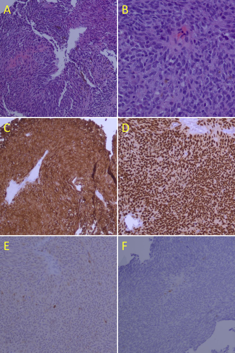
Observations: The amelanotic choroidal tumor, suspected to be a granuloma, failed initial steroid treatment. Full-thickness chorioretinal biopsy demonstrated histologic presence of uveal melanoma and tumor genetics via GEP analysis demonstrated a PRAME negative, Class 1A lesion. The amelanotic choroidal melanoma was treated successfully with I-125 plaque brachytherapy.
Conclusion and importance: Despite a systemic diagnosis which predisposes a patient to uveal granuloma, amelanotic choroidal melanomas can still occur and should be considered. The association of uveal melanoma and sarcoidosis remains rare and of unclear significance.
Keywords: Eye; Sarcoidosis; Tumor; Uveal melanoma.
© 2020 The Authors.
Conflict of interest statement
The following authors have no financial disclosures: STB, ALB, DAR.
Figures

References
-
- Egan K.M., Seddon J.M., Glynn R.J., Gragoudas E.S., Albert D.M. Epidemiologic aspects of uveal melanoma. Surv Ophthalmol. 1988;32:239–251. - PubMed
-
- Singh A.D., Turell M.E., Topham A.K. Uveal melanoma: trends in incidence, treatment, and survival. Ophthalmol. 2011;118:1881–1885. - PubMed
-
- Shields C.L., Furuta M., Thangappan A. Metastasis of uveal melanoma millimeter-by-millimeter in 8033 consecutive eyes. Arch Ophthalmol. 2009;127:989–998. - PubMed
-
- Welch R.J., Newman J.H., Honig S.E. Choroidal amelanotic tumours: clinical differentiation of benign from malignant lesions in 5586 cases. Br J Ophthalmol. 2020;104(2):194–201. - PubMed
Publication types
LinkOut - more resources
Full Text Sources
