

Imaging diagnosis for intervertebral disc
- PMID: 32211585
- PMCID: PMC7084050
- DOI: 10.1002/jsp2.1066
Imaging diagnosis for intervertebral disc
Abstract
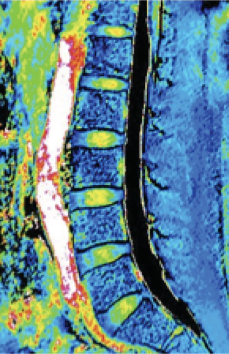
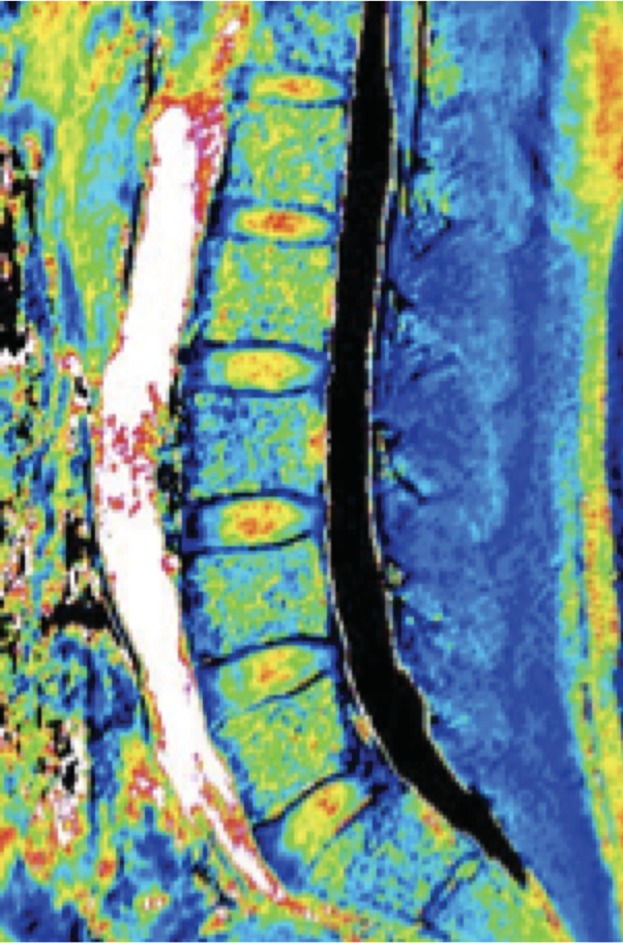
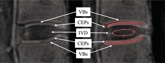
Various functional magnetic resonance imaging (MRI) techniques have been investigated in recent years and are being used in clinical practice for the patients with low back pain (LBP). MRI is an important modality for diagnosing intervertebral disc (IVD) degeneration. In recent years, there have been several reported attempts to use MRI T2 mapping and MRI T1ρ mapping to quantify lumbar disc degeneration. MRI T2 mapping involves digitizing water content, proteoglycan content, and collagen sequence breakdown as relaxation times (T2 values) at each site. These digitized values are used to create a map, that is, then used to quantitatively evaluate the metabolite concentrations within IVD tissues. MRI T2 mapping utilizes the T2 relaxation time to quantify moisture content and the collagen sequence breakdown. MRI T1ρ mapping digitizes water molecule dispersion within the cartilaginous matrix to evaluate the degree of cartilaginous degeneration. Magnetic resonance spectroscopy is a less-invasive diagnostic test that provides biochemical information. Adequate analysis of the IVD has not yet been performed, although there are indications of a relationship between the adipose content of the multifidus muscle in the low back and LBP. The ultra short TE technique has been recently used to investigate lumbar cartilaginous endplates. Unlike diagnosis based on contrast-enhanced images of the IVD, which depends on the recurrence of pain that is determined subjectively, MRI-based diagnosis is less-invasive and based on objective imaging findings. It is therefore expected to play a key role in the diagnostic imaging of IVD conditions in the future.
Keywords: imaging diagnosis; intervertebral disc; magnetic resonance imaging.
© 2019 The Authors. JOR Spine published by Wiley Periodicals, Inc. on behalf of Orthopaedic Research Society.
Conflict of interest statement
The authors declare no potential conflict of interests.
Figures
References
-
- Deyo RA, Weinstein JN. Low back pain. N Engl J Med. 2001;344(5):363‐370. - PubMed
-
- Hurri H, Karppinen J. Discogenic pain. Pain. 2004;112(3):225‐228. - PubMed
-
- Boden SD, Davis DO, Dina TS, Patronas NJ, Wiesel SW. Abnormal magnetic‐resonance scans of the lumbar spine in asymptomatic subjects. A prospective investigation. J Bone Joint Surg Am. 1990;72(3):403‐408. - PubMed
-
- Palmgren T, Gronblad M, Virri J, Kaapa E, Karaharju E. An immunohistochemical study of nerve structures in the anulus fibrosus of human normal lumbar intervertebral discs. Spine (Phila Pa 1976). 1999;24(20):2075‐2079. - PubMed
-
- Cavanaugh JM, Ozaktay AC, Yamashita T, Avramov A, Getchell TV, King AI. Mechanisms of low back pain: a neurophysiologic and neuroanatomic study. Clin Orthop Relat Res. 1997;335:166‐180. - PubMed
Publication types
LinkOut - more resources
Full Text Sources
Miscellaneous
