. 2019 Oct 24:5:100032.
doi: 10.1016/j.mtbio.2019.100032. eCollection 2020 Jan.

Ceramics with the signature of wood: a mechanical insight

Affiliations

Ceramics with the signature of wood: a mechanical insight

D Bigoni et al. Mater Today Bio. .

Abstract

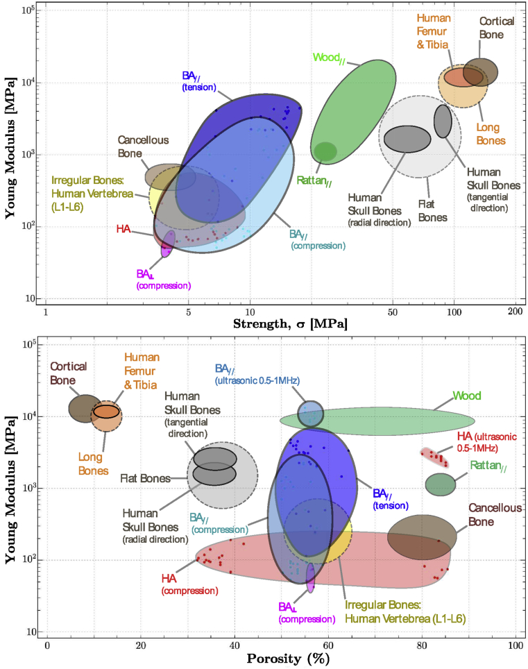
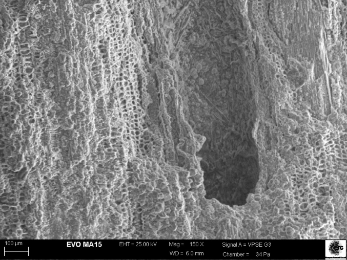
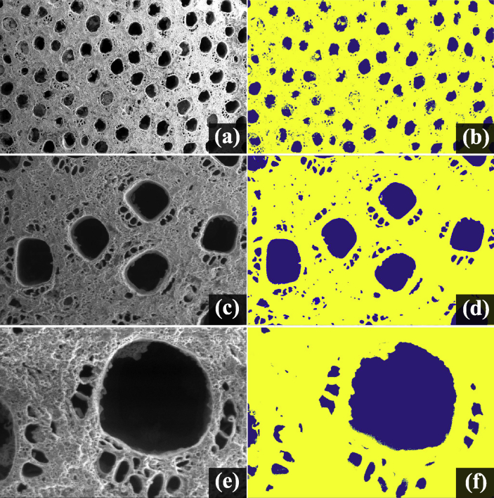
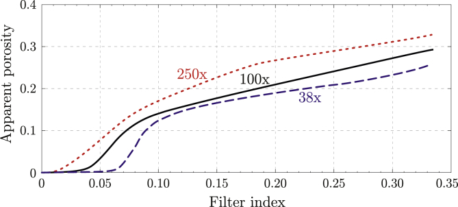
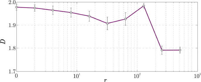
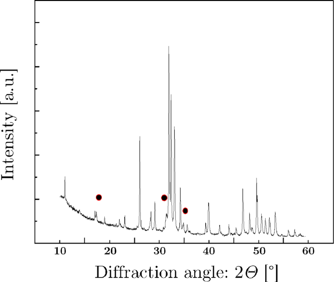
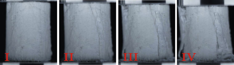
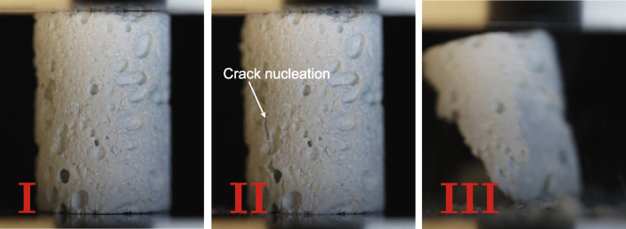
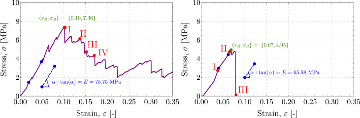
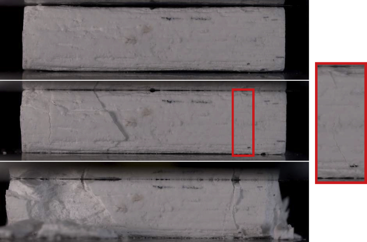
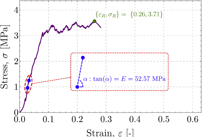
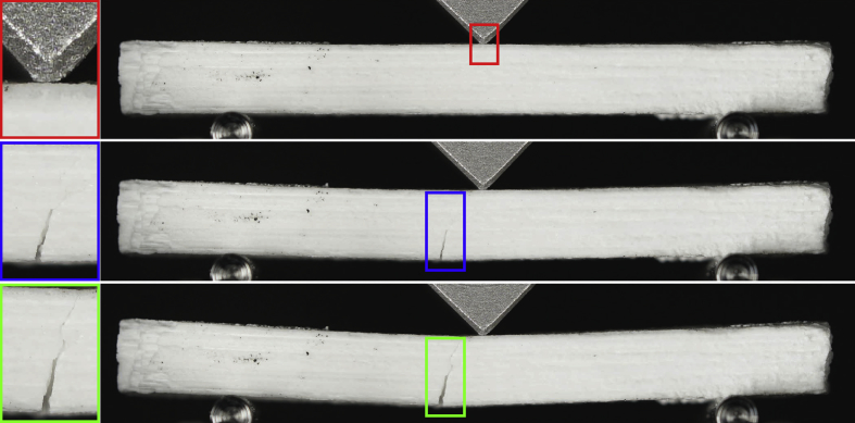
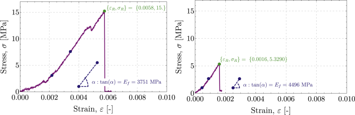
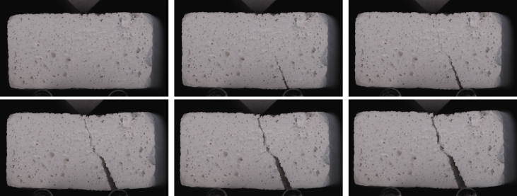
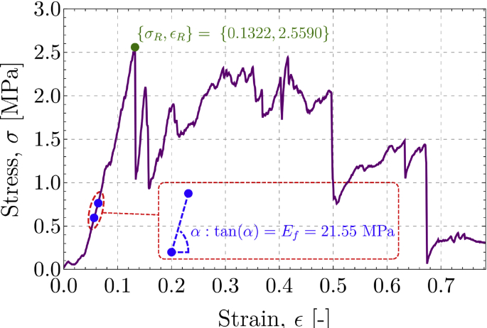
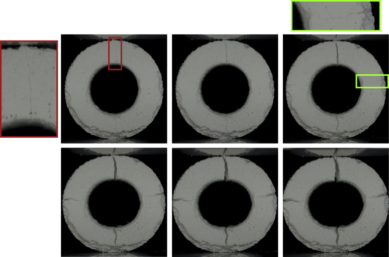
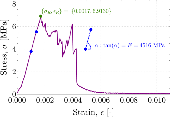
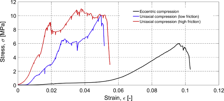
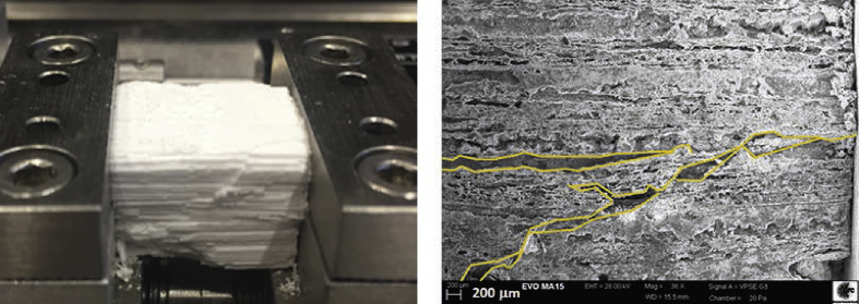
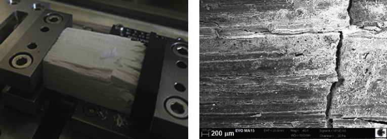
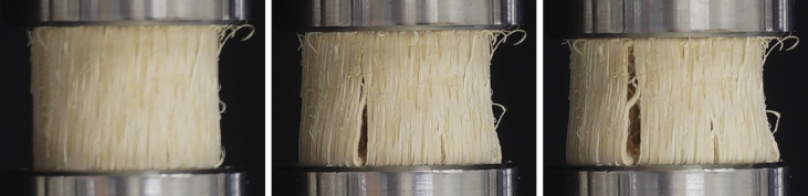
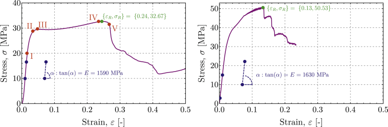
In an attempt to mimic the outstanding mechanical properties of wood and bone, a 3D heterogeneous chemistry approach has been used in a biomorphic transformation process (in which sintering is avoided) to fabricate ceramics from rattan wood, preserving its hierarchical fibrous microstructure. The resulting material (called biomorphic apatite ​[BA] henceforth) possesses a highly bioactive composition and is characterised by a multiscale hierarchical pore structure, based on nanotwinned hydroxyapatite lamellae, which is shown to display a lacunar fractal nature. The mechanical properties of BA are found to be exceptional (when compared with usual porous hydroxyapatite and other ceramics obtained from wood through sintering) and unique ​as they occupy a zone in the Ashby map previously free from ceramics, but not far from wood and bone. Mechanical tests show the following: (i) the strength in tension may exceed that in compression, (ii) failure in compression involves complex exfoliation patterns, thus resulting in high toughness, (iii) unlike in sintered porous hydroxyapatite, fracture does not occur 'instantaneously,' ​but its growth may be observed, and it exhibits tortuous patterns that follow the original fibrillar structure of wood, thus yielding outstanding toughness, (iv) the anisotropy of the elastic stiffness and strength show unprecedented values when situations of stresses parallel and orthogonal to the main channels are compared. Despite being a ceramic material, BA displays a mechanical behavior similar on the one hand to the ligneous material from which it was produced (therefore behaving as a 'ceramic with the signature of wood') and on the other hand to the cortical/spongy osseous complex constituting the structure of compact bone.

Keywords: Fractal porosity; Fracture; Hydroxyapatite; Mechanical properties; Mechanical tests; Strength.


Conflict of interest statement

The authors declare that they have no known competing financial interests or personal relationships that could have appeared to influence the work reported in this paper.

Figures

Fig. 1
Fig. 1
Ashby charts reporting Young modulus vs strength (upper part) and vs porosity (lower part) for biomorphic apatite, loaded parallel (BA//) and perpendicular (BA) to the microtubule structure (‘grain’ in the following), for rattan wood (from which BA was obtained), and for bones. BA, biomorphic apatite; HA, hydroxyapatite.
Fig. 2
Fig. 2
An scanning electron microscopy image of the microstructure of biomorphic apatite showing long open channels surrounded by finer alveolar closed pores.
Fig. 3
Fig. 3
Scanning electron microscopy images of cross sections of biomorphic apatite ​(BA) and the corresponding apparent porosity identified on the basis of the application of an ad hoc image analysis software with a filter index of 0.2. In particular, (a) 38× scanning electron microscopy image of BA; (b) at 38×, the porosity is detected to be 14.6%; (c) 100× scanning electron microscopy image of BA; (d) at 100×, the porosity is detected to be 19.0%; (e) 250× scanning electron microscopy image of BA; (f) at 250×, the porosity is detected to be 27.0%. The image correlation software shows that porosity is increasing with the magnification, and this reveals the lacunar fractality of the cross section domain. BA, biomorphic apatite.
Fig. 4
Fig. 4
Increase of apparent porosity for BA with the filter index at different magnification scales, showing the lacunar fractal nature of BA. BA, biomorphic apatite.
Fig. 5
Fig. 5
Local fractal dimension D (which quantifies the porosity) as a function of r (the lateral side division of the image in the box-counting algorithm).
Fig. 6
Fig. 6
XRD (x-ray diffraction) pattern of one sample of bioapatite used for mechanical tests, where the dots point to reflections addressing the β-TCP phase. All the unmarked peaks point to the HA phase. HA, hydroxyapatite; β-TCP, beta-tricalcium phosphate.
Fig. 7
Fig. 7
A sequence of photos taken during a uniaxial compression test of a cylindrical sample of BA. The instances when the photos have been taken are marked on the stress/strain curve (Fig. 9 on the left) reported in the following. Note the progressive exfoliation of the sample, strongly enhancing toughness and related to the peaks in the stress/strain diagram. BA, biomorphic apatite.
Fig. 8
Fig. 8
A sequence of photos taken during a uniaxial compression test of hydroxyapatite samples. The instances when the photos have been taken are marked on the stress/strain curve (Fig. 9 on the right) reported in the following. Note the abrupt failure of the specimen immediately after the appearance of a splitting crack.
Fig. 9
Fig. 9
Stress/strain behavior of a BA sample (#1, on the left) and of a HA sample (#1, on the right) subject to uniaxial compression, parallel to the grain for BA. The green spot identifies the peak strength, and a straight line drawn through the two indicated blue spots was used to evaluate the Young modulus E=tanα. The superior toughness of BA is evidenced by the slow load fall, contrasting with the sharp jump to zero displayed by HA.
Fig. 10
Fig. 10
A sequence of photos showing progressive failure during uniaxial compression orthogonal to the grain of a BA prismatic sample (from set #4, geometrical and mechanical properties reported in Table 4). BA, biomorphic apatite.
Fig. 11
Fig. 11
Stress/strain behavior of a BA sample compressed parallel to the grain. The green spot identifies the peak strength, and a straight line drawn through the two indicated blue spots was used to evaluate the Young modulus E=tanα. BA, biomorphic apatite.
Fig. 12
Fig. 12
A sequence of photos taken during a three-point bending test on the prismatic sample of biomorphic apatite (BA set # 1), tested in the direction parallel to the grain. The growth of a tensile fracture is clearly documented. (See the insets marked blue and green; the inset marked red shows a detail of the loading blade.) BA, biomorphic apatite.
Fig. 13
Fig. 13
Tensile stress/strain behavior of BA (set #1, on the left) and of HA (set # 2, on the right) from a three-point bending test, parallel to the grain for BA. The green spot identifies the peak strength, and a straight line drawn through the two indicated blue spots was used to evaluate the Young modulus E=tanα. The superior toughness of BA is highlighted by the failure strain much higher than in the HA sample. BA, biomorphic apatite; HA, hydroxyapatite.
Fig. 14
Fig. 14
A sequence of photos taken during a three-point bending test on biomorphic apatite prismatic samples (set #4) tested in the direction orthogonal to the grain. Note the growth of a tensile fracture.
Fig. 15
Fig. 15
Tensile stress/strain behavior of BA (set #4) from a three-point bending test, with tensile stress parallel to the grain. The green spot identifies the peak strength, and a straight line drawn through the two indicated blue spots was used to evaluate the Young modulus E=tanα. The high toughness of BA is highlighted by the slow load fall. BA, biomorphic apatite.
Fig. 16
Fig. 16
A sequence of photos taken during a ring compression test on biomorphic apatite (BA) tubular samples (set #3) tested in the direction orthogonal to the grain. The growth of tensile fractures is clearly documented (see the insets).
Fig. 17
Fig. 17
Tensile stress/strain behavior of BA (Set #3) from a ring compression test, with tensile stress parallel to the grain. The green spot identifies the peak strength, and a straight line drawn through the two indicated blue spots was used to evaluate the Young modulus E=tanα. The high toughness of BA is highlighted by the slow load fall. BA, biomorphic apatite.
Fig. 18
Fig. 18
Amplitude of the signal versus time for two ultrasonic tests on a BA sample (set #1, on the left) and on a HA sample (set #1, on the right) at two different frequencies (0.5 and 1.0 ​MHz). Note that the points that have been identified as the representative of the time of flight tv have been identified in agreement with the standards [60] so that the arrival of the P-wave has been marked at the instance when the leading edge of the first peak was reached at 80%. (The evaluation of the time of flight was performed automatically using a Mathematica routine ad hoc developed.) ​BA, biomorphic apatite; HA, hydroxyapatite.
Fig. 19
Fig. 19
Stress/strain curve of BA specimens tested in situ using a SEM under uniaxial compression at two levels of friction (at the specimen/platen contacts) and for eccentric loading. BA, biomorphic apatite; SEM, scanning electron microscope.
Fig. 20
Fig. 20
Specimen tested under uniaxial compression with high friction. On the left: view of the specimen at failure; on the right: cracks at failure.
Fig. 21
Fig. 21
Specimen tested under uniaxial compression by scanning electron microscopy under conditions of low friction. On the left: view of the specimen at failure (note the high-density polyethylene layer in black colour); on the right: cracks at failure.
Fig. 22
Fig. 22
BA specimen tested under eccentric compression in situ using a SEM and shown at failure on the left (note the paperboard layers in contact with the sample); cracks at failure are detailed on the right. BA, biomorphic apatite; SEM, scanning electron microscope.
Fig. 23
Fig. 23
A sequence of photos showing progressive failure (involving exfoliation by fiber buckling) of a rattan wood specimen during uniaxial compression.
Fig. 24
Fig. 24
Stress/strain behavior of a rattan wood sample subject to uniaxial compression (on the left) and three-point bending (on the right), parallel to the grain. The red spots (in the compression plot) identify different stress levels corresponding to different definitions of failure. A straight line drawn through the two indicated blue spots was used to evaluate the Young modulus E=tanα.

References

    1. Fratzl P., Weinkamer R. Nature's hierarchical materials. Prog. Mater. Sci. 2007;52:1263–1334.
    1. Weinkamer R., Fratzl P. Mechanical adaptation of biological materials - the examples of bone and wood. Mater. Sci. Eng. C-Mater. Biol. Appl. 2011;31:1164–1173.
    1. Hueuer A.H., Fink D.J., Laraia V.J., Arias J.L., Calvert P.D., Kendall K., Messing G.L., Blackwell J., Rieke P.C., Thompson D.H., Wheeler A.P., Veis A., Caplan A.I. Innovative materials processing strategies: a biomimetic approach. Science. 1992;255:1098–1105. - PubMed
    1. Bryne C.E., Nagle D.C. Cellulose derived composites - a new method for materials processing. Mater. Res. Innov. 1997;1:137–144.
    1. Greil P. Biomorphous ceramics from lignocellulosics. J. Eur. Ceram. Soc. 2001;21:105–118.

LinkOut - more resources