

Structural and functional footprint of visual snow syndrome
- PMID: 32211752
- PMCID: PMC7534145
- DOI: 10.1093/brain/awaa053
Structural and functional footprint of visual snow syndrome
Abstract
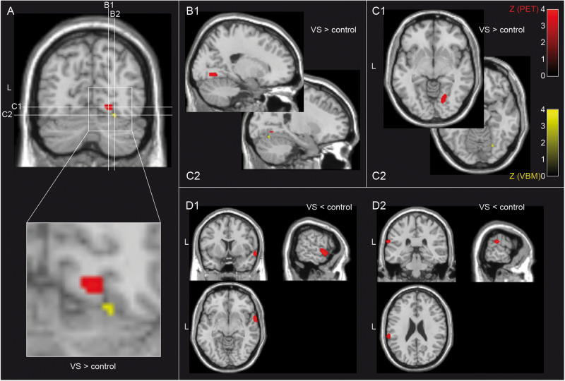
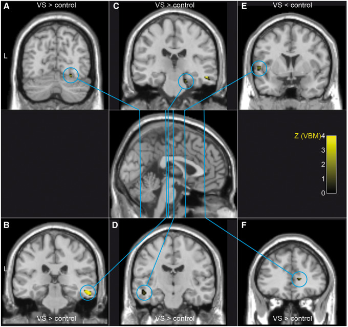
Patients with visual snow syndrome suffer from a continuous pan-field visual disturbance, additional visual symptoms, tinnitus, and non-perceptional symptoms. The pathophysiology of visual symptoms might involve dysfunctional visual cortex. So far, the extra-visual system has not been investigated. We aimed at identifying structural and functional correlates for visual and non-visual symptoms in visual snow syndrome. Patients were compared to age- and sex-matched controls using 18F-2-fluoro-2-deoxy-d-glucose PET (n = 20 per group) and voxel-based morphometry (n = 17 per group). Guided by the PET results, region of interest analysis was done in voxel-based morphometry to identify structural-functional correspondence. Grey matter volume was assessed globally. Patients had corresponding hypermetabolism and cortical volume increase in the extrastriate visual cortex at the junction of the right lingual and fusiform gyrus. There was hypometabolism in the right superior temporal gyrus and the left inferior parietal lobule. Patients had grey matter volume increases in the temporal and limbic lobes and decrease in the superior temporal gyrus. The corresponding structural and functional alterations emphasize the relevance of the visual association cortex for visual snow syndrome. The broad structural and functional footprint, however, confirms the clinical impression that the disorder extends beyond the visual system.
Keywords: FDG PET; migraine; non-visual symptoms; visual snow; voxel-based morphometry.
© The Author(s) (2020). Published by Oxford University Press on behalf of the Guarantors of Brain.
Figures
References
-
- Aurora SK, Barrodale PM, Tipton RL, Khodavirdi A.. Brainstem dysfunction in chronic migraine as evidenced by neurophysiological and positron emission tomography studies. Headache 2007; 47: 996–1003; discussion 4–7. - PubMed
-
- Brett M, Anton J-L, Valabregue R, Poline J-B. Region of interest analysis using an SPM toolbox [abstract]. 8th International Conference on Functional Mapping of the Human Brain. Sendai, Japan; 2002.
-
- Carreiras M, Seghier ML, Baquero S, Estevez A, Lozano A, Devlin JT, et al. An anatomical signature for literacy. Nature 2009; 461: 983–6. - PubMed
-
- Chehadi O, Rusu AC, Konietzny K, Schulz E, Koster O, Schmidt-Wilcke T, et al. Brain structural alterations associated with dysfunctional cognitive control of pain in patients with low back pain. Eur J Pain 2018; 22: 745–55. - PubMed
