Review
. 2020 Apr 6;217(4):e20190062.
doi: 10.1084/jem.20190062.

The blood-brain barrier in health and disease: Important unanswered questions

Affiliations
Review

The blood-brain barrier in health and disease: Important unanswered questions

Caterina P Profaci et al. J Exp Med. .

Abstract

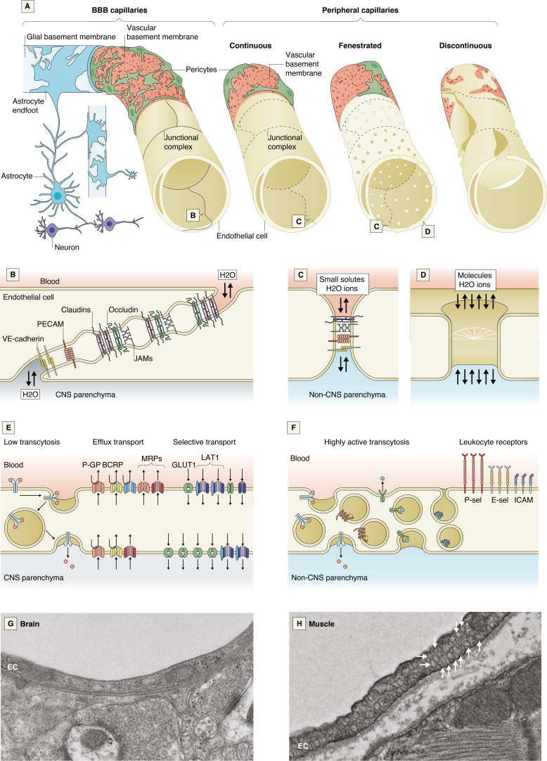
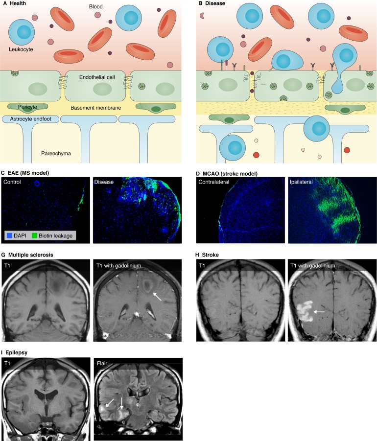
The blood vessels vascularizing the central nervous system exhibit a series of distinct properties that tightly control the movement of ions, molecules, and cells between the blood and the parenchyma. This "blood-brain barrier" is initiated during angiogenesis via signals from the surrounding neural environment, and its integrity remains vital for homeostasis and neural protection throughout life. Blood-brain barrier dysfunction contributes to pathology in a range of neurological conditions including multiple sclerosis, stroke, and epilepsy, and has also been implicated in neurodegenerative diseases such as Alzheimer's disease. This review will discuss current knowledge and key unanswered questions regarding the blood-brain barrier in health and disease.


Conflict of interest statement

Disclosures: The authors declare no competing interests exist.

Figures

Figure 1.
Figure 1.
Cellular and molecular properties of the BBB. (A) A schematic comparison of the BBB capillaries with the continuous nonfenestrated, continuous fenestrated, and discontinuous capillaries found in peripheral organs. (B–F) Schematics of the molecular composition of junctional complexes of BBB ECs (B) and of ECs in peripheral organs (C), peripheral endothelial fenestra (D), and transport mechanisms in CNS ECs (E) and peripheral ECs (F). (G and H) Electron micrographs of a mouse brain EC (G) and a mouse muscle EC, which is densely packed with vesicles (arrows; H). BCRP, breast cancer resistance protein; GLUT1, glucose transporter 1; ICAM, intercellular adhesion molecule; JAM, junctional adhesion molecule; LAT1, L-type amino acid transporter 1; MRP, multidrug resistance-associated protein; PECAM-1, platelet and EC adhesion molecule 1; P-GP, p-glycoprotein; VE-cadherin, vascular endothelial cadherin; P-sel, P-selectin; E-sel, E-selectin.
Figure 2.
Figure 2.
Dysfunction of the BBB in disease. (A and B) Schematic representation of the NVU in health and disease. (A) In health, CNS ECs exhibit TJs, low rates of transcytosis, and low expression of leukocyte adhesion molecules. Pericytes embedded in the BM help to maintain the barrier, and astrocyte endfeet contact the BM. (B) In disease, TJs are internalized or down-regulated, rates of transcytosis increase, increased leukocyte adhesion molecule expression leads to increased leukocyte extravasation, the BM degrades, and pericytes and astrocytes less tightly cover the ECs. Made with BioRender. (C–F) BBB disruption in models of MS, traumatic brain injury, and stroke. Sections showing BBB leakage to a sulfo-N-hydroxysulfosuccinimide-biotin tracer (green) in three disease models. (C and D) A section of spinal cord from a healthy mouse (C) and from the EAE model of MS (D). (E and F) The contralateral (E) and ipsilateral (F) hemispheres in a coronal section of the middle-cerebral artery occlusion model of ischemic stroke. (GI) BBB leakage and edema in human cases of MS, stroke, and epilepsy. T1 weighted MRI images with gadolinium enhancement to show BBB leakage in (G) MS lesions and (H) stroke infarct. (I) T1 weighted and fluid attenuation inversion recovery (FLAIR) MRI images showing edema in epilepsy. Images courtesy of Dr. John Hesselink, University of California, San Diego, San Diego, CA.

References

    1. Abadier M., Haghayegh Jahromi N., Cardoso Alves L., Boscacci R., Vestweber D., Barnum S., Deutsch U., Engelhardt B., and Lyck R.. 2015. Cell surface levels of endothelial ICAM-1 influence the transcellular or paracellular T-cell diapedesis across the blood-brain barrier. Eur. J. Immunol. 45:1043–1058. 10.1002/eji.201445125 - DOI - PubMed
    1. Abbott N.J., Pizzo M.E., Preston J.E., Janigro D., and Thorne R.G.. 2018. The role of brain barriers in fluid movement in the CNS: is there a ‘glymphatic’ system? Acta Neuropathol. 135:387–407. 10.1007/s00401-018-1812-4 - DOI - PubMed
    1. Aird W.C. 2007. Phenotypic heterogeneity of the endothelium: II. Representative vascular beds. Circ. Res. 100:174–190. 10.1161/01.RES.0000255690.03436.ae - DOI - PubMed
    1. Alafuzoff I., Adolfsson R., Bucht G., and Winblad B.. 1983. Albumin and immunoglobulin in plasma and cerebrospinal fluid, and blood-cerebrospinal fluid barrier function in patients with dementia of Alzheimer type and multi-infarct dementia. J. Neurol. Sci. 60:465–472. 10.1016/0022-510X(83)90157-0 - DOI - PubMed
    1. Alafuzoff I., Adolfsson R., Grundke-Iqbal I., and Winblad B.. 1987. Blood-brain barrier in Alzheimer dementia and in non-demented elderly. An immunocytochemical study. Acta Neuropathol. 73:160–166. 10.1007/BF00693782 - DOI - PubMed

MeSH terms