. 2020 Mar 2;219(3):e201911126.
doi: 10.1083/jcb.201911126.

Heparan sulfate is a clearance receptor for aberrant extracellular proteins

Affiliations

Heparan sulfate is a clearance receptor for aberrant extracellular proteins

Eisuke Itakura et al. J Cell Biol. .

Abstract

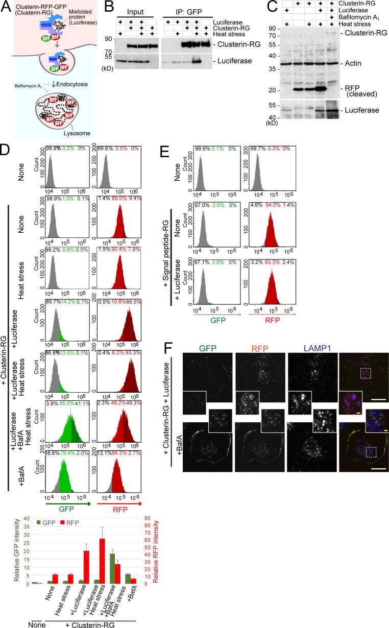
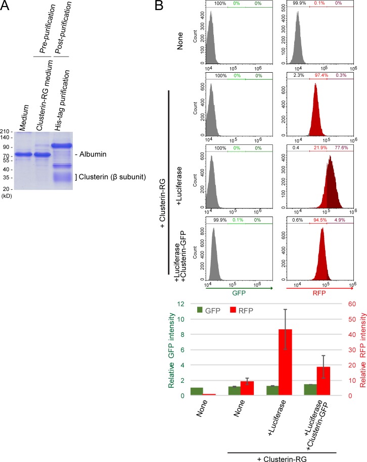
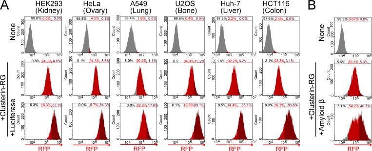
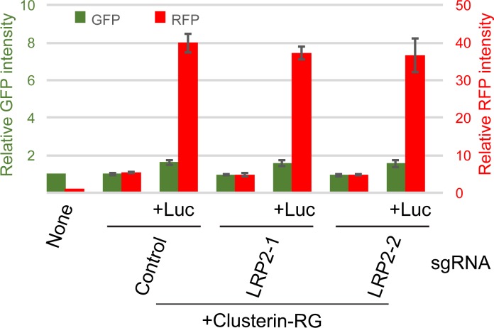
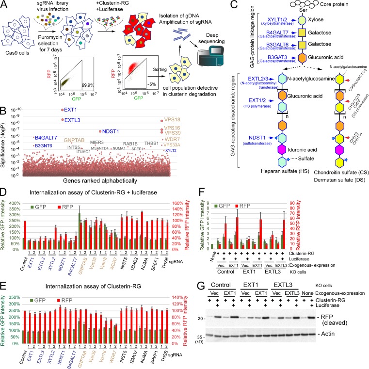
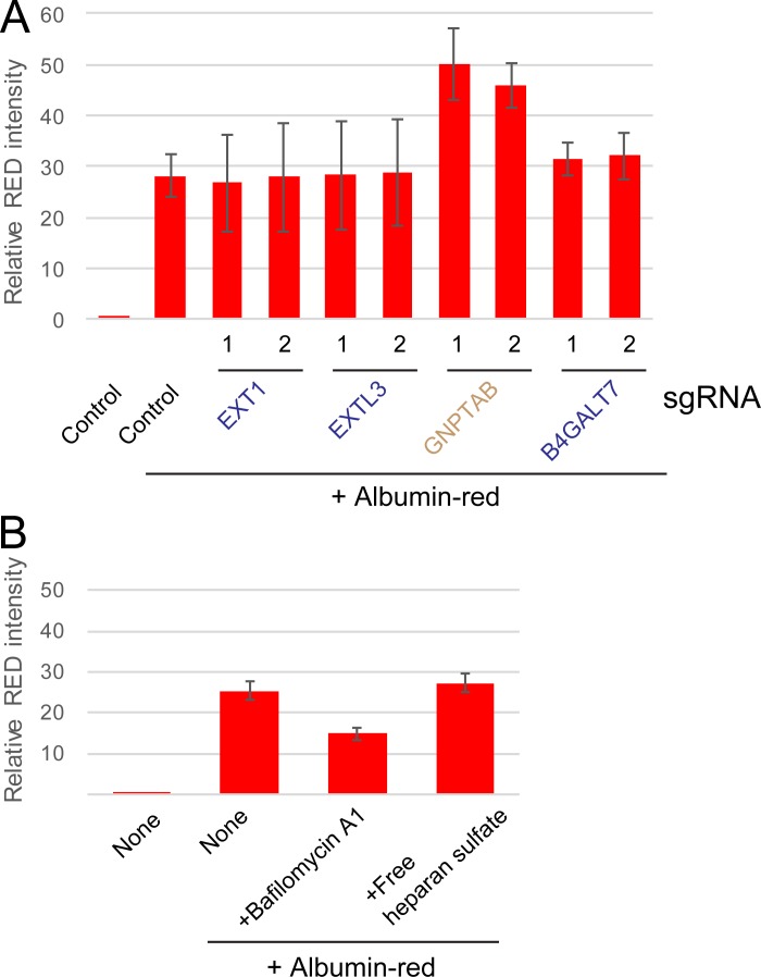
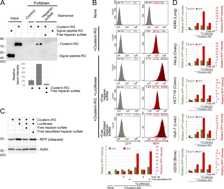
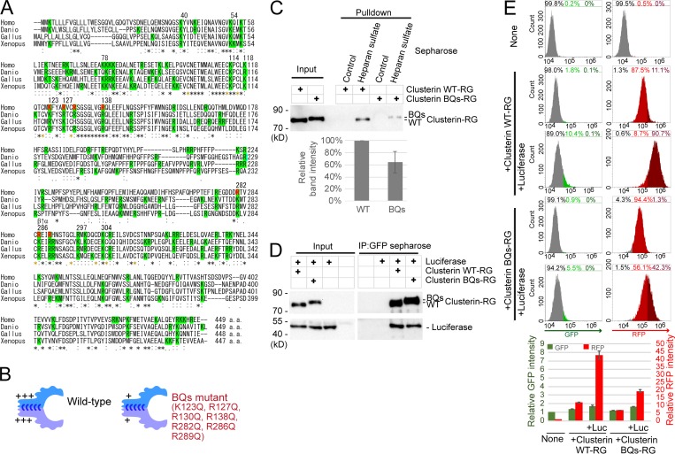
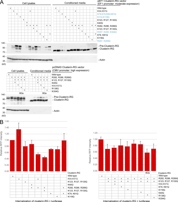
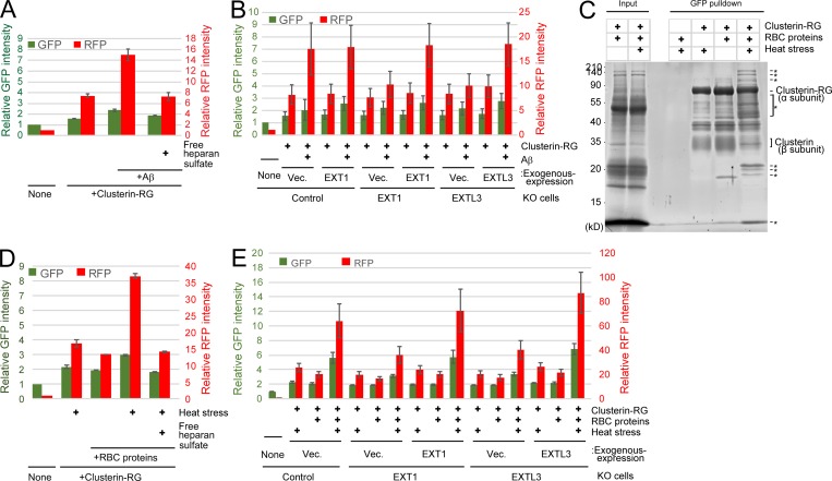
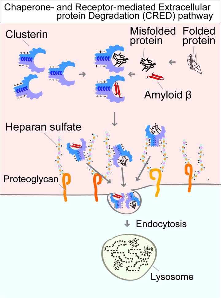
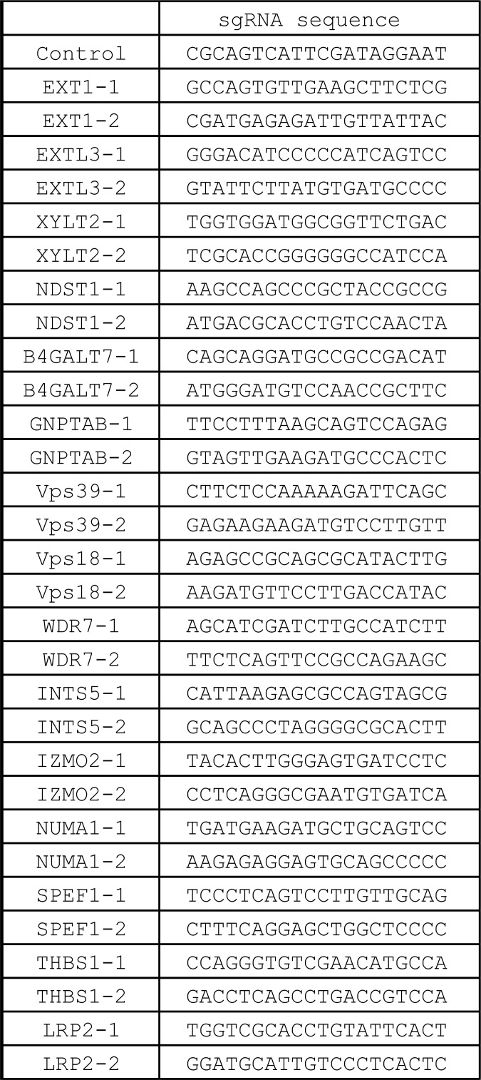
The accumulation of aberrant proteins leads to various neurodegenerative disorders. Mammalian cells contain several intracellular protein degradation systems, including autophagy and proteasomal systems, that selectively remove aberrant intracellular proteins. Although mammals contain not only intracellular but also extracellular proteins, the mechanism underlying the quality control of aberrant extracellular proteins is poorly understood. Here, using a novel quantitative fluorescence assay and genome-wide CRISPR screening, we identified the receptor-mediated degradation pathway by which misfolded extracellular proteins are selectively captured by the extracellular chaperone Clusterin and undergo endocytosis via the cell surface heparan sulfate (HS) receptor. Biochemical analyses revealed that positively charged residues on Clusterin electrostatically interact with negatively charged HS. Furthermore, the Clusterin-HS pathway facilitates the degradation of amyloid β peptide and diverse leaked cytosolic proteins in extracellular space. Our results identify a novel protein quality control system for preserving extracellular proteostasis and highlight its role in preventing diseases associated with aberrant extracellular proteins.


Figures

Figure 1.
Figure 1.
A novel method to monitor cumulative lysosomal degradation of an extracellular protein. (A) Schematic of the Clusterin-RFP-GFP (Clusterin-RG/CluRG) internalization assay. After internalization, lysosomal enzymes degrade Clusterin and GFP, whereas RFP, which is resistant to proteases and acidic pH, accumulates in lysosomes. The blue C-shaped structures in the Clusterin structure represent disulfide bonds between the α and β subunits. (B) Clusterin selectively binds to a misfolded protein. Purified CluRG was mixed with or without recombinant Luc in serum-free medium and preincubated at 4°C or 42°C for 20 min (heat stress). Samples were subjected to immunoprecipitation (IP) with anti-GFP Sepharose at 4°C. (C) Increased lysosomal degradation of Clusterin by misfolded proteins. Purified CluRG was mixed with or without recombinant Luc in serum-free medium and preincubated at 4°C or 42°C for 20 min (heat stress). HEK293 cells were cultured in the medium with or without Bafa for 14 h at 37°C and analyzed by immunoblotting. (D) Misfolded protein–dependent Clusterin internalization. Cells were treated as in C and analyzed by flow cytometry. The bar graph shows the relative fluorescence intensities (GFP, green left axis; RFP, red right axis) in a cell normalized to those in nontreated cells (n = 3). The data are presented as the mean ± SEM. Note that a 14-h incubation at 37°C moderately induces the misfolding of Luc without preheat stress. (E) Misfolded protein-independent RFP-GFP internalization. Purified SP-RG was mixed with or without recombinant Luc in serum-free medium and preincubated at 42°C for 20 min. HEK293 cells were cultured in the medium for 14 h at 37°C and analyzed by flow cytometry. (F) Lysosomal accumulation of Clusterin. Purified CluRG was mixed with recombinant Luc in serum-free medium and preincubated at 42°C for 20 min. HEK293 cells were cultured in the medium with or without BafA for 14 h at 37°C, immunolabeled for LAMP1 (a lysosomal marker), and imaged by confocal microscopy. Main scale bar, 10 µm. Inset scale bar, 2 µm.
Figure S1.
Figure S1.
Competitive inhibition of Clusterin internalization. (A) Recombinant CluRG (His-tagged) was purified from mammalian cells (see Materials and methods). Conditioned medium was collected from HEK293 cells expressing CluRG (prepurification). CluRG in the conditioned medium was affinity purified via the His tag (postpurification) and analyzed by Coomassie brilliant blue staining. (B) Purified CluRG was mixed with or without recombinant Luc in serum-free medium and preincubated at 42°C for 20 min. HEK293 cells were cultured in the medium with or without a 10-fold excess of Clusterin-GFP with Luc for 14 h at 37°C and analyzed by flow cytometry. The bar graph shows the relative fluorescence intensities (GFP, green left axis; RFP, red right axis) in a cell normalized to those in nontreated cells (n = 3). The data are presented as the mean ± SEM.
Figure 2.
Figure 2.
Selective extracellular protein degradation is ubiquitous in diverse tissues and for Aβ. (A) Clusterin-mediated degradation in various cell lines. Purified CluRG was mixed with or without recombinant Luc in serum-free medium and preincubated at 42°C for 20 min. The cells were cultured in medium for 14 h at 37°C and analyzed by flow cytometry. (B) Aβ-dependent Clusterin internalization. Purified CluRG was mixed with or without recombinant Aβ in serum-free medium and preincubated at 37°C. HEK293 cells were cultured in medium with or without free HS for 14 h at 37°C and analyzed by flow cytometry.
Figure S2.
Figure S2.
LRP2 is not involved in the internalization of the Clusterin–misfolded protein complex. Purified CluRG was mixed with or without recombinant Luc in serum-free medium and incubated at 42°C for 20 min. HEK293 cells expressing Cas9 with control or LRP2 sgRNA were cultured in the mixtures for 14 h at 37°C and analyzed by flow cytometry. The bar graph shows the relative fluorescence intensities (GFP, green left axis; RFP, red right axis) in a cell normalized to those in nontreated cells (n = 3). The data are presented as the mean ± SEM.
Figure 3.
Figure 3.
HS biosynthesis enzymes are essential for the degradation of the Clusterin-substrate complex. (A) Schematic representation of the screening. The GeCKO v2 sgRNA library was delivered into Cas9-expressing HEK293 cells by lentiviral infection. After 1 wk of culture, KO cells were treated with heat-stressed CluRG–Luc complex for 14 h, and those in the bottom 5% of the RFP/GFP ratio (cell population defective in Clusterin degradation) were sorted using a cell sorter. After this enrichment process was repeated twice, PCR amplification of the sgRNA coding sequence integrated into the chromosomes was conducted for next-generation sequencing. (B) CRISPR screening of Clusterin degradation-deficient cells. A modified robust rank aggregation algorithm was used to rank sgRNAs based on P values. HS biosynthesis enzyme genes (blue) and endolysosomal genes (brown) are indicated (B–E). (C) Schematic representation of the synthesis of the GAG backbones of HS or CS/DS chains. (D and E) HS synthesis enzymes are essential for misfolded protein–dependent Clusterin internalization. Purified CluRG was mixed with recombinant Luc in serum-free medium and preincubated at 42°C for 20 min. HEK293 cells expressing Cas9 with the indicated sgRNAs (two per candidate) were cultured in the medium for 14 h at 37°C and analyzed by flow cytometry (D). Cells were treated as in D except without Luc and analyzed by flow cytometry (E). The bar graph shows the relative fluorescence intensities (GFP, green left axis; RFP, red right axis) in a cell normalized to those in control cells (n = 3). The data are presented as the mean ± SEM. (F and G) Loss of Clusterin internalization in EXT1 or EXTL3 KO cells. Purified CluRG was mixed with or without recombinant Luc in serum-free medium and preincubated at 42°C for 20 min. Control, EXT1 KO, or EXTL3 KO HEK293 cells (generated by CRISPR) introduced with EXT1, EXTL3 or empty vector were cultured in the medium for 14 h at 37°C and analyzed by flow cytometry. The bar graph shows the relative fluorescence intensities (GFP, green left axis; RFP, red right axis) in a cell normalized to those in nontreated cells (n = 3). The data are presented as the mean ± SEM (F). Cells were treated as in F and analyzed by immunoblotting (G).
Figure S3.
Figure S3.
HS synthesis enzymes are not required for endocytosis. (A) HEK293 cells expressing Cas9 with the indicated sgRNAs (two per candidate) were cultured with or without albumin-red for 14 h and analyzed by flow cytometry. The bar graph shows the relative fluorescence intensities in a cell normalized to those in nontreated cells (n = 3). The data are presented as the mean ± SEM. (B) HS is not required for albumin endocytosis. HEK293 cells were cultured with or without albumin-red in the presence of BafA or free HS for 14 h and analyzed by flow cytometry. The bar graph shows the relative fluorescence intensities in a cell normalized to those in nontreated cells (n = 3). The data are presented as the mean ± SEM.
Figure 4.
Figure 4.
Clusterin directly interacts with HS. (A) In vitro binding of Clusterin and HS. Purified CluRG or SP-RG in the presence or absence of free HS was subjected to a pulldown assay with HS-conjugated or control Sepharose at 4°C. The bar graph shows the relative band intensity normalized to that of CluRG by HS-conjugated Sepharose pulldown (n = 3). The data are presented as the mean ± SEM. (B and C) Competitive inhibition of Clusterin internalization by free HS. Purified CluRG was mixed with or without recombinant Luc in serum-free medium and preincubated at 42°C for 20 min. HEK293 cells were cultured in the medium with or without free HS or free desulfated HS for 14 h at 37°C and analyzed by flow cytometry. The bar graph shows the relative fluorescence intensities (GFP, green left axis; RFP, red right axis) in a cell normalized to those in nontreated cells (n = 3). The data are presented as the mean ± SEM (B). HEK293 cells were treated as in B and analyzed by immunoblotting (C). (D) The Clusterin–HS pathway in various cell lines. Purified Clusterin WT-RG was mixed with or without recombinant Luc in serum-free medium and preincubated at 42°C for 20 min. The cells were cultured in medium with or without free HS for 14 h at 37°C and analyzed by flow cytometry. The bar graph shows the relative fluorescence intensities (GFP, green left axis; RFP, red right axis) in the cells normalized to that in nontreated cells (n = 3). The data are presented as mean ± SEM.
Figure 5.
Figure 5.
Electrostatic interactions between HS and Clusterin. (A) Multiple sequence alignment of different vertebrate Clusterin sequences. Identical residues in all sequences are indicated by asterisks (*), conserved substitutions are indicated by colons (:), and conserved cysteine residues for disulfide bonds are indicated by yellow asterisks. Basic amino acid resides are highlighted in green. Residues for BQs mutant are indicated by red. Homo, Homo sapiens; Danio, Danio rerio; Gallus, Gallus gallus; Xenopus, Xenopus laevis. The arrow between β and α indicates the cleavage site producing the β and α subunits. (B) Diagrams of WT Clusterin and the BQs mutant. The BQs mutant exhibited decreased positive charges. (C) Weak binding of Clusterin BQs to HS. Purified Clusterin WT-RG or Clusterin-BQs-RG was subjected to pulldown assays with HS-conjugated or control Sepharose at 4°C. The bar graph shows the relative band intensity normalized to that of Clusterin WT-RG by HS-conjugated Sepharose pulldown (n = 3). The data are presented as the mean ± SEM. (D) Normal binding of Clusterin BQs to misfolded proteins. Purified Clusterin WT-RG or Clusterin BQs-RG was mixed with recombinant Luc and incubated at 42°C for 20 min. Samples were subjected to immunoprecipitation with anti-GFP Sepharose at 4°C. (E) Inefficient internalization of Clusterin BQs. Purified Clusterin WT-RG or Clusterin BQs-RG was mixed with or without recombinant Luc in serum-free medium and preincubated at 42°C for 20 min. HEK293 cells were cultured in the medium for 14 h at 37°C and analyzed by flow cytometry. The bar graph shows the relative fluorescence intensities (GFP, green left axis; RFP, red right axis) in a cell normalized to those in nontreated cells (n = 4). The data are presented as the mean ± SEM.
Figure S4.
Figure S4.
The conserved basic amino acids in Clusterin for secretion and internalization. (A) Some basic residues in Clusterin are required for secretion. HEK293 cells transiently transfected with the indicated CluRG vectors were cultured for 3 d, and conditioned medium and cell lysates were analyzed by immunoblotting. Clusterin mutants colored in blue showed reduced cleavage efficiency and secretion. Note that the expression of CluRG by the cytomegalovirus (CMV) promoter increased pre-Clusterin expression in the cell due to overexpression. (B) The indicated Clusterin-RG mutants were mixed with recombinant Luc in serum-free medium and incubated at 42°C for 20 min. HEK293 cells were cultured in the mixtures for 14 h at 37°C and analyzed by flow cytometry. The bar graph shows the relative RFP intensity in a cell normalized to that of Clusterin WT-RG (n = 3). The data are presented as the mean ± SEM. The combination of the R282Q, R286Q, R289Q, K123Q, R127Q, R130Q, and R138Q mutations (BQs) most significantly reduced Clusterin internalization.
Figure 6.
Figure 6.
The Clusterin–HS pathway is a universal degradation system for a wide variety of substrates. (A) Clusterin–HS pathway–dependent Aβ degradation. Purified CluRG was mixed with or without recombinant Aβ in serum-free medium and preincubated at 37°C. HEK293 cells were cultured in medium with or without free HS for 14 h at 37°C and analyzed by flow cytometry. (B) Medium was prepared as in A. Control, EXT1 KO, or EXTL3 KO HEK293 cells (generated by CRISPR) introduced with EXT1, EXTL3, or empty vector (Vec.) were cultured in medium for 14 h at 37°C and analyzed by flow cytometry. The bar graph shows the relative fluorescence intensities (GFP, green left axis; RFP, red right axis) in the cells normalized to that in nontreated cells (n = 3). The data are presented as mean ± SEM. (C) Coimmunoprecipitation of CluRG with various RBC proteins. Purified CluRG was mixed with or without RBC proteins and preincubated at 4°C or 50°C for 60 min (heat stress). Samples were subjected to a pulldown assay using an anti-GFP sepharose antibody at 4°C, separated by SDS-PAGE, and detected with SYPRO-Ruby stain. The asterisks indicate heat stress–dependent Clusterin-binding proteins. (D and E) HS is essential for internalization of Clusterin–RBC protein complexes. Purified CluRG was mixed with or without RBC proteins in serum-free medium and preincubated at 50°C for 60 min (heat stress). Control (D), EXT1 KO, or EXTL3 KO (E) HEK293 cells introduced with EXT1, EXTL3, or empty vector were cultured in medium with or without free HS for 14 h at 37°C and analyzed by flow cytometry. The bar graph shows the relative fluorescence intensity (GFP, green left axis; RFP, red right axis) in the cells normalized to that in nontreated cells (n = 3). The data are presented as mean ± SEM.
Figure 7.
Figure 7.
Model of the CRED pathway. Stresses induce the generation of extracellular aberrant proteins, which selectively interact with Clusterin. Positively charged residues on Clusterin electrostatically bind to the negatively charged HS chain on proteoglycan. The Clusterin–protein complex on the HS chain is then delivered into the lysosome via endocytosis and is digested into amino acids.
Figure S5.
Figure S5.
sgRNA sequences used in this study.

Comment in

Similar articles

Cited by

References

    1. Bell R.D., Sagare A.P., Friedman A.E., Bedi G.S., Holtzman D.M., Deane R., and Zlokovic B.V.. 2007. Transport pathways for clearance of human Alzheimer’s amyloid beta-peptide and apolipoproteins E and J in the mouse central nervous system. J. Cereb. Blood Flow Metab. 27:909–918. 10.1038/sj.jcbfm.9600419 - DOI - PMC - PubMed
    1. Bettens K., Brouwers N., Engelborghs S., Lambert J.C., Rogaeva E., Vandenberghe R., Le Bastard N., Pasquier F., Vermeulen S., Van Dongen J., et al. . 2012. Both common variations and rare non-synonymous substitutions and small insertion/deletions in CLU are associated with increased Alzheimer risk. Mol. Neurodegener. 7:3 10.1186/1750-1326-7-3 - DOI - PMC - PubMed
    1. Billings P.C., and Pacifici M.. 2015. Interactions of signaling proteins, growth factors and other proteins with heparan sulfate: mechanisms and mysteries. Connect. Tissue Res. 56:272–280. 10.3109/03008207.2015.1045066 - DOI - PMC - PubMed
    1. Boland B., Yu W.H., Corti O., Mollereau B., Henriques A., Bezard E., Pastores G.M., Rubinsztein D.C., Nixon R.A., Duchen M.R., et al. . 2018. Promoting the clearance of neurotoxic proteins in neurodegenerative disorders of ageing. Nat. Rev. Drug Discov. 17:660–688. 10.1038/nrd.2018.109 - DOI - PMC - PubMed
    1. Byun K., Gil S.Y., Namkoong C., Youn B.S., Huang H., Shin M.S., Kang G.M., Kim H.K., Lee B., Kim Y.B., and Kim M.S.. 2014. Clusterin/ApoJ enhances central leptin signaling through Lrp2-mediated endocytosis. EMBO Rep. 15:801–808. 10.15252/embr.201338317 - DOI - PMC - PubMed

Publication types

MeSH terms