

Coronavirus disease 2019: initial chest CT findings
- PMID: 32211963
- PMCID: PMC7095437
- DOI: 10.1007/s00330-020-06816-7
Coronavirus disease 2019: initial chest CT findings
Abstract
Objectives: To systematically analyze CT findings during the early and progressive stages of natural course of coronavirus disease 2019 and also to explore possible changes in pulmonary parenchymal abnormalities during these two stages.
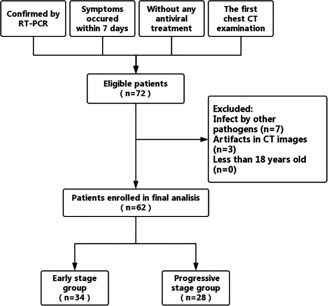
Methods: We retrospectively reviewed the initial chest CT data of 62 confirmed coronavirus disease 2019 patients (34 men, 28 women; age range 20-91 years old) who did not receive any antiviral treatment between January 21 and February 4, 2020, in Chongqing, China. Patients were assigned to the early-stage group (onset of symptoms within 4 days) or progressive-stage group (onset of symptoms within 4-7 days) for analysis. CT characteristics and the distribution, size, and CT score of pulmonary parenchymal abnormalities were assessed.
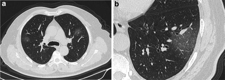
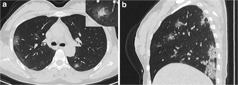
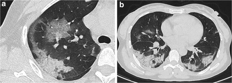
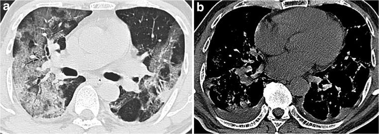
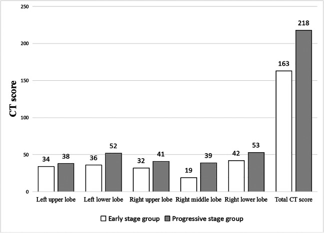
Results: In our study, the major characteristic of coronavirus disease 2019 was ground-glass opacity (61.3%), followed by ground-glass opacity with consolidation (35.5%), rounded opacities (25.8%), a crazy-paving pattern (25.8%), and an air bronchogram (22.6%). No patient presented cavitation, a reticular pattern, or bronchial wall thickening. The CT scores of the progressive-stage group were significantly greater than those of the early-stage group (p = 0.004).
Conclusions: Multiple ground-glass opacities with consolidations in the periphery of the lungs were the primary CT characteristic of coronavirus disease 2019. CT score can be used to evaluate the severity of the disease. If these typical alterations are found, then the differential diagnosis of coronavirus disease 2019 must be considered.
Key points: • Multiple GGOs with consolidations in the periphery of the lungs were the primary CT characteristic of COVID-19. • The halo sign may be a special CT feature in the early-stage COVID-19 patients. • Significantly increased CT score may indicate the aggravation of COVID-19 in the progressive stage.
Keywords: COVID-19; Coronavirus; Pneumonia; SARS-CoV-2; Spiral CT scan.
Conflict of interest statement
The authors of this manuscript declare no relationships with any companies whose products or services may be related to the subject matter of the article.
Figures
References
-
- World Health Organization . Statement on novel coronavirus in Thailand. Geneva: World Health Organization; 2020.
-
- World Health Organization . WHO confirms first cases of novel coronavirus (2019-nCoV) in the Eastern Mediterranean Region. Geneva: World Health Organization; 2020.
-
- World Health Organization . Readiness is the key to detect, combat spread of the new coronavirus. Geneva: World Health Organization; 2020.
MeSH terms
Grants and funding
LinkOut - more resources
Full Text Sources
Other Literature Sources
Medical
Miscellaneous
