. 2021 Mar;24(1):107-114.
doi: 10.1007/s40477-020-00452-z. Epub 2020 Mar 25.

Pediatrician performed point-of-care ultrasound for the detection of ingested foreign bodies: case series and review of the literature

Affiliations

Pediatrician performed point-of-care ultrasound for the detection of ingested foreign bodies: case series and review of the literature

Danilo Buonsenso et al. J Ultrasound. 2021 Mar.

Abstract

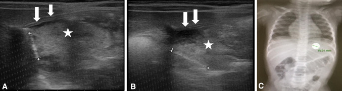
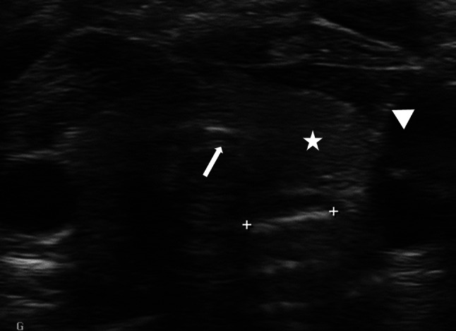
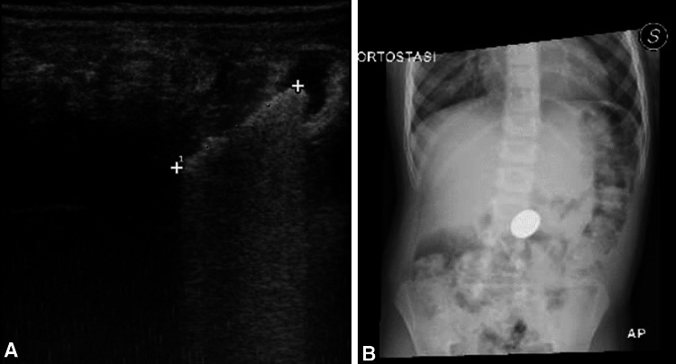
Purpose: Foreign body (FB) ingestions represent a common problem in children. History and physical examination are commonly not enough to diagnose a foreign body ingestion; therefore, conventional radiography is routinely used to detect them. Point-of-care ultrasound is widely used in the emergency department for several diagnostic applications but there are few articles describing the possibility to use point-of-care ultrasound to detect ingested foreign bodies, and the necessary training to get competent in this application. The main objective of this paper is to illustrate the use of point-of-care ultrasound (POCUS) to detect ingested foreign bodies. The secondary objective is to describe a limited training, necessary for emergency pediatricians, to obtain this skill.

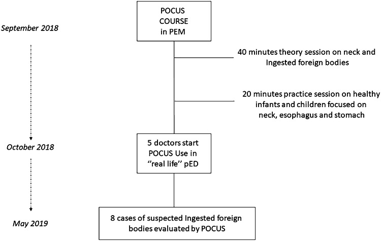
Methods: This is a case series of eight pediatric patients who presented to the pediatric Emergency Department (ED), with suspected ingestion of FB, and were assessed with POCUS. Physician sonographers were two pediatricians and three residents in pediatrics working in two Italian Pediatric EDs. All sonographers participated in a 2-day POCUS workshop which included the most common pediatric POCUS applications.

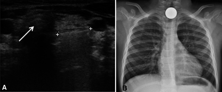
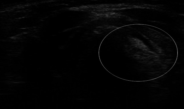
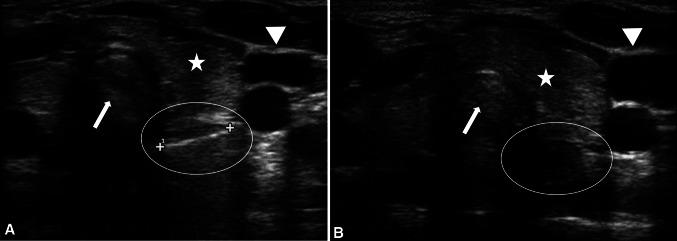
Results: POCUS, performed by emergency pediatricians who participated to a limited training, allowed to always identify the foreign bodies ingested.

Conclusions: We demonstrate that an appropriate and limited training allows pediatric emergency physicians to correctly identify foreign body in the esophagus or stomach. Point-of-care ultrasound in foreign body ingestion in the Emergency Department may allow to prioritize the escalation of care in children and it can contribute to reduce the time to endoscopic management when needed.

Keywords: Coin ingested; Emergency department; Foreign body; Point-of-care ultrasound.


Conflict of interest statement

The authors declare that they have no competing interests.

Figures

Fig. 1
Fig. 1
Study flow
Fig. 2
Fig. 2
Ultrasound image a shows the foreign body (between the two crosses) in the esophagus, at the level of the thoracic inlet, adjacent to the trachea (white arrow). The coin appears as a hyperechoic lesion with back acoustic shadowing. Chest X-ray (b) confirming a coin (radiopaque oval object) in the cervico-thoracic tract of the esophagus
Fig. 3
Fig. 3
Ultrasound image shows an oval structure of mixed echogenicity contents, mainly hyperechoic with small hypoechoic bubbles inside (white circle)
Fig. 4
Fig. 4
Ultrasound image a shows foreign body (between the two crosses) in the esophagus, at the level of the thoracic inlet, adjacent to the trachea. The coin appears as a hyperechoic lesion with back acoustic shadowing. Chest X-ray (b, c) confirming a coin (radiopaque oval object) in the cervico-thoracic tract of the esophagus
Fig. 5
Fig. 5
Ultrasound image a shows the foreign body (between the two crosses) within the esophagus (white circle), at the level of the thoracic inlet, adjacent to the trachea (white arrow) and main vessels (white arrowhead), behind the thyroid (white star). The coin appears as a hyperechoic lesion with back acoustic shadowing. Ultrasound image after coin removal b shows the esophagus (white circle) surrounded by the same structures, without the hyperecogenic line inside
Fig. 6
Fig. 6
Ultrasound images a and b show foreign body (between the two crosses) within the stomach; the stomach wall is defined by the white arrows, while its content by the white star. The coin appears as a hyperechoic lesion with back acoustic shadowing. Chest–abdominal radiograph confirming a coin (radiopaque oval object) in the stomach (c)
Fig. 7
Fig. 7
Ultrasound image a shows the foreign body (between the two crosses) in the esophagus, at the level of the thoracic inlet, adjacent to the trachea (white arrow) main vessels (white arrowhead), behind the thyroid (white star). The coin appears as a hyperechoic lesion with back acoustic shadowing. Video 1, supplementary file, shows a clip of the same case
Fig. 8
Fig. 8
Ultrasound images a showing a hyperechoic line with shadowing within the bowel. X-ray confirmed the presence of the coin in the abdomen b

References

    1. Gurevich Y, Sahn B, Weinstein T. Foreign body ingestion in pediatric patients. Curr Opin Pediatr. 2018;30:677–682. doi: 10.1097/MOP.0000000000000670. - DOI - PubMed
    1. Seguin J, Kwan C. Novel use of point-of-care ultrasound for pediatric foreign bodies: an emergency department cases series. J Emerg Med. 2018;55:530–533. doi: 10.1016/j.jemermed.2018.06.011. - DOI - PubMed
    1. Little DC, Shah SR, St Peter SD, et al. Esophageal foreign bodies in the pediatric population: our first 500 cases. J Pediatr Surg. 2006;41:914–918. doi: 10.1016/j.jpedsurg.2006.01.022. - DOI - PubMed
    1. Kramer RE, Lerner DG, Lin T, et al. Management of ingested foreign bodies in children: a clinical report of the NASPGHAN Endoscopy Committee. J Pediatr Gastroenterol Nutr. 2015;60:562–574. doi: 10.1097/MPG.0000000000000729. - DOI - PubMed
    1. Marin JR, Abo AM, Arroyo AC, et al. Pediatric emergency medicine point-of-care ultrasound: summary of the evidence. Crit Ultrasound J. 2016;8:16. doi: 10.1186/s13089-016-0049-5. - DOI - PMC - PubMed