. 2020 May;51(5):1570-1577.
doi: 10.1161/STROKEAHA.120.029071. Epub 2020 Mar 26.

PERK (Protein Kinase RNA-Like ER Kinase) Branch of the Unfolded Protein Response Confers Neuroprotection in Ischemic Stroke by Suppressing Protein Synthesis

Affiliations

PERK (Protein Kinase RNA-Like ER Kinase) Branch of the Unfolded Protein Response Confers Neuroprotection in Ischemic Stroke by Suppressing Protein Synthesis

Ya-Chao Wang et al. Stroke. 2020 May.

Abstract

Background and Purpose- Ischemic stroke impairs endoplasmic reticulum (ER) function, causes ER stress, and activates the unfolded protein response. The unfolded protein response consists of 3 branches controlled by ER stress sensor proteins, which include PERK (protein kinase RNA-like ER kinase). Activated PERK phosphorylates eIF2α (eukaryotic initiation factor 2 alpha), resulting in inhibition of global protein synthesis. Here, we aimed to clarify the role of the PERK unfolded protein response branch in stroke. Methods- Neuron-specific and tamoxifen-inducible PERK conditional knockout (cKO) mice were generated by cross-breeding Camk2a-CreERT2 with Perkf/f mice. Transient middle cerebral artery occlusion was used to induce stroke. Short- and long-term stroke outcomes were evaluated. Protein synthesis in the brain was assessed using a surface-sensing-of-translation approach. Results- After tamoxifen-induced deletion of Perk in forebrain neurons was confirmed in PERK-cKO mice, PERK-cKO and control mice were subjected to transient middle cerebral artery occlusion and 3 days or 3 weeks recovery. PERK-cKO mice had larger infarcts and worse neurological outcomes compared with control mice, suggesting that PERK-induced eIF2α phosphorylation and subsequent suppression of translation protects neurons from ischemic stress. Indeed, better stroke outcomes were observed in PERK-cKO mice that received postischemic treatment with salubrinal, which can restore the ischemia-induced increase in phosphorylated eIF2α in these mice. Finally, our data showed that post-treatment with salubrinal improved functional recovery after stroke. Conclusions- Here, we presented the first evidence that postischemic suppression of translation induced by PERK activation promotes recovery of neurological function after stroke. This confirms and further extends our previous observations that recovery of ER function impaired by ischemic stress critically contributes to stroke outcome. Therefore, future research should include strategies to improve stroke outcome by targeting unfolded protein response branches to restore protein homeostasis in neurons.

Keywords: animals; mice; neurons; neuroprotection; proteostasis.


Figures

Figure 1.
Figure 1.. Characterization of PERK conditional knockout (PERK-cKO) mice.
A) Schematic depicting the timeline for the generation of conditional Perk deletion mice for the subsequent experiments. Perkf/f;Camk2a-CreERT2 (PERK-cKO) mice were identified by PCR genotyping and treated with tamoxifen daily for 5 days. Three weeks later, mice were used for experiments. B) Verification of Perk deletion in the brain of PERK-cKO. PCR analysis on brain DNA samples was used to confirm deletion of Perk exons in the brain of PERK-cKO mice. WT, wild-type. C) Quantitative reverse transcription PCR analysis on brain RNA samples was used to confirm deficiency in Perk mRNA expression in PERK-cKO mouse brains (n = 6). D) Western blot analysis. Control and PERK-cKO mice were subjected to 30 minutes MCAO and 1 hour reperfusion. Brain cortex samples from the contralateral (Contra) and ipsilateral (Ipsi) hemispheres were collected and evaluated by Western blotting. Intensities of each band were measured and normalized to β-actin (n = 3). The mean values in contralateral samples of control mice were set to 1.0. Data are presented as mean ± SD. *, p < 0.05; **, p < 0.01.
Figure 2.
Figure 2.. Deletion of Perk in forebrain neurons worsens both short- and long-term outcomes after ischemic stroke.
A, B) Short-term stroke outcomes (3-day). PERK-cKO (cKO) and littermate control mice (n = 9–10/group) were subjected to 30 minutes MCAO. After 24 hours reperfusion, mice were evaluated for neurologic deficit scores and by tight rope test (A). On day 3 after stroke, infarct volumes were measured (B; shown are representative TTC-stained brain slices). C) Long-term stroke outcomes (3-week). PERK-cKO and control mice (n = 7–8/group) were subjected to 30 minutes MCAO. Body weight was monitored over time. Tight rope and rotarod tests were evaluated pre-surgery (day 0) for baselines and then weekly for 3 weeks. The open field test was performed on day 21 after stroke. Horizontal bars represent median value of neurologic scores, and other data are presented as mean ± SD. *, p < 0.05; **, p < 0.01; ***, p < 0.001; ****, p < 0.0001.
Figure 3.
Figure 3.. PERK-cKO mice exhibit protein synthesis suppression in the brain to a lesser extent than control mice after brain ischemia/reperfusion.
Mice were subjected to sham surgery or 15 minutes forebrain ischemia. After 15 minutes reperfusion, puromycin was administered, and 2 hours later, hippocampal samples were collected for Western blot analysis. A) Suppression of protein synthesis after brain ischemia/reperfusion. Levels of newly synthesized proteins labeled with puromycin were markedly lower in the post-ischemic brains than in the sham brains (n = 3). B) The marked post-ischemic suppression of protein synthesis was reversed by Perk deletion (n = 3). C) The effect of Perk deletion on post-ischemic suppression of protein synthesis was largely nullified by salubrinal (Sal) treatment (n=7). Data are presented as mean ± SD. *, p < 0.05.
Figure 4.
Figure 4.. Post-treatment with salubrinal improves stroke outcome in PERK-cKO mice.
PERK-cKO mice were subjected to 30 minutes MCAO, and at 30 minutes reperfusion, vehicle or salubrinal was intraperitoneally injected. After 24 hours reperfusion, animals were evaluated for neurologic deficit scores (A), and then for infarct volume (B). Horizontal bars represent median value of neurologic scores. Infarct volumes are presented as mean ± SD (n = 6–7/group). **, p < 0.01.
Figure 5.
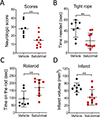
Figure 5.. Stroke outcome in C57Bl/6 mice is improved after post-treatment with salubrinal.
C57Bl/6 mice (n = 8–10/group) were subjected to 30 minutes MCAO, and at 30 minutes reperfusion, vehicle or salubrinal was intraperitoneally injected. After 24 hours reperfusion, the animals were subjected to neurologic scoring (A), and to tight rope (B) and rotarod (C) tests. On day 3 after stroke, infarct volumes were assessed. Horizontal bars represent median value of neurologic scores, and other data are presented as mean ± SD. *, p < 0.05; **, p < 0.01.

References

    1. Kleihues P, Hossmann KA. Protein synthesis in the cat brain after prolonged cerebral ischemia. Brain Res. 1971;35:409–418 - PubMed
    1. Thilmann R, Xie Y, Kleihues P, Kiessling M. Persistent inhibition of protein synthesis precedes delayed neuronal death in postischemic gerbil hippocampus. Acta Neuropathol. 1986;71:88–93 - PubMed
    1. Paschen W, Proud CG, Mies G. Shut-down of translation, a global neuronal stress response: Mechanisms and pathological relevance. Curr Pharm Des. 2007;13:1887–1902 - PubMed
    1. Cooper HK, Zalewska T, Kawakami S, Hossmann KA, Kleihues P. The effect of ischaemia and recirculation on protein synthesis in the rat brain. J Neurochem. 1977;28:929–934 - PubMed
    1. Burda J, Martin ME, Garcia A, Alcazar A, Fando JL, Salinas M. Phosphorylation of the alpha subunit of initiation factor 2 correlates with the inhibition of translation following transient cerebral ischaemia in the rat. Biochem J. 1994;302 ( Pt 2):335–338 - PMC - PubMed

Publication types

MeSH terms