. 2020 Jun;8(5):594-606.
doi: 10.1177/2050640620905183. Epub 2020 Feb 19.

Pancreatic cancer-derived organoids - a disease modeling tool to predict drug response

Affiliations

Pancreatic cancer-derived organoids - a disease modeling tool to predict drug response

Pierre-Olivier Frappart et al. United European Gastroenterol J. 2020 Jun.

Erratum in

  • Corrigendum.
    [No authors listed] [No authors listed] United European Gastroenterol J. 2020 Oct;8(8):987. doi: 10.1177/2050640620944750. United European Gastroenterol J. 2020. PMID: 32981490 Free PMC article. No abstract available.

Abstract

Background: Organotypic cultures derived from pancreatic ductal adenocarcinoma (PDAC) termed pancreatic ductal cancer organoids (PDOs) recapitulate the primary cancer and can be derived from primary or metastatic biopsies. Although isolation and culture of patient-derived pancreatic organoids were established several years ago, pros and cons for individualized medicine have not been comprehensively investigated to date.

Methods: We conducted a feasibility study, systematically comparing head-to-head patient-derived xenograft tumor (PDX) and PDX-derived organoids by rigorous immunohistochemical and molecular characterization. Subsequently, a drug testing platform was set up and validated in vivo. Patient-derived organoids were investigated as well.

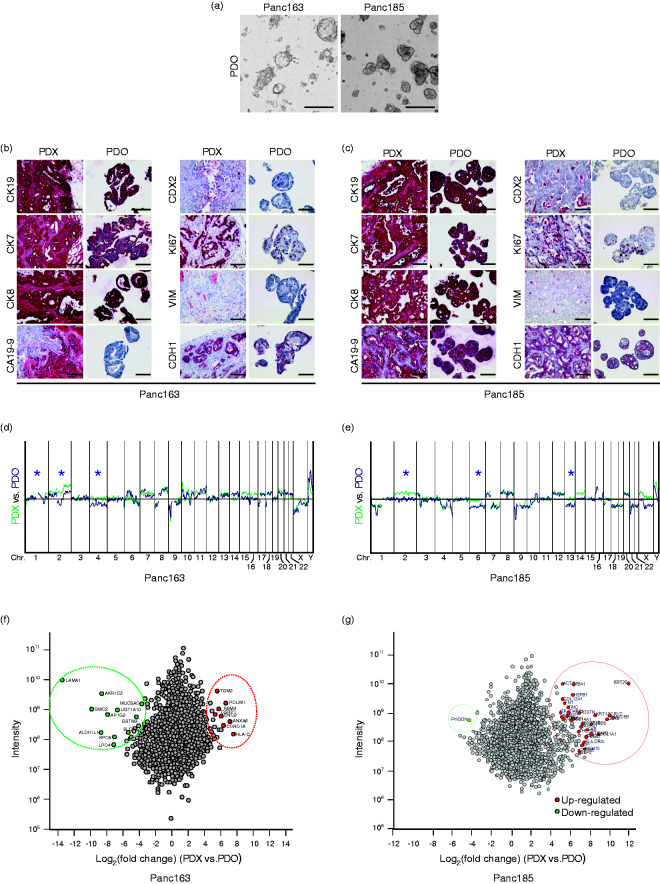
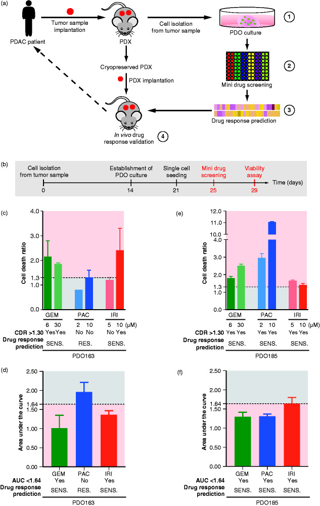
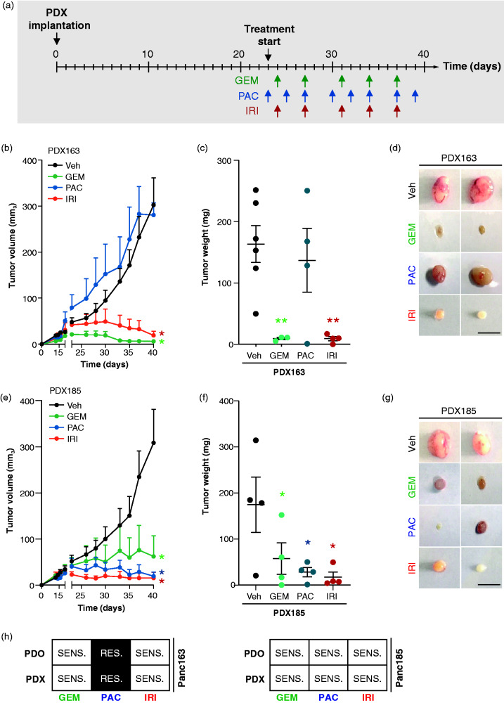
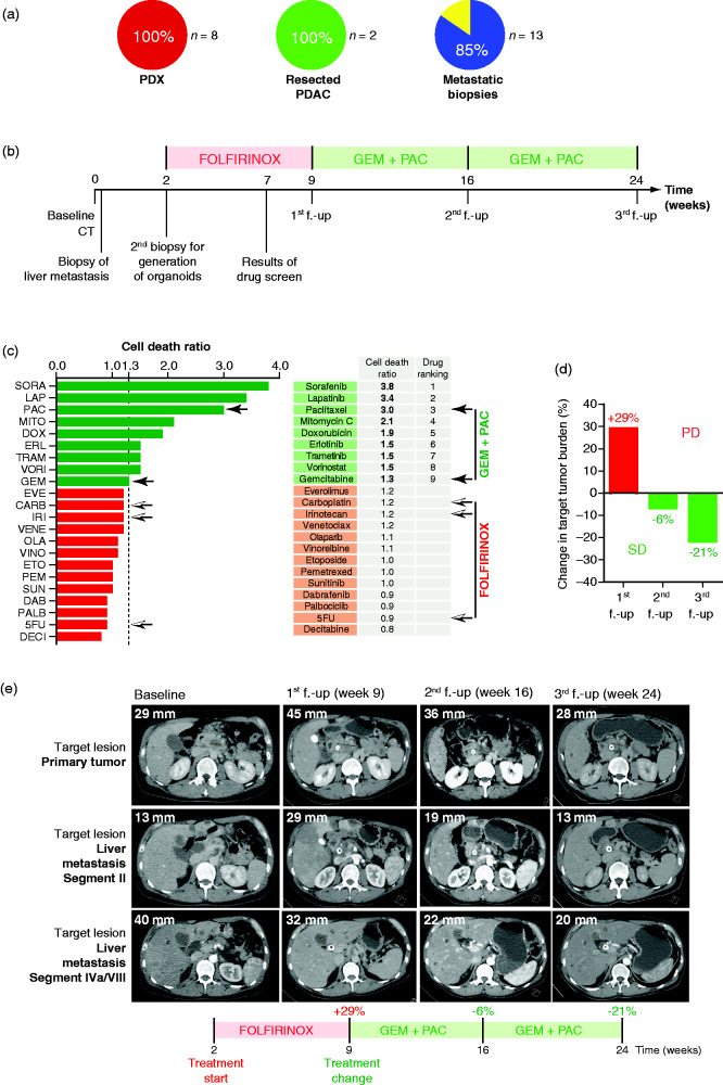
Results: First, PDOs faithfully recapitulated the morphology and marker protein expression patterns of the PDXs. Second, quantitative proteomes from the PDX as well as from corresponding organoid cultures showed high concordance. Third, genomic alterations, as assessed by array-based comparative genomic hybridization, revealed similar results in both groups. Fourth, we established a small-scale pharmacotyping platform adjusted to operate in parallel considering potential obstacles such as culture conditions, timing, drug dosing, and interpretation of the results. In vitro predictions were successfully validated in an in vivo xenograft trial. Translational proof-of-concept is exemplified in a patient with PDAC receiving palliative chemotherapy.

Conclusion: Small-scale drug screening in organoids appears to be a feasible, robust and easy-to-handle disease modeling method to allow response predictions in parallel to daily clinical routine. Therefore, our fast and cost-efficient assay is a reasonable approach in a predictive clinical setting.

Keywords: PDAC; drug response prediction; organoids.


Figures

Figure 1.
Figure 1.
Pancreatic ductal organoid vs. patient-derived xenograft comparison. (a) Representative images of pancreatic ductal organoids (PDO) isolated from human PDAC. Scale bars represent 400 µm. Immunohistochemical characterization of patient-derived xenograft (PDX) and PDO from (b) Panc163 and (c) from Panc185. Scale bars represent 100 µm. Array-Comparative Genomic Hybridization analysis showing genomic rearrangements (*) in PDO vs. PDX of Panc163 (d) and of Panc185 (e). Proteome comparison between PDO vs. PDX of Panc163 (f) and of Panc185 (g).
Figure 2.
Figure 2.
A pancreatic cancer-derived organoids tool to predict drug response. (A) Schematic representation of our pancreatic ductal organoid (PDO)-derived tool. (1) PDO and patient-derived xenograft (PDX) models were generated from cryopreserved xenografts of two patients with pancreatic ductal adenocarcinoma (PDAC). Several FDA-approved drugs were screened either single or in combination (2) and validated on the established organoid cultures (3). Drugs which showed sensitivity in PDO were selected for further small-scale drug screenings and validation in the PDXs (4). (B) Schematic representation of an organoid-based drug screening. (C) Cell death ratio (CDR) analysis of a viability assay and (D) area under the curve (AUC) analysis of a dose-response assay conducted on PDO163. (E) CDR analysis of a viability assay and (F) AUC analysis of a dose-response assay conducted on PDO185. Drug response prediction using CDR and AUC are provided below each bar graph. Yes and No indicate respectively a positive drug response and a lack of drug response. PDO is considered to be sensitive when CDR of one of treatment conditions >1.30 and if AUC < 1.64. GEM (gemcitabine); IRI (irinotecan); PAC (paclitaxel); RES., resistant; SENS., sensitive.
Figure 3.
Figure 3.
Drug response validation in patient-derived xenograft (PDX). (A) Illustration of the experimental in vivo set-up. (B) Time-dependent development (over the course of 40 days) of subcutaneously engrafted PDX163 treated with GEM (125 mg/kg; n = 3; green lines), PAC (12 mg/kg; n = 4; blue lines), IRI (50 mg/kg; n = 4; red lines), and vehicle (n = 6; black lines). (C) Quantification of tumor weight and (D) representative macroscopic pictures of resected PDXs from subcutaneous assay shown in (B). Scale bars represent 1 cm. (E) Time-dependent development (over the course of 40 days) of subcutaneously engrafted PDX185 treated or not with GEM (125 mg/kg; n = 4; green lines), PAC (12 mg/kg; n = 4; blue lines), IRI (50 mg/kg; n = 4; red lines), and vehicle (n = 4; black lines). (F) Quantification of tumor weight and (G) representative macroscopic pictures of resected PDXs from subcutaneous assay shown in (E). Scale bars represent 1 cm. Error bars indicate SEM. (H) Summary of the prediction efficiency of the mini drug screening on PDOs vs. PDXs. GEM (gemcitabine); IRI (irinotecan); PAC (paclitaxel); Veh, vehicle; RES., resistant; SENS., sensitive. *p < 0.05; **p < 0.001.
Figure 4.
Figure 4.
Reproducibility and power of the assay. (A) Schematic representation of PDO163 and PDO163W/R-free isolation and propagation using pancreatic ductal organoid media respectively supplemented or not with WNT3A/RSPOI. (B) Survival analysis of a viability assay conducted on PDO163 and PDO163W/R-free. (C) Schematic representation of the comparison of multiple independent experiments vs. single experiment of drug screening. Cell death ratio (CDR) analysis of multiple viability assays (n = 5) vs. a single experiment conducted on (D) PDO163 (n = 5) and (E) PDO185 (n = 3). Drug response prediction using CDR is provided below each bar graph. Yes and No indicate respectively a positive drug response and a lack of drug response. PDO is considered to be sensitive when CDR of one of treatment conditions >1.30. GEM (gemcitabine); IRI (irinotecan); PAC (paclitaxel); RES., resistant; SENS., sensitive.
Figure 5.
Figure 5.
Organoid-based tailored therapies. (A) Take rate of pancreatic ductal organoid (PDO) isolation from PDX (n = 8), resected primary pancreatic ductal adenocarcinoma (PDAC) (n = 2) and ultrasound-guided biopsy of the primary cancer or liver metastases (n = 13). (B) Scheme of the course of treatment of the patient. (C) Twenty-two drug viability assay screening performed on patient PDO (one dose per drug). Black arrows show components of GEM + PAC therapy. Black-and-white arrows show components of FOLFIRINOX therapy. (D) Waterfall plot showing percent change in tumor burden determined based on RECIST 1.1. criteria at first follow-up (f.-up) with an increase of 29% corresponding to progressive disease (PD). At second f.-up and third f.-up, there was a decrease of, respectively, 6% and 21%, corresponding to stable disease (SD). (E) Representative pictures of CT scan. Shown is the longest dimension of target lesion of the primary tumor and target lesion of a liver metastasis segment II and segment IVa/VIII at baseline (time of initial diagnosis), at first f.-up (week 9) after treatment with FOLFIRINOX, at second f.-up (week 16) after treatment with gemcitabine/nab-paclitaxel, and at third f.-up (week 24) after continued therapy with gemcitabine/nab-paclitaxel. 5FU: 5-fluorouracil; CARB: carboplatin; DAB: dabrafenib; DECI: decitabine; DOX: doxorubicin; ERL: erlotinib; ETO: etoposide; EVE: everolimus; GEM: gemcitabine; IRI: irinotecan; LAP: lapatinib; MITO: mitomycin C; OLA: olaparib; PAC: paclitaxel; PALB: palbociclib; PEM: pemetrexed; SORA: sorafenib; SUN: sunitinib; TRAM: trametinib; VENE: venetoclax; VINO: vinorelbine tartrate; VORI: vorinostat.

References

    1. Kleger A, Perkhofer L, Seufferlein T. Smarter drugs emerging in pancreatic cancer therapy. Ann Oncol 2014; 25: 1260–1270. - PubMed
    1. Russell R, Perkhofer L, Liebau S, et al. Loss of ATM accelerates pancreatic cancer formation and epithelial-mesenchymal transition. Nat Commun 2015; 6: 7677–7677. - PMC - PubMed
    1. Mueller S, Engleitner T, Maresch R, et al. Evolutionary routes and KRAS dosage define pancreatic cancer phenotypes. Nature 2018; 554: 62–68. - PMC - PubMed
    1. Waddell N, Pajic M, Patch AM, et al. Whole genomes redefine the mutational landscape of pancreatic cancer. Nature 2015; 518: 495–501. - PMC - PubMed
    1. Seufferlein T, Kleger A. Organoidomics – falling star or new galaxy in pancreatic cancer? Nat Rev Gastroenterol Hepatol 2018; 15: 586–587. - PubMed

Publication types

MeSH terms

Substances