Review
. 2020 Mar 27;126(7):926-941.
doi: 10.1161/CIRCRESAHA.119.314681. Epub 2020 Mar 26.

Mitochondrial Determinants of Doxorubicin-Induced Cardiomyopathy

Affiliations
Review

Mitochondrial Determinants of Doxorubicin-Induced Cardiomyopathy

Kendall B Wallace et al. Circ Res. .

Abstract

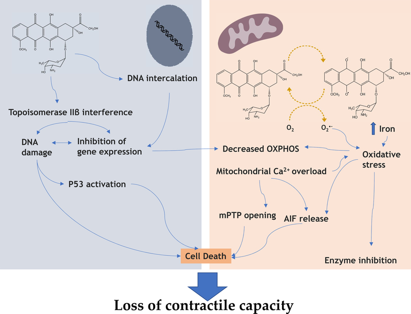
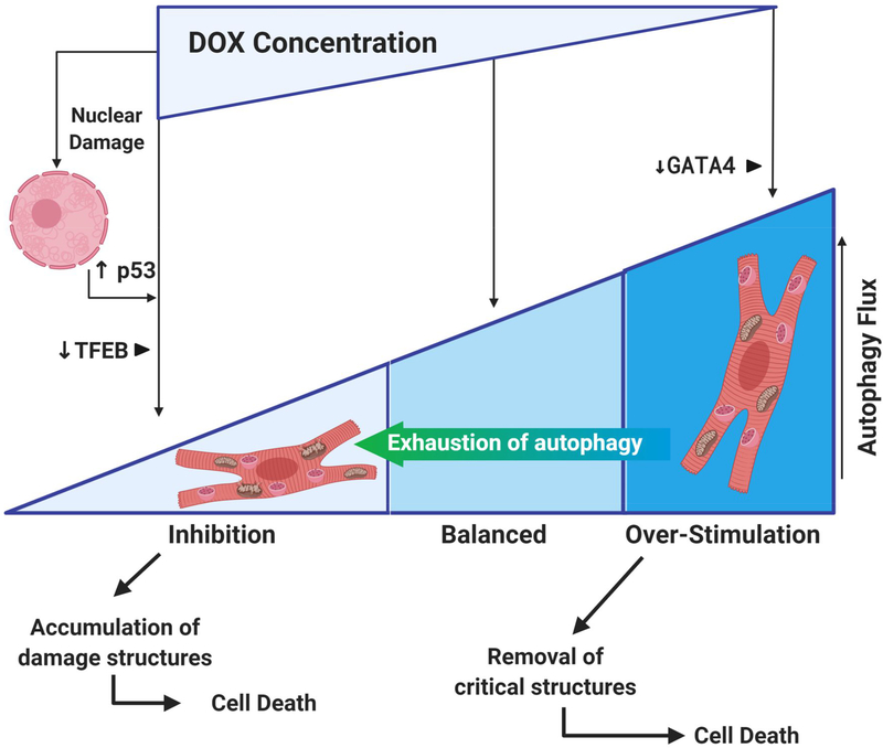
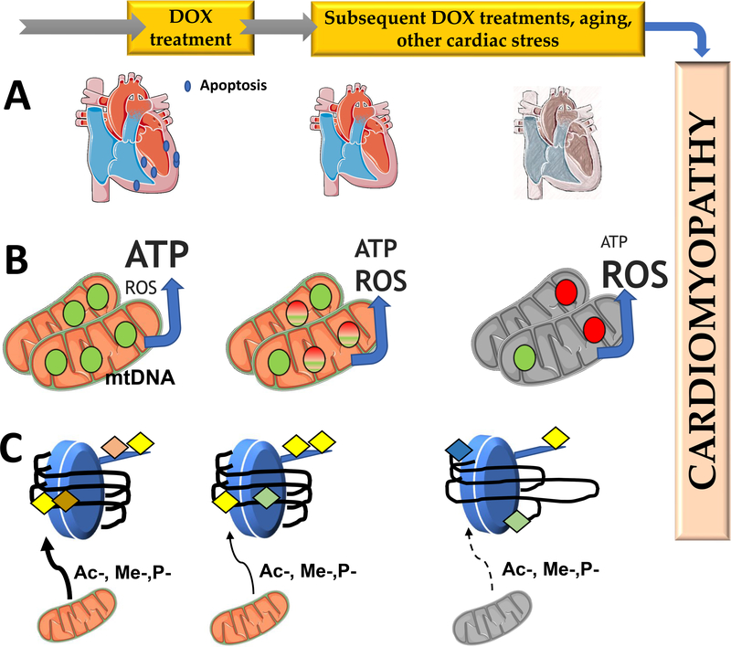
Anthracycline-based chemotherapy can result in the development of a cumulative and progressively developing cardiomyopathy. Doxorubicin is one of the most highly prescribed anthracyclines in the United States due to its broad spectrum of therapeutic efficacy. Interference with different mitochondrial processes is chief among the molecular and cellular determinants of doxorubicin cardiotoxicity, contributing to the development of cardiomyopathy. The present review provides the basis for the involvement of mitochondrial toxicity in the different functional hallmarks of anthracycline toxicity. Our objective is to understand the molecular determinants of a progressive deterioration of functional integrity of mitochondria that establishes a historic record of past drug treatments (mitochondrial memory) and renders the cancer patient susceptible to subsequent regimens of drug therapy. We focus on the involvement of doxorubicin-induced mitochondrial oxidative stress, disruption of mitochondrial oxidative phosphorylation, and permeability transition, contributing to altered metabolic and redox circuits in cardiac cells, ultimately culminating in disturbances of autophagy/mitophagy fluxes and increased apoptosis. We also suggest some possible pharmacological and nonpharmacological interventions that can reduce mitochondrial damage. Understanding the key role of mitochondria in doxorubicin-induced cardiomyopathy is essential to reduce the barriers that so dramatically limit the clinical success of this essential anticancer chemotherapy.

Keywords: cardiomyopathy; cardiotoxicity; doxorubicin; mitochondria; oxidative stress.


Figures

Figure 1 –
Figure 1 –
Bioactivation of Doxorubicin (DOX) (A) by mitochondrial complex I (mitochondrial respiratory chain) at the expense of NADH or by the cytochrome P450 system, using reducing equivalents from NADPH. NAD(P) H acts as an electron donor for the reduction of DOX producing a semiquinone radical (B) which rapidly re-oxidizes in the presence of molecular oxygen to generate superoxide anion free radicals and the parent DOX. This process is known as DOX redox-cycle and increases the production of superoxide anion which can directly damage lipids and proteins or converted to other ROS, including hydrogen peroxide. Alternatively, in the absence of molecular oxygen, DOX semiquinone can be simply converted to secondary metabolism doxorubicinol (C) or can undergo aglycosylation (D) forming a C7 radical (DNA alkylation) (E) which can form dimmers (F) or be converted to a 7-deoxyaglycone (G) (DNA intercalation). Legend: SOD: Superoxide dismutase; O2•−: superoxide anion, H2O2: hydrogen peroxide, HO: hydroxyl radical, ROOH: lipid peroxides, ROO: peroxy radical.
Figure 2 –
Figure 2 –
Summary of published data demonstrating the interplay between nuclear and mitochondrial effects of Doxorubicin (DOX). DOX directly inhibit mitochondrial function by direct interaction with Complex I and other complexes of the respiratory chain, as well as other proteins involved in oxidative phosphorylation, and by inactivation through increased reactive oxygen species (ROS) generation. ROS can play a dual role, depending on the species produced or magnitude or oxidative stress. Excessive ROS production, also resulting from mitochondrial free iron accumulation caused by DOX, if not counteracted by antioxidant defenses, can lead to cell death, while milder production can lead to an oxidative disruption of enzyme activity in cardiomyocytes. Excessive ROS production, combined with augmented cytosolic calcium leads to the opening of the mitochondrial permeability transition pore, which can result in cell death. Increased mitochondrial permeability is also responsible for the release of the apoptosis-inducing factor (AIF) which is involved in caspase-independent cell death. On the other hand, DOX accumulates in the nuclei of cardiac cells and intercalates into DNA and interferes with topoisomerase IIβ. Interaction of the complex DOX/ topoisomerase IIβ can lead to inhibition of mitochondrial biogenesis and gene expression resulting in secondary OXPHOS inhibition. DNA damage caused by DOX can also lead to overexpression of p53, a consequence of which can be an increased expression of downstream pro-apoptotic targets, activating cell death.
Figure 3 –
Figure 3 –
Anthracyclines alter autophagic fluxes in cardiac cells. The literature reports apparent contradictory observations, with some indicating that anthracyclines stimulate autophagy/mitophagy, while others suggesting the opposite. The apparent contradictory results may stem not only from different experimental conditions used in in vitro studies, but also from the cumulative DOX concentration used, and the different oxidative damage caused. Extensive oxidative and DNA damage may lead to block of mitophagy/autophagy mediated by p53 over-expression, which binds to Parkin and inhibits its translocation to mitochondria. Arrest of autophagic fluxes may follow protective autophagy if the insult continues and autophagy capacity is exhausted. The accumulation of damaged structures can lead to cardiomyocyte death, which is also a consequence of anthracycline activation of the intrinsic and extrinsic cell death pathways amplified by stress responses following nuclear DNA damage. Alterations induced by DOX on different autophagy regulators TFEB and GATA4 can also drive autophagy fluxes towards inhibition or hyper-activation, respectively.
Figure 4 –
Figure 4 –
Possible mechanisms for DOX cardiotoxicity memory. The figure shows three proposed mechanisms by which a cardiac memory of DOX toxicity results in delayed and cumulative nature of DOX cardiotoxicity. Panel A represents removal of cardiomyocytes by cell death processes, which is more extensive in pediatric patients, to the higher activity of the apoptotic machinery in young hearts, when compared with adults. In panel B, DOX treatment results in oxidation of mtDNA, inhibiting its ability for replication and expression. With time, this leads to a reduction in mtDNA copy number and bioenergetic collapse with decrease mitochondrial ATP production and increased reactive oxygen species (ROS) production due to reduced transcription of critical OXPHOS subunits which can develop into cardiomyopathy. In panel C, DOX alters the nuclear epigenetic landscape in cardiomyocytes, either from direct interaction with DNA and histones, or indirectly by disturbing mitochondrial metabolism, altering the availability of methyl, acetyl or phosphate donors for epigenetic regulation. This persistent alteration of the epigenetic landscape would result in different profiles of gene expression, which can cause disruption of metabolism in cardiomyocytes and increase the susceptibility of the cardiomyocyte to subsequent DOX treatments or cardiac stress events.
Figure 5:
Figure 5:
Interventions aimed to decrease DOX cardiotoxicity through mitochondrial direct or indirect effects. Represented are compounds that prevent mitochondrial oxidative stress (including by chelating excess iron, as in the case for carvedilol), including MitoQ, carvedilol and berberine, with secondary protection of DNA damage. Dexrazoxane, the only Food and Drug Administration agent approved to decrease anthracycline toxicity which may act through two different mechanisms, iron chelation and inhibition of topoisomerase 2-beta. Both caloric restriction and physical activity are described to act through similar mechanisms, namely by activating metabolic modulators such as AMPK, SIRT1, PGC-1α, and in the case of physical activity by increasing cell defenses such as heat-shock proteins and antioxidant networks. It has also been described that physical activity decreases the amount of DOX accumulated by cardiomyocytes, thus indirectly reducing cardiac damage.

References

    1. Weiss RB. The anthracyclines: Will we ever find a better doxorubicin? Semin. Oncol 1992;19:670–686 - PubMed
    1. Bonadonna G, Monfardini S, De Lena M, Fossati-Bellani F. Clinical evaluation of adriamycin, a new antitumour antibiotic. Br. Med. J 1969;3:503–506 - PMC - PubMed
    1. Bonadonna G, Monfardini S, De Lena M, Fossati-Bellani F, Beretta G. Phase i and preliminary phase ii evaluation of adriamycin (nsc 123127). Cancer Res. 1970;30:2572–2582 - PubMed
    1. Hortobagyi GN. Anthracyclines in the treatment of cancer. An overview. Drugs 1997;54 Suppl 4:1–7 - PubMed
    1. Aubel-Sadron G, Londos-Gagliardi D. Daunorubicin and doxorubicin, anthracycline antibiotics, a physicochemical and biological review. Biochimie. 1984;66:333–352 - PubMed

Publication types

MeSH terms