. 2020 Mar 25;19(1):65.
doi: 10.1186/s12943-020-01152-2.

Autophagy-associated circRNA circCDYL augments autophagy and promotes breast cancer progression

Affiliations

Autophagy-associated circRNA circCDYL augments autophagy and promotes breast cancer progression

Gehao Liang et al. Mol Cancer. .

Abstract

Background: Although both circular RNAs (circRNAs) and autophagy are associated with the function of breast cancer (BC), whether circRNAs regulate BC progression via autophagy remains unknown. In this study, we aim to explore the regulatory mechanisms and the clinical significance of autophagy-associated circRNAs in BC.

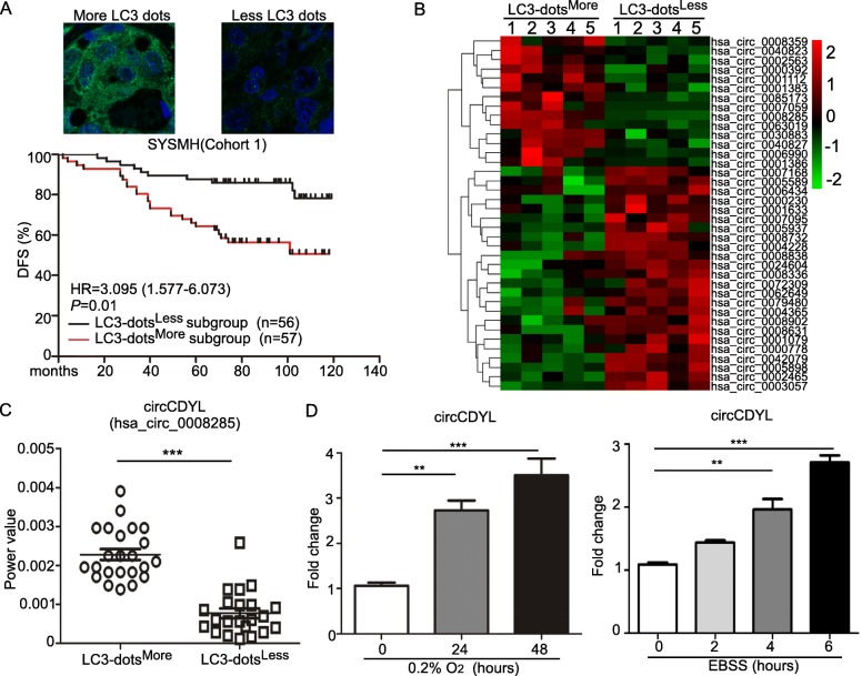
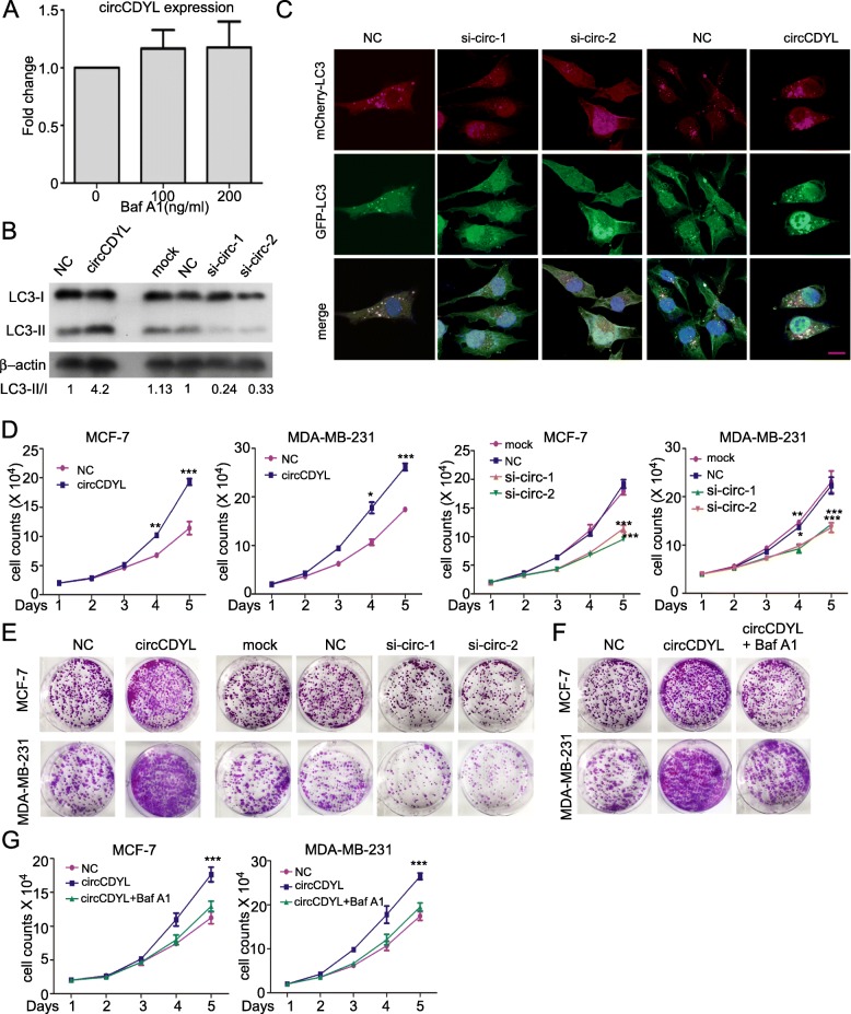
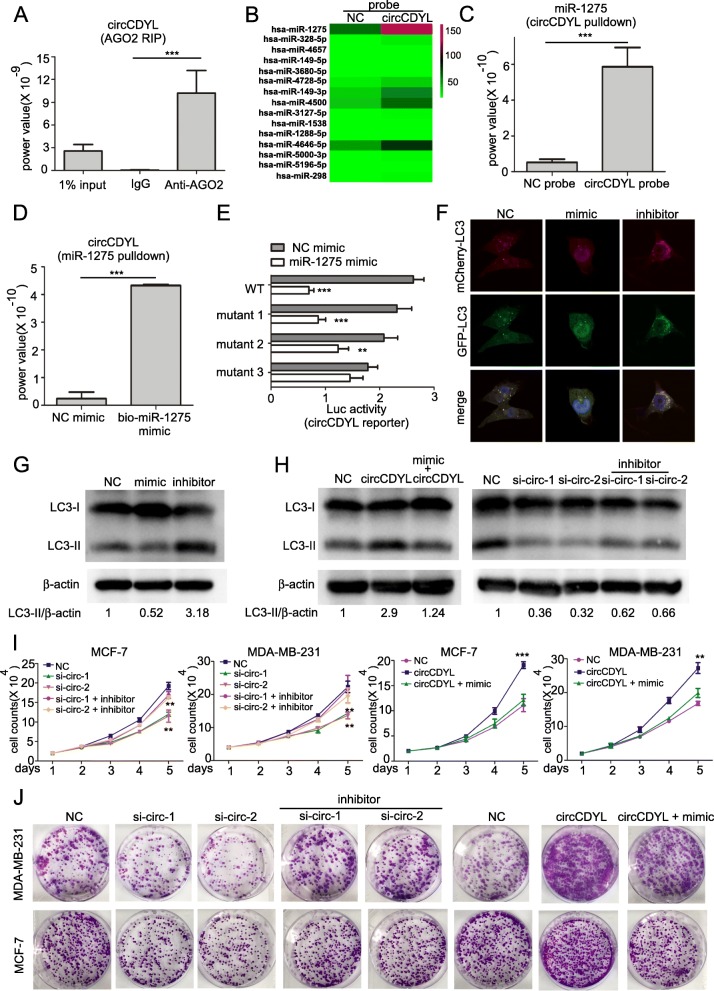
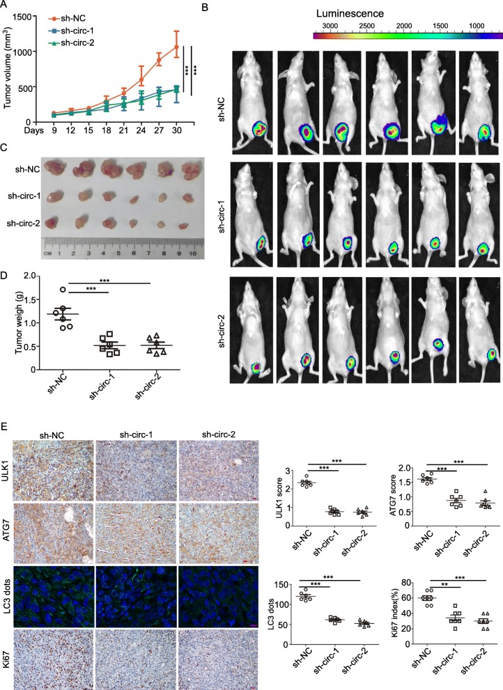
Methods: Autophagy associated circRNAs were screened by circRNAs deep sequencing and validated by qRT-PCR in BC tissues with high- and low- autophagic level. The biological function of autophagy associated circRNAs were assessed by plate colony formation, cell viability, transwells, flow cytometry and orthotopic animal models. For mechanistic study, RNA immunoprecipitation, circRNAs pull-down, Dual luciferase report assay, Western Blot, Immunofluorescence and Immunohistochemical staining were performed.

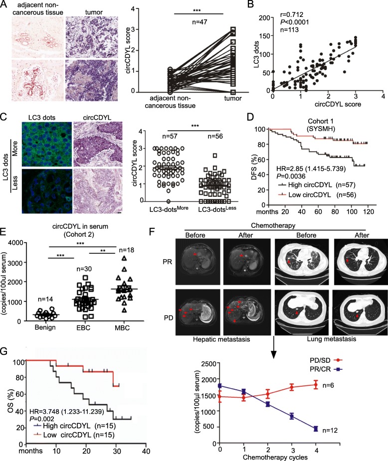
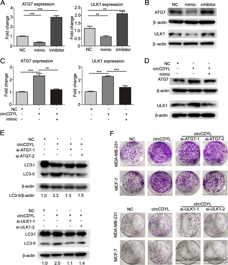
Results: An autophagy associated circRNA circCDYL was elevated by 3.2 folds in BC tissues as compared with the adjacent non-cancerous tissues, and circCDYL promoted autophagic level in BC cells via the miR-1275-ATG7/ULK1 axis; Moreover, circCDYL enhanced the malignant progression of BC cells in vitro and in vivo. Clinically, increased circCDYL in the tumor tissues and serum of BC patients was associated with higher tumor burden, shorter survival and poorer clinical response to therapy.

Conclusions: circCDYL promotes BC progression via the miR-1275-ATG7/ULK1-autophagic axis and circCDYL could act as a potential prognostic and predictive molecule for breast cancer patients.

Keywords: Autophagy; Breast cancer; circCDYL; miRNA sponge.


Conflict of interest statement

The authors declare that they have no competing interests.

Figures

Fig. 1
Fig. 1
Autophagy-associated circRNA (circCDYL) expression profile in breast cancer. a The association between LC3 dots and disease-free survival (DFS) in 113 breast cancer patients from Cohort 1 of SYSMH. b circRNA expression profile in breast cancer patients with more and less LC3 dots by circRNA deep sequencing. c Expression of circCDYL in breast cancer tissue with more (n = 23) and less (n = 22) LC3 dots by qRT-PCR. d The relative expression of circCDYL under hypoxic condition (0.2% O2) at various time points (0, 24, 48 h) (left) and EBSS starvation induction at various time points (0, 2, 4, 6 h) (right) in MDA-MB-231 cells
Fig. 2
Fig. 2
Clinical significance of circCDYL in breast cancer. a Comparison of circCDYL expression between breast cancer tissues and paired adjacent non-cancerous tissues by qRT-PCR (n = 47) and their representative images by ISH. Scale bar = 50 μm. b Correlations of circCDYL expression and the number of LC3 dots in breast cancer tumors, analyzed by Pearson analysis. c Representative images of LC3 dots and circCDYL expression by ISH in breast cancer tissues (left) and quantitative analysis of circCDYL expression in cancer tissues with more and less LC3 dots. Scale bar = 50 μm. d Kaplan-Meier analysis of the correlation between circCDYL expression and disease-free survival (DFS). e circCDYL abundance in serum of patients detected by ddPCR (n = 62, Cohort 2). EBC: early breast cancer, MBC: metastatic breast cancer.f Representative images in cases with lung and liver metastasis before and after chemotherapy and real time kinetics of serum circCDYL during chemotherapy in MBC patients (n = 18, Cohort 2). PR: partial response; CR: complete response; PD: progressive disease; SD: stable disease. g Kaplan-Meier analysis of the correlation between serum circCDYL expression and overall survival (OS) in MBC patients (Cohort 3). *P <  0.05, **P < 0.01, ***P < 0.005. Error bars indicate Standard Error of Mean (S.E.M)
Fig. 3
Fig. 3
circCDYL promotes proliferation of breast cancer cells via autophagy. a qRT-PCR analysis of circCDYL in MDA-MB-231 cells after Baf A1 (Bafilomycin A1) treatment. b LC3 expression in MDA-MB-231 after circCDYL silencing via siRNA or over-expressing circCDYL by over-expressing plasmid, as detected by Western Blot. LC3-II/I, the gray value ratio of LC3-II and LC3-I, normalized to NC group. c Autophagosomes in mCherry-GFP-LC3 labeled MDA-MB-231 after silencing circCDYL via siRNA or over-expressing circCDYL by over-expressing plasmid, as detected by confocal microscopy. Scale bar = 50 μm. d, e The proliferation of MDA-MB-231 and MCF-7 cells after silencing circCDYL viasiRNA or over-expressing circCDYL via over-expressing plasmid, as detected by cell viability assay (d) and plate colony formation (e). f, g Proliferation of MDA-MB-231 and MCF-7, as detected by plate colony formation (f) and cell viability assay (g) after transfection with circCDYL over-expressing plasmid or co-treatment with Baf A1. siNC: negative control siRNA. All experiments above were repeated at least 5 times. *P < 0.05, **P < 0.01, ***P < 0.005. Error bars indicate S.E.M
Fig. 4
Fig. 4
circCDYL functions as miR-1275 sponge. a qRT-PCR analysis of circCDYL in RNA sample after RIP assay by AGO2 antibody. b Heatmap of miRNA microarray of RNA sample by circCDYL pull-down. c qRT-PCR analysis of miR-1275 in RNA sample by circCDYL pull-down. d qRT-PCR analysis of circCDYL in RNA sample by miR-1275 miRNA pull-down. e Dual luciferase assay of HEK-293 T cells co-transfected with miR-1275 mimic and luciferase reporter containing full length of circCDYL with or without miR-1275 binding site mutant. NC: negative control; WT: wild-type luciferase reporter containing full length of circCDYL. f LC3 dots in mCherry-GFP-LC3-labeled MDA-MB-231 cells. g LC3-II expression in MDA-MB-231 after treatments with miR-1275 mimic or inhibitor, as detected by Western Blot. h Western Blot analysis of LC3-II expression in MDA-231 co-transfected with circCDYL over-expressing plasmid and miR-1275 mimic or co-transfected with circCDYL specific siRNA and miR-1275 inhibitor. i-j. Proliferation level of MCF-7 and MDA-MB-231 detected by cell viability assay (i) and plate colony formation (j) after co-transfection with circCDYL over-expressing plasmid and miR-1275 mimic or co-transfection with circCDYL specific siRNA and miR-1275 inhibitor. All data are shown as the mean ± S.E.M. *P < 0.05, **P < 0.01, ***P < 0.005
Fig. 5
Fig. 5
circCDYL promotes proliferation of breast cancer cells via miR-1275-ATG7/ ULK1-autophagic axis. a ATG7 and ULK1 expression of MDA-MB-231 after transfection with miR-1275 mimic or inhibitor, as detected by qRT-PCR. b ATG7 and ULK1 expression of MDA-MB-231 after transfection with miR-1275 mimic or inhibitor, as detected by Western Blot. c, d ATG7 and ULK1 expression in MDA-MB-231 after transfection with circCDYL over-expressing plasmid and ATG7 or ULK1 siRNA, as detected by qRT-PCR (C) and Western Blot (D). e Western Blot analysis of LC3-II expression in MDA-MB-231 after co-transfection with circCDYL over-expressing plasmids and ATG7 or ULK1 siRNA. f Proliferation level of MCF-7 and MDA-MB-231 after co-transfection with circCDYL over-expressing plasmids and ATG7 or ULK1 siRNA, as detected by plate colony formation
Fig. 6
Fig. 6
circCDYL promotes progression of breast cancer in vivo. a Tumor size derived from MDA-MB-231 cell line was measured in Balb/c nude mice with orthotopic tumors in the fourth left mammary fat pad. b Live imaging of the animals prior to euthanasia, and photon intensities are indicated above the picture. c, d Tumor size (C) and tumor weight (D) measured after tumor excision. e IHC staining of ATG7, ULK1 and Ki67, and IF detection of LC3 dots in orthotopic tumors derived from MDA-MB-231 cell line. Scale bar = 50 μm

References

    1. Kalimutho M, Nones K, Srihari S, Duijf PHG, Waddell N, Khanna KK. Patterns of genomic instability in breast Cancer. Trends Pharmacol Sci. 2019;40:198–211. - PubMed
    1. Eskelinen E. Autophagy: supporting cellular and organismal homeostasis by self-eating. Int J Biochem Cell Biol. 2019;111:1–10. - PubMed
    1. Mizushima N. A brief history of autophagy from cell biology to physiology and disease. Nat Cell Biol. 2018;20:521–527. - PubMed
    1. Vera-Ramirez L, Vodnala SK, Nini R, Hunter KW, Green JE. Autophagy promotes the survival of dormant breast cancer cells and metastatic tumour recurrence. Nat Commun. 2018;9:1944. - PMC - PubMed
    1. Liu H, Ma Y, He H, Wang J, Jiang J, Shao R. SLC9A3R1 stimulates autophagy via BECN1 stabilization in breast cancer cells. Autophagy. 2015;11:2323–2334. - PMC - PubMed

Publication types

MeSH terms