

Image analysis and artificial intelligence in infectious disease diagnostics
- PMID: 32213317
- PMCID: PMC7508855
- DOI: 10.1016/j.cmi.2020.03.012
Image analysis and artificial intelligence in infectious disease diagnostics
Abstract
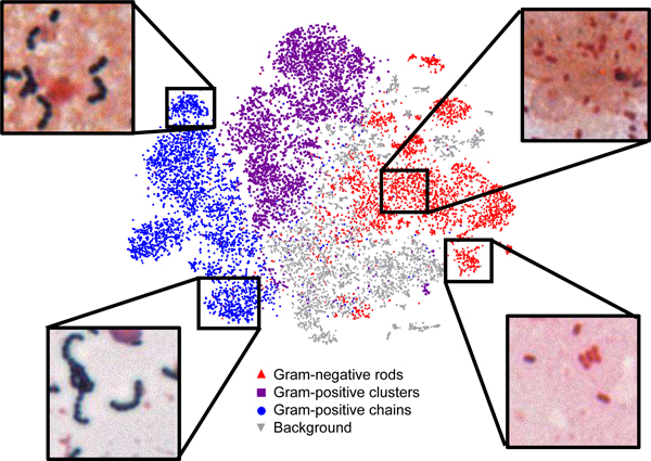
Background: Microbiologists are valued for their time-honed skills in image analysis, including identification of pathogens and inflammatory context in Gram stains, ova and parasite preparations, blood smears and histopathologic slides. They also must classify colony growth on a variety of agar plates for triage and assessment. Recent advances in image analysis, in particular application of artificial intelligence (AI), have the potential to automate these processes and support more timely and accurate diagnoses.
Objectives: To review current AI-based image analysis as applied to clinical microbiology; and to discuss future trends in the field.
Sources: Material sourced for this review included peer-reviewed literature annotated in the PubMed or Google Scholar databases and preprint articles from bioRxiv. Articles describing use of AI for analysis of images used in infectious disease diagnostics were reviewed.
Content: We describe application of machine learning towards analysis of different types of microbiologic image data. Specifically, we outline progress in smear and plate interpretation as well as the potential for AI diagnostic applications in the clinical microbiology laboratory.
Implications: Combined with automation, we predict that AI algorithms will be used in the future to prescreen and preclassify image data, thereby increasing productivity and enabling more accurate diagnoses through collaboration between the AI and the microbiologist. Once developed, image-based AI analysis is inexpensive and amenable to local and remote diagnostic use.
Keywords: Artificiall intelligence; Deep learning; Gram stain; Machine learning.
Copyright © 2020 European Society of Clinical Microbiology and Infectious Diseases. Published by Elsevier Ltd. All rights reserved.
Figures
References
-
- Garcia E, Kundu I, Ali A, Soles R, The American Society for Clinical Pathology’s 20162017 Vacancy Survey of Medical Laboratories in the United States, Am J Clin Pathol 149(5) (2018) 387–400. - PubMed
-
- LeCun Y, Bengio Y, Hinton G, Deep learning, Nature 521(7553) (2015) 436–44. - PubMed
-
- Ehteshami Bejnordi B, Mullooly M, Pfeiffer RM, Fan S, Vacek PM, Weaver DL, Herschorn S, Brinton LA, van Ginneken B, Karssemeijer N, Beck AH, Gierach GL, van der Laak J, Sherman ME, Using deep convolutional neural networks to identify and classify tumor-associated stroma in diagnostic breast biopsies, Modern pathology : an official journal of the United States and Canadian Academy of Pathology, Inc 31(10) (2018) 15021512. - PMC - PubMed
Publication types
MeSH terms
Grants and funding
LinkOut - more resources
Full Text Sources
Medical
