. 2020 May 5;31(5):920-936.e7.
doi: 10.1016/j.cmet.2020.03.004. Epub 2020 Mar 25.

Glutathione Restricts Serine Metabolism to Preserve Regulatory T Cell Function

Affiliations

Glutathione Restricts Serine Metabolism to Preserve Regulatory T Cell Function

Henry Kurniawan et al. Cell Metab. .

Abstract

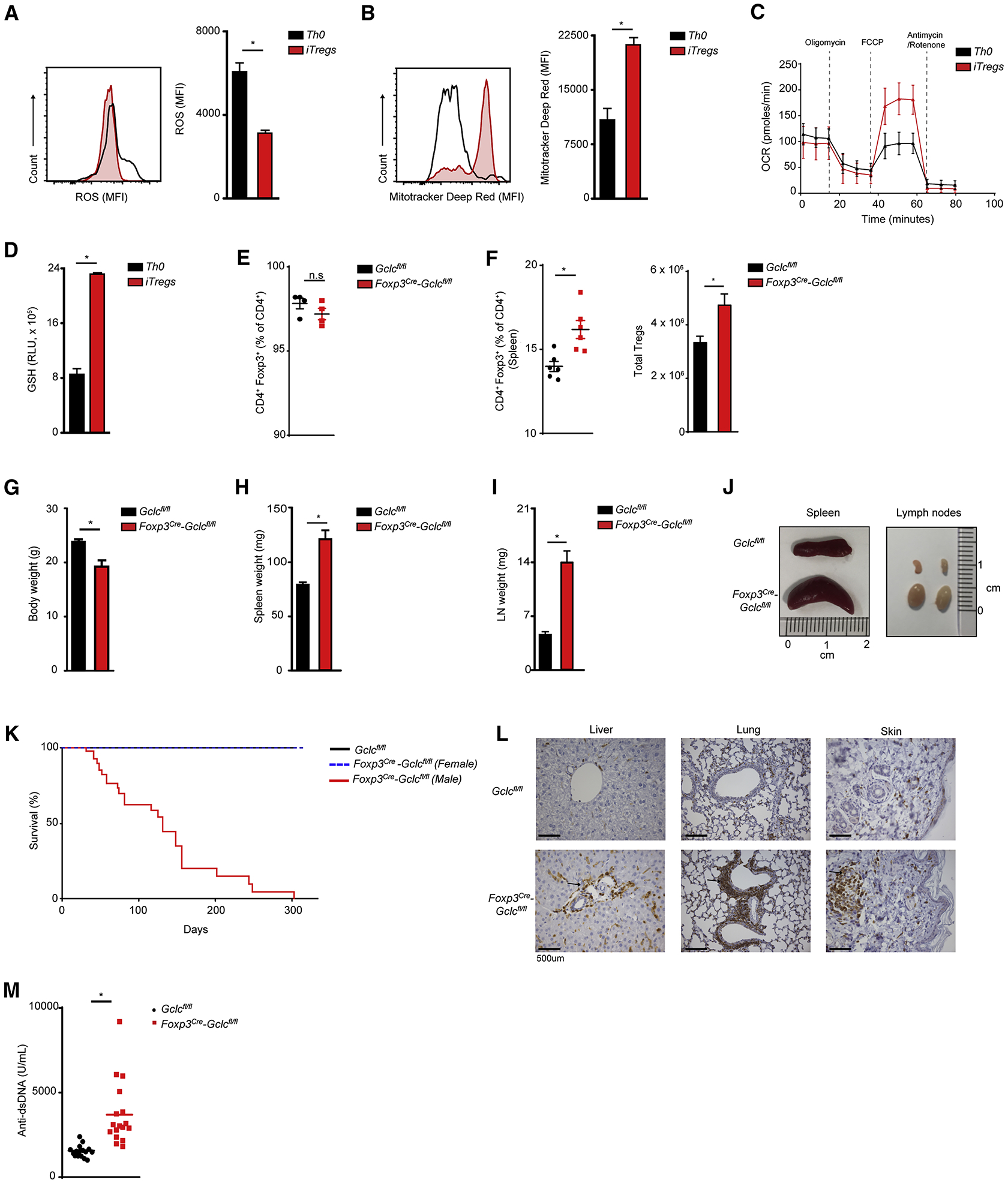
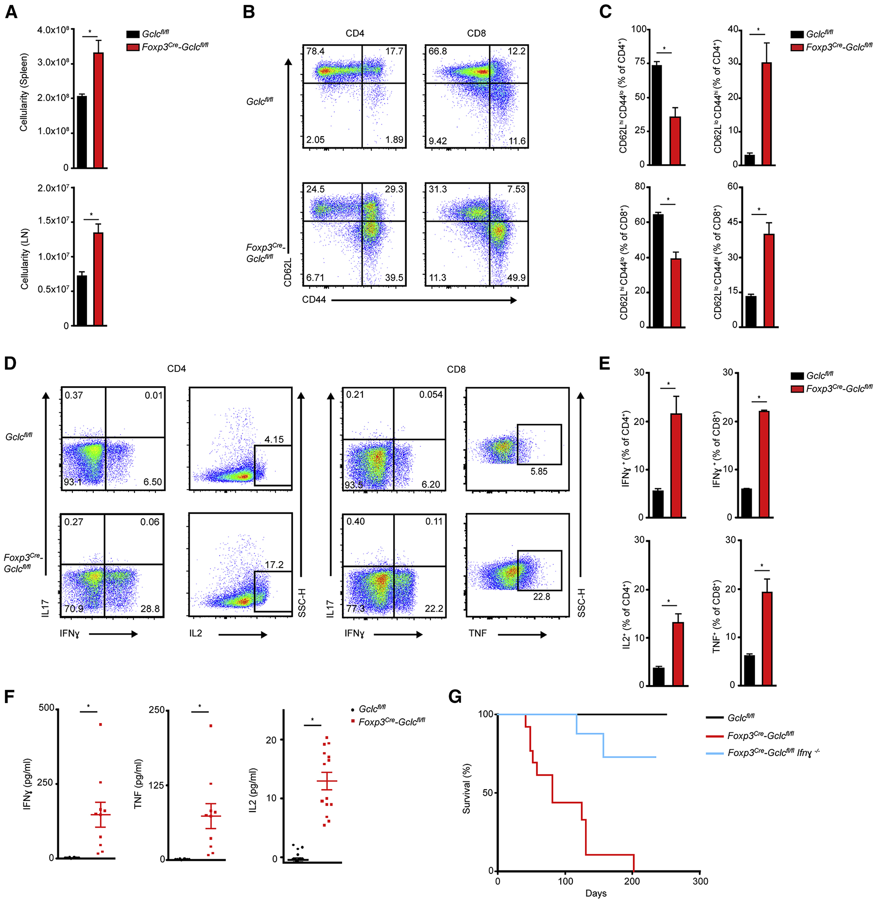
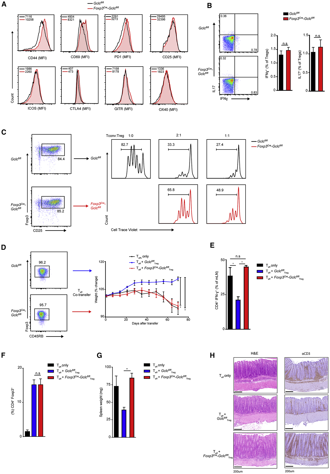
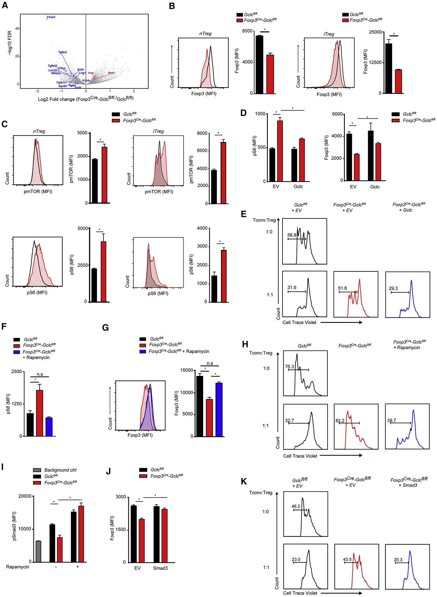
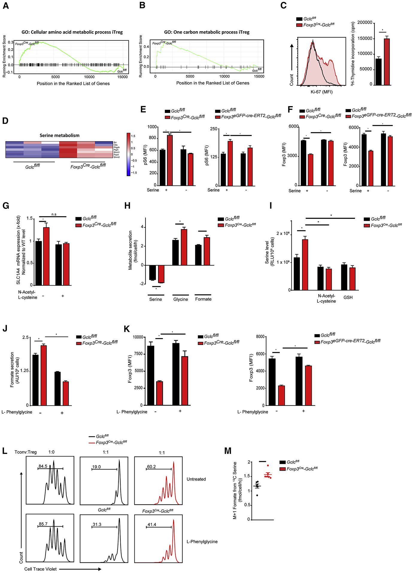
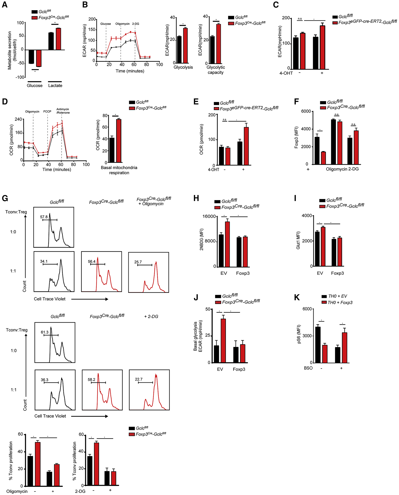
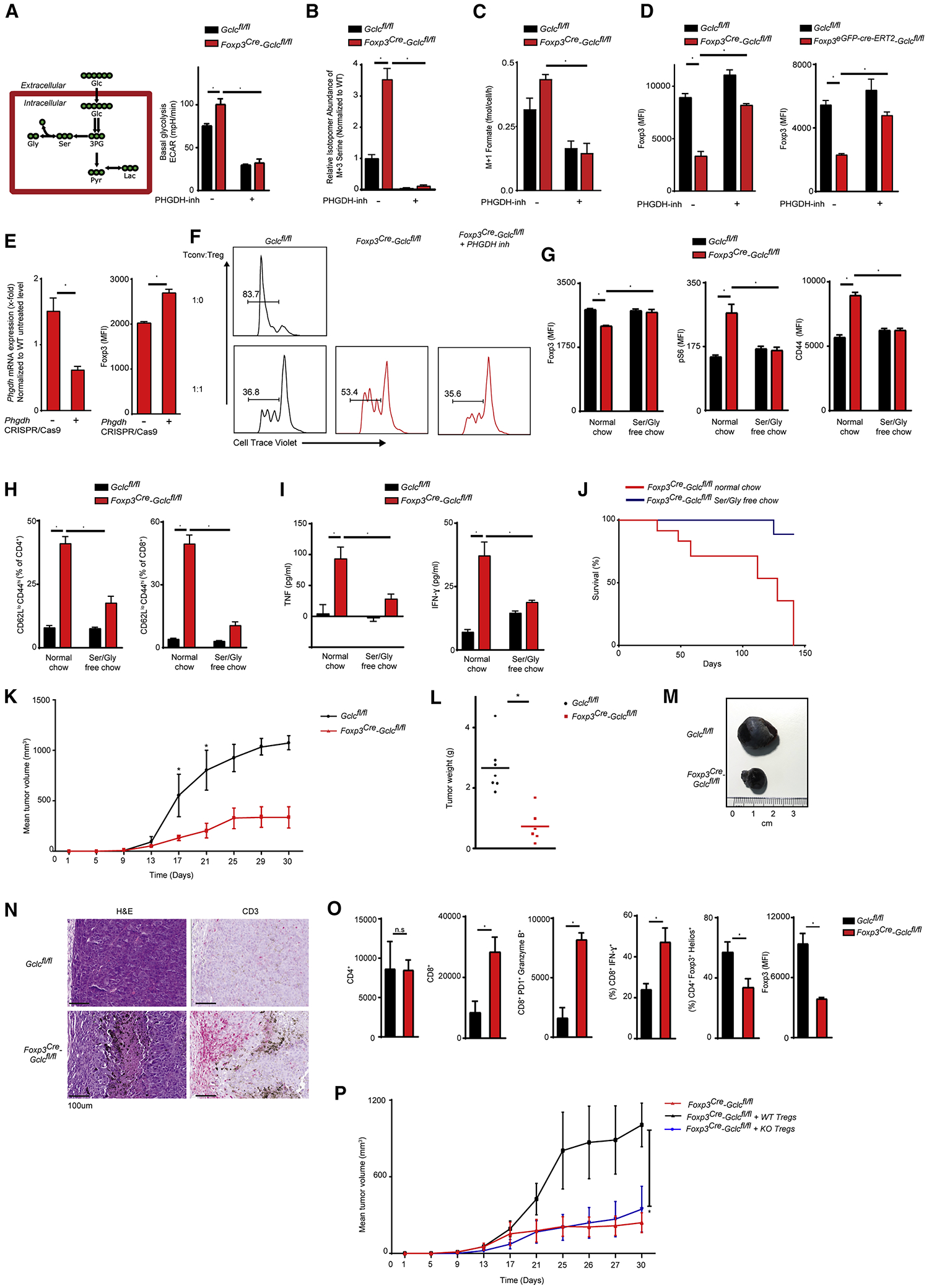
Regulatory T cells (Tregs) maintain immune homeostasis and prevent autoimmunity. Serine stimulates glutathione (GSH) synthesis and feeds into the one-carbon metabolic network (1CMet) essential for effector T cell (Teff) responses. However, serine's functions, linkage to GSH, and role in stress responses in Tregs are unknown. Here, we show, using mice with Treg-specific ablation of the catalytic subunit of glutamate cysteine ligase (Gclc), that GSH loss in Tregs alters serine import and synthesis and that the integrity of this feedback loop is critical for Treg suppressive capacity. Although Gclc ablation does not impair Treg differentiation, mutant mice exhibit severe autoimmunity and enhanced anti-tumor responses. Gclc-deficient Tregs show increased serine metabolism, mTOR activation, and proliferation but downregulated FoxP3. Limitation of cellular serine in vitro and in vivo restores FoxP3 expression and suppressive capacity of Gclc-deficient Tregs. Our work reveals an unexpected role for GSH in restricting serine availability to preserve Treg functionality.

Keywords: FoxP3; ROS; Treg; autoimmunity; cancer; diet; glutamate cysteine ligase; glutathione; one carbon metabolism; serine metabolism.


Conflict of interest statement

Declaration of Interests The authors declare no competing interests.

Figures

Figure 1.
Figure 1.. Gclc deficiency does not affect Treg homeostasis but does lead to multi-organ inflammation.
(A-D) Splenic naïve T cells from C57/BL6 mice were treated with αCD3+αCD28+IL2, with/without TGFβ, to generate iTreg or Th0 cells, respectively. Cells were stained with (A) DCF-DA to detect ROS, or (B) Mitotracker Deep Red to assess mitochondrial function, followed by flow cytometry (FC). (C) Seahorse determination of OCR. (D) Luminescence-based quantification of intracellular GSH. Data are mean±SEM (n=3) and representative of 3 independent trials. (E) FC quantitation of CD4+Foxp3+ iTregs among splenic naïve T cells isolated from Gclcfl/fl (control) or Foxp3cre-Gclcfl/fl mice and treated in vitro with αCD3+αCD28+IL2+TGF-β. Data are mean±SEM (n=3); 5 trials. (F) Flow cytometric analysis (FCA) of CD4+Foxp3+ nTregs from spleen of Gclcfl/fl and Foxp3cre-Gclcfl/fl mice. Data are mean±SEM (n=3); 5 trials. (G-I) Weights of (G) whole body, (H) spleen, and (I) LN from Gclcfl/fl and Foxp3cre-Gclcfl/fl mice at age 8–12wk. Data are mean±SEM (n=13). (J) Images of spleens and LN from Gclcfl/fl and Foxp3cre-Gclcfl/fl mice (8wk). (K) Survival of Gclcfl/fl (n=48) and Foxp3cre-Gclcfl/fl (male n=38, female=21) mice. (L) Histology of the indicated tissues resected from one Gclcfl/fl and one Foxp3cre-Gclcfl/fl mouse and stained with αCD3. Scale bars, 500 μm. Results are representative of 5 mice/group; 2 trials. (M) ELISA of anti-dsDNA Ab in serum of Gclcfl/fl (n=17) and Foxp3cre-Gclcfl/fl (n=17) mice (8–12 wk). *p<0.05.
Figure 2.
Figure 2.. Treg-specific Gclc deletion impairs homeostasis.
(A) Total lymphocyte numbers in spleen and LN of Gclcfl/fl and Foxp3cre-Gclcfl/fl mice. Data are mean±SEM (n=6); 2 trials. (B, C) FCA of naïve (CD44loCD62Lhi), central memory (CD44hiCD62Lhi), and effector (CD44hiCD62Llo) subsets (B), and quantification within the CD4+ population (top) and CD8+ population (bottom) (C), from Gclcfl/fl and Foxp3cre-Gclcfl/fl mice as in (A). Data are mean±SEM (n=3); 5 trials. (D, E) Intracellular staining and FCA of IFNγ, IL17, IL2 and TNF production by purified CD4+ (left) and CD8+ (right) splenic Gclcfl/fl and Foxp3cre-Gclcfl/fl T cells re-stimulated in vitro with 50 ng PMA + 750 ng Iono for 6hr. Data are mean±SEM (n=3); 3 trials. (F) ELISA of IFNγ, TNF and IL2 in serum of Gclcfl/fl and Foxp3cre-Gclcfl/fl mice (8–12wk). Each symbol = individual mouse. Data are mean±SEM (Gclcfl/fl n=10; Foxp3cre-Gclcfl/fl n=17); 2 trials. (G) Survival of Gclcfl/fl (n=23), Foxp3cre-Gclcfl/fl (n=13), and Foxp3cre-Gclcfl/fl × Ifnγ−/− (n=14) mice. *p<0.05.
Figure 3.
Figure 3.. GSH modulates Treg functionality in vitro and in vivo.
(A) FCA of the indicated surface markers on splenic nTregs from Gclcfl/fl and Foxp3cre-Gclcfl/fl mice (n=3); 4 trials. (B) FC quantification of intracellular IFNγ and IL17 in splenic nTregs of Gclcfl/fl and Foxp3cre-Gclcfl/fl mice re-stimulated in vitro with PMA+Iono for 6hr. Data are mean±SEM (n=3); 3 trials. (C) In vitro suppression assay of splenic nTregs from Gclcfl/fl and Foxp3cre-Gclcfl/fl mice incubated at the indicated ratios with Tconv labeled with 5 μM Cell-Trace Violet (CTV). Suppresion was determined by FC as a decrease in Tconv proliferation; 5 trials. (D) In vivo assay of Treg suppression using a T cell adoptive transfer-based induced colitis model. Rag1−/− mice received WT Teff (CD4+CD45RBhigh) alone or together with FACS-sorted nTregs (CD4+ CD45RBlow) from Gclcfl/fl or Foxp3cre-Gclcfl/fl mice. Results are presented as post-transfer body weight relative to initial weight of recipients. Data are mean±SEM (n=4); 2 trials. (E) Intracellular staining and FCA of IFNγ produced by CD4+ Teff isolated from mesenteric LN of the mice in (D) at day70 post-transfer and re-stimulated in vitro with PMA+Iono. Data are mean±SEM (n=4); 2 trials. (F) Quantification of nTregs in peripheral blood of Rag1−/− recipients treated as in (D) at 50 days post-transfer. Data are mean±SEM (n=2–3). 2 trails. (G) Quantification of spleen weights of the Rag1−/− recipients in (D) at experimental endpoint. Data are mean±SEM (n=4); 2 trials. (H) Histology of large intestine of the mice in (D) after staining with H&E or αCD3. Scale bars, 200 μm. Data are mean±SEM (n=4); 2 trials. *p<0.05.
Figure 4.
Figure 4.. Lack of GSH alters mTOR signaling and impairs FoxP3 expression.
(A) Volcano plot comparing mRNAs of the indicated Treg-associated genes in Foxp3cre-Gclcfl/fl and Gclcfl/fl iTregs. Downregulated (blue) and upregulated (red) transcripts are shown. (B) FCA of FoxP3 in Foxp3cre-Gclcfl/fl and Gclcfl/fl Tregs isolated from spleen (left) or induced in vitro (right). Data are mean±SEM (n=3); 5 trials. (C) Intracellular staining and FCA of pmTOR and pS6 in Gclcfl/fl and Foxp3cre-Gclcfl/fl Tregs as in (B). Data are mean±SEM (n=3); 5 trials. (D) Intracellular staining and FCA of pS6 and Foxp3 in Gclcfl/fl and Foxp3cre-Gclcfl/fl iTregs transduced with retrovirus expressing EV or Gclc. Data are mean±SEM (n=3); 3 trials. (E) In vitro suppression assay of Gclcfl/fl and Foxp3cre-Gclcfl/fl iTregs transduced with retrovirus expressing EV or Gclc. Transduced Tregs were FACS-sorted and incubated with CTV-labeled Tconv at the indicated ratios; 2 trials. *p<0.05. (F) FCA of pS6 in Foxp3cre-Gclcfl/fl and Gclcfl/fl iTregs incubated with/without Rap. Data are mean±SEM (n=3); 3 trials. (G) Intracellular staining and FCA of FoxP3 in Foxp3cre-Gclcfl/fl and Gclcfl/fl iTregs incubated with/wihout 100 nM Rap. Data are mean±SEM (n=3); 3 trials. (H) In vitro suppression assay of splenic nTregs from Gclcfl/fl and Foxp3cre-Gclcfl/fl mice that were incubated with/without Rap for 24hr and mixed with Tconv at the indicated ratios; 5 trials. (I) Intracellular staining and FCA of pSmad3 in Foxp3cre-Gclcfl/fl and Gclcfl/fl iTregs incubated with/without Rap. Data are mean±SEM (n=3); 2 trials. (J) Intracellular staining and FCA of Foxp3 in Gclcfl/fl and Foxp3cre-Gclcfl/fl iTregs transduced with retrovirus expressing EV or Smad3. Data are mean±SEM (n=3); 3 trials. (K) In vitro suppression assay of Gclcfl/fl and Foxp3cre-Gclcfl/fl iTregs transduced with retrovirus expressing EV or Smad3. Transduced Tregs were FACS-sorted and incubated with CTV-labeled Tconv at the indicated ratios; 2 trials. *p<0.05.
Figure 5.
Figure 5.. GSH-mediated regulation of the serine pool is required for Treg function.
(A) Barcode enrichment plot of KEGG pathway GO:0006520 (Cellular amino acid metabolic processes) for Gclcfl/fl and Foxp3cre-Gclcfl/fl iTregs. (B) Barcode enrichment plot of KEGG pathway GO:0006730 (1CMet) for Gclcfl/fl and Foxp3cre-Gclcfl/fl iTregs. (C) Determination of proliferation of Gclcfl/fl and Foxp3cre-Gclcfl/fl iTregs by Ki-67 staining (left) and 3H-thymidine incorporation (right). Data are mean±SEM (n=3); 3 trials. (D) Heat map showing normalized differential gene expression patterns of genes associated with serine metabolism in Gclcfl/fl vs. Foxp3cre-Gclcfl/fl iTregs. (E, F) Intracellular staining and FCA of (E) pS6 and (F) Foxp3 in Gclcfl/fl vs Foxp3cre-Gclcfl/fl (left) and Gclcfl/fl vs Foxp3eGPF-cre-ERT2-Gclcfl/fl (right) iTregs cultured in normal or serine-deficient medium. Gclcfl/fl vs Foxp3eGPF-cre-ERT2-Gclcfl/fl iTregs were co-incubated with 1μM 4-OHT. Data are mean±SEM (n=3); 3 trials. (G) RT-qPCR of ASCT1 mRNA in Gclcfl/fl and Foxp3cre-Gclcfl/fl iTregs. Data are mean±SEM (n=7); 2 trials. (H) LC/MS quantification of serine uptake from, and glycine and formate secretion into, culture medium of Gclcfl/fl and Foxp3cre-Gclcfl/fl iTregs. Data are mean±SEM (n=3); 2 trials. (I) Quantification of intracellular serine in Gclcfl/fl and Foxp3cre-Gclcfl/fl iTregs treated with/without 200 μM NAC or 0.5 mM GSH. Data are mean±SEM (n=3); 2 trials. (J) Quantification of formate secretion into culture medium of Gclcfl/fl and Foxp3cre-Gclcfl/fl iTregs treated with/without 50 μM L-phenylglycine. Data are mean±SEM (n=3); 2 trials. (K) Intracellular staining and FCA of Foxp3 in Gclcfl/fl vs Foxp3cre-Gclcfl/fl (left), and Gclcfl/fl vs Foxp3eGPF-cre-ERT2-Gclcfl/fl (right), iTregs treated with/without L-phenylglycine. Gclcfl/fl vs Foxp3eGPF-cre-ERT2-Gclcfl/fl iTregs were co-incubated with 4-OHT. Data are mean±SEM (n=3); 3 trials. (L) In vitro suppression assay of nTregs that were isolated from Gclcfl/fl and Foxp3cre-Gclcfl/fl mice, incubated with/without L-phenylglycine, and mixed in vitro with Tconv at the indicated ratios; 3 trials. (M) Mass isotopomer distribution of M+1 formate following incubation of Gclcfl/fl and Foxp3cre-Gclcfl/fl iTregs with [U-13C3]-serine for 24hr. Data are mean±SEM (n=3); 2 trials. *p<0.05.
Figure 6.
Figure 6.. Gclc expression is required to modulate Treg metabolism supporting Treg function.
(A) Quantification of glucose uptake from and lactate secretion into culture medium of Gclcfl/fl and Foxp3cre-Gclcfl/fl iTregs. Data are mean±SEM (n=3); 3 trials. (B) Quantification of ECAR of Gclcfl/fl vs Foxp3cre-Gclcfl/fl iTregs. Data are mean±SEM (n=4); 4 trials. (C) Quantification of ECAR of Gclcfl/fl vs Foxp3eGPF-cre-ERT2-Gclcfl/fl iTregs incubated with 4-OHT. Data are mean±SEM (n=3); 2 trials. (D) Quantification of OCR of Gclcfl/fl vs Foxp3cre-Gclcfl/fl iTregs. Data are mean±SEM (n=4); 4 trials. (E) Quantification of OCR of Gclcfl/fl vs Foxp3eGPF-cre-ERT2-Gclcfl/fl iTregs incubated with 4-OHT. Data are mean±SEM (n=3); 2 trials. (F) Intracellular staining and FCA of FoxP3 in Gclcfl/fl vs Foxp3cre-Gclcfl/fl iTregs treated with suboptimal doses of 0.5 nM oligomycin or 100 μM 2-DG for 24hr. Data are mean±SEM (n=3); 3 trials. (G) In vitro suppression assay of Gclcfl/fl vs Foxp3cre-Gclcfl/fl iTregs treated (or not) with suboptimal doses of oligomycin (top) or 2-DG (bottom) prior to incubation with CTV-labeled Tconv at the indicated ratios; 3 trials. (H, I) FCA of (H) 2-NBDG uptake and (I) Glut-1 expression by Gclcfl/fl vs. Foxp3cre-Gclcfl/fl iTregs transduced with retrovirus expressing EV or Foxp3. (J) Quantification of ECAR for the iTregs in (H) as determined in (B). For (H-J), data are the mean±SEM (n=3); 2 trials. (K) Intracellular staining and FCA of pS6 in activated WT Tconv transduced with retrovirus expressing EV or FoxP3 as in (H) and treated with BSO for 48hr. Data are mean±SEM (n=3); 3 trials. *p<0.05.
Figure 7.
Figure 7.. Glutathione restricts de novo serine synthesis and enhances anti-tumor immune responses.
(A) Diagram of serine synthesis pathway (left), and quantification of ECAR of Gclcfl/fl and Foxp3cre-Gclcfl/fl iTregs cultured with/without 10 μM PHGDH inhibitor (right). Data are mean±SEM (n=3); 2 trials. (B) Mass isotopomer distribution of M+3 serine in the cells in (A) following incubation with [U-13C6]-glucose for 24hr. (C) Mass isotopomer distribution of M+1 formate in the cells in (A) following incubation with [U-13C6]-glucose for 24hr. (D) Intracellular staining and FCA of FoxP3 in Gclcfl/fl vs Foxp3cre-Gclcfl/fl (left) and Gclcfl/fl vs Foxp3eGPF-cre-ERT2-Gclcfl/fl (right) iTregs treated with/without PHGDH inhibitor. Gclcfl/fl vs Foxp3eGPF-cre-ERT2-Gclcfl/fl iTregs were co-incubated with 4-OHT. Data are mean±SEM (n=3); 3 trials. (E) Phgdh mRNA expression (left) and intracellular staining and FCA of FoxP3 (right) in Gclcfl/fl vs Foxp3cre-Gclcfl/fl iTregs that were nucelofected with sgRNAs specific for Phgdh or controls. Data are mean±SEM (n=3); 2 trials. (F) In vitro suppression assay of Gclcfl/fl vs Foxp3cre-Gclcfl/fl iTregs treated with PHGDH inhibitor prior to incubation with CTV-labeled Tconv at the indicated ratios; 3 trials. (G) Intracellular staining and FCA of FoxP3, pS6 and CD44 in splenic Tregs of Gclcfl/fl vs Foxp3cre-Gclcfl/fl mice (12wk) fed with normal chow or a serine/glycine-deficient diet for 9wk. Data are mean±SEM (n=4–11); 2 trials. (H) FCA and quantification of Teff (CD44hiCD62Llo) within CD4+ (left) and CD8+ (right) T cell populations in blood of the mice in (G). Data are mean±SEM (n=4–11); 2 trials. (I) ELISA of IFNγ and TNF in serum of the mice in (G). Data are mean±SEM (n=4–11); 2 trials. (J) Survival of Foxp3cre-Gclcfl/fl mice on normal chow (n=12) or a serine/glycine-deficient diet (n=9); (K-N) Gclcfl/fl and Foxp3cre-Gclcfl/fl mice (8wk) were transplanted s.c. with B16F10 melanoma cells. (K) Mean tumor volumes determined at the indicated times. (L) Quantification of tumor weights at time of sacrifice. Each dot = individual mouse. (M) Representative macroscopic images of tumors from transplanted Gclcfl/fl and Foxp3cre-Gclcfl/fl mice. (N) Histology of tumor sections from the mice in (F) stained with H&E or αCD3. Scale bars, 100 μm. Results are representative of 4 mice/group; 2 trials. (O) Quantification of the indicated TIL subsets in tumors of Gclcfl/fl and Foxp3cre-Gclcfl/fl mice treated as in (K). Data are mean±SEM (n=5); (P) Mean tumor volumes at the indicated times in Foxp3cre-Gclcfl/fl mice (8wk) transplanted s.c. with B16F10 melanoma cells and injected intravenously with nTregs from Gclcfl/fl (WT) or Foxp3cre-Gclcfl/fl (KO) mice at day 0. Data are mean±SEM (n=4) 2 trials. *p<0.05.

References

    1. Almeida L, Lochner M, Berod L, and Sparwasser T (2016). Metabolic pathways in T cell activation and lineage differentiation. Semin Immunol 28, 514–524. - PubMed
    1. Apostolidis SA, Rodriguez-Rodriguez N, Suarez-Fueyo A, Dioufa N, Ozcan E, Crispin JC, Tsokos MG, and Tsokos GC (2016). Phosphatase PP2A is requisite for the function of regulatory T cells. Nat Immunol 17, 556–564. - PMC - PubMed
    1. Ben-Sahra I, Hoxhaj G, Ricoult SJH, Asara JM, and Manning BD (2016). mTORC1 induces purine synthesis through control of the mitochondrial tetrahydrofolate cycle. Science 351, 728–733. - PMC - PubMed
    1. Bhattacharyya S, and Saha J (2015). Tumour, Oxidative Stress and Host T Cell Response: Cementing the Dominance. Scand J Immunol 82, 477–488. - PubMed
    1. Bothur E, Raifer H, Haftmann C, Stittrich AB, Brustle A, Brenner D, Bollig N, Bieringer M, Kang CH, Reinhard K, et al. (2015). Antigen receptor-mediated depletion of FOXP3 in induced regulatory T-lymphocytes via PTPN2 and FOXO1. Nat Commun 6, 8576. - PMC - PubMed

Publication types

LinkOut - more resources