. 2020 May;10(5):674-687.
doi: 10.1158/2159-8290.CD-20-0215. Epub 2020 Mar 25.

HER2-Mediated Internalization of Cytotoxic Agents in ERBB2 Amplified or Mutant Lung Cancers

Affiliations

HER2-Mediated Internalization of Cytotoxic Agents in ERBB2 Amplified or Mutant Lung Cancers

Bob T Li et al. Cancer Discov. 2020 May.

Abstract

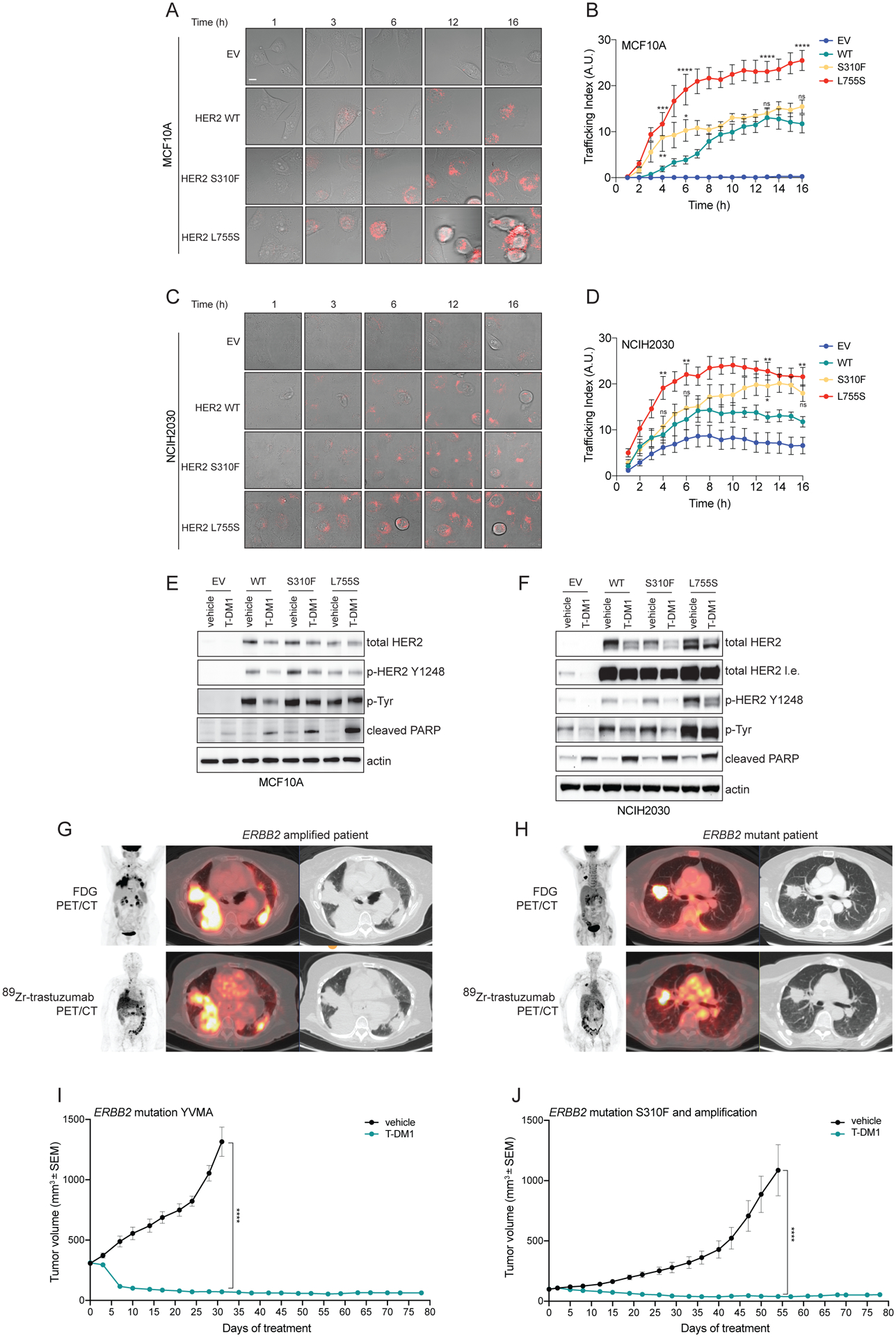
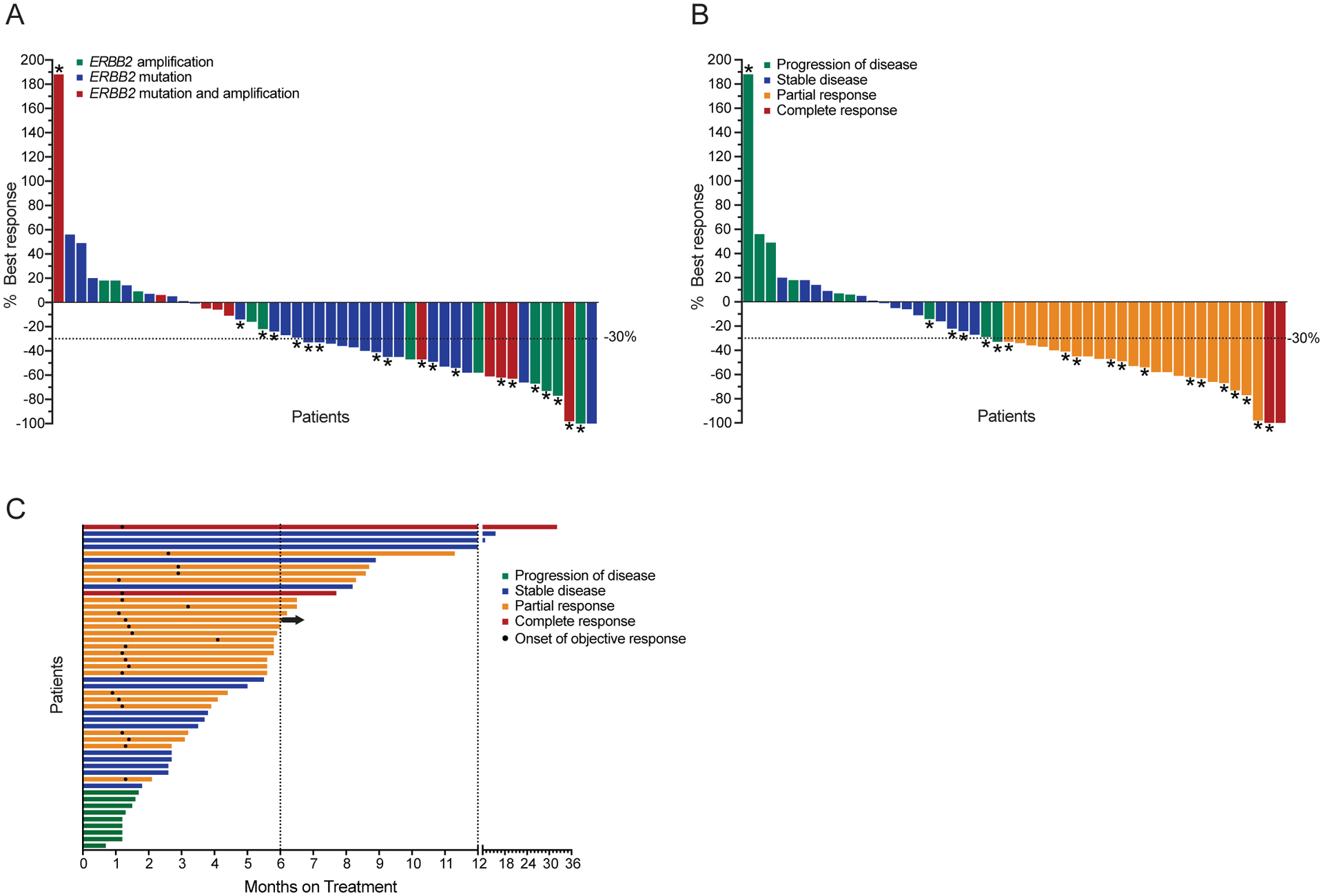
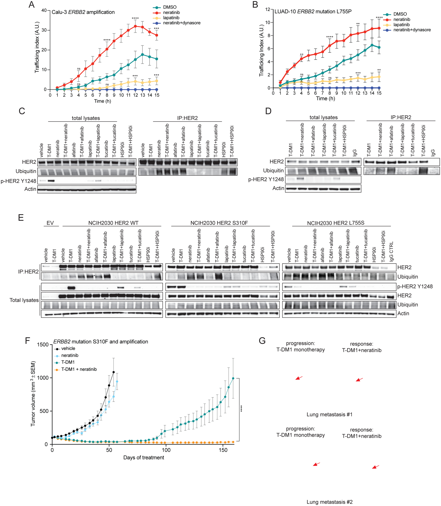
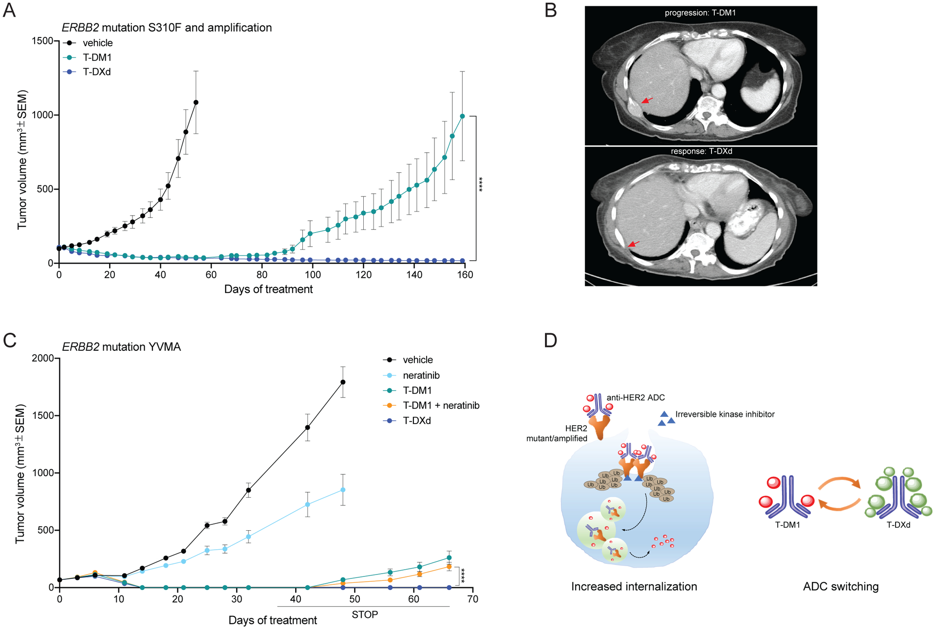
Amplification of and oncogenic mutations in ERBB2, the gene encoding the HER2 receptor tyrosine kinase, promote receptor hyperactivation and tumor growth. Here we demonstrate that HER2 ubiquitination and internalization, rather than its overexpression, are key mechanisms underlying endocytosis and consequent efficacy of the anti-HER2 antibody-drug conjugates (ADC) ado-trastuzumab emtansine (T-DM1) and trastuzumab deruxtecan (T-DXd) in lung cancer cell lines and patient-derived xenograft models. These data translated into a 51% response rate in a clinical trial of T-DM1 in 49 patients with ERBB2-amplified or -mutant lung cancers. We show that cotreatment with irreversible pan-HER inhibitors enhances receptor ubiquitination and consequent ADC internalization and efficacy. We also demonstrate that ADC switching to T-DXd, which harbors a different cytotoxic payload, achieves durable responses in a patient with lung cancer and corresponding xenograft model developing resistance to T-DM1. Our findings may help guide future clinical trials and expand the field of ADC as cancer therapy. SIGNIFICANCE: T-DM1 is clinically effective in lung cancers with amplification of or mutations in ERBB2. This activity is enhanced by cotreatment with irreversible pan-HER inhibitors, or ADC switching to T-DXd. These results may help address unmet needs of patients with HER2-activated tumors and no approved targeted therapy.See related commentary by Rolfo and Russo, p. 643.This article is highlighted in the In This Issue feature, p. 627.


Conflict of interest statement

Competing interests

M.S. has received research funds from AstraZeneca, Puma Biotechnology, Daiichi Sankyo, Immunomedics, Targimmune and Menarini Ricerche. He is in the scientific advisory board (SAB) of Menarini Ricerche and Bioscience Institute and is a cofounder of Medendi.org. D.B.S. has served as consulted/received honoraria from Pfizer, Loxo Oncology, Illumina, Lilly Oncology and Vivideon Therapeutics. S.M. consulted for Boehringer-Ingelheim. M.S. and B.T.L. have two pending institutional patents at Memorial Sloan Kettering Cancer Center (US62/685,057, US62/514,661). B.T.L. has served as a consultant/advisory board member for Roche/Genentech, Biosceptre International, Thermo Fisher Scientific, Mersana Therapeutics, Hengrui Therapeutics, Guardant Health and has received research funds to his institution from Roche/Genentech, Daiichi Sankyo, Hengrui Therapeutics, Illumina, Guardant Health, BioMed Valley Discoveries, AstraZeneca, GRAIL, MORE Health, Amgen and Lilly. C.M.R. is a consultant/advisory board member for AbbVie, Amgen, Ascentage, AstraZeneca, Bristol-Myers Squibb, Celgene, Daiichi Sankyo, Genentech/Roche, Ipsen, Loxo, PharmaMar, Bridge Medicine, and Harpoon. R.A.F. receives research funds from Puma Biotechnology and Eisai Pharmaceuticals. N.U.L. has received research support (to institution) from Pfizer, Genentech, Seattle Genetics, and Merck, and has been a consultant for Puma, Daichii, and Seattle Genetics. D.M.H. reports receiving commercial research grants from Loxo, PUMA Biotechnology, AstraZeneca, and Bayer Pharmaceuticals, and is a consultant/advisory board member for Chugai Pharma, CytomX Therapeutics, Boehringer Ingelheim, AstraZeneca, Pfizer, Bayer Pharmaceuticals, and Genentech/Roche. G.A.U. is a consultant for Sanofi and receives research support for Sanofi, Novartis, Genentech, and Puma Biotechnology. M.G.K. has received consulting fees from AstraZeneca, Pfizer, and Regeneron. He has received research funding from The National Cancer Institute (USA), The Lung Cancer Research Foundation, Genentech Roche, and PUMA Biotechnology and honoraria He has received honoraria for participation in educational programs from WebMD, OncLive, Physicians Education Resources, Prime Oncology, Intellisphere, Creative Educational Concepts, Peerview, i3 Health, Paradigm Medical Communications, AXIS, Carvive Systems, AstraZeneca, and Research to Practice. N.R. is on the SAB and receives research funding from Chugai, on the SAB and owns equity in Beigene, and Fortress. N.R. is also on the SAB of Daiichi-Sankyo, Astra-Zeneca-MedImmune, and F-Prime, and is a past SAB member of Millenium-Takeda, Kadmon, Kura, and Araxes. N.R. is a consultant to Novartis, Boehringer Ingelheim, Tarveda, and Foresight and consulted in the last three years with Eli Lilly, Merrimack, Kura Oncology, Araxes, and Kadman. N.R. owns equity in ZaiLab, Kura Oncology, Araxes, and Kadman. N.R. also collaborates with Plexxikon. M.F.B. declares consulting/advisory board activities for Roche. J.S.R.-F. is a consultant of Goldman Sachs Merchant Bank and REPARE Therapeutics, a member of the SAB of Volition RX and Paige.AI, and an ad hoc member of the SABs of Roche Tissue Diagnostics, Ventana, InVicro, Novartis and Genentech. D.R.J. is a senior consultant for Diffusions Pharmaceuticals, Consultant for AstraZeneca, and Merck. M.O. is a consultant/advisory board member for PharmaMar, Novartis and Targeted Oncology. He received research funds from Bristol-Myers Squibb (Inst) and Merck Sharp & Dohme (Inst). J.M.I. owns equity in LumaCyte, LLC. F.C. is an AstraZeneca employee. S.T., W-L.L. and A.B. are mProbe Inc. employees.

Memorial Sloan Kettering has an institutional agreement with IBM for Watson For Oncology and receives royalties from IBM.

No potential conflicts of interests were disclosed by the other authors.

Figures

Figure 1.
Figure 1.. Internalization and efficacy of T-DM1 depends on HER2 mutational status.
A) Isogenic breast epithelial cells MCF10A ectopically expressing either wild-type (WT) or mutant (S310F or L755S) HER2 or transduced with an empty vector (EV) control were incubated with T-DM1 conjugated to a red fluorescent pH-sensitive dye (pHrodo-T-DM1, 1 μg/mL) for 30 minutes at 4°C. Cells were then released at 37°C and Z-stack imaged every hour over 16 hours on a confocal microscope. Representative images of merged bright-field and Z-projected pHrodo signals depict intracellular red fluorescent dots, corresponding to T-DM1 reaching the endolysosomal compartments. Scale bar, 10 μm. B) Quantification of the experiment described in (A). Data are shown as number of normalized pHrodo dots per cell (Trafficking Index) over time. The error bars indicate SEM. Groups were compared to WT for each time point using 2-way ANOVA test. pValue *=<0.05, **=<0.01, ***=<0.001, ****=<0.0001 at the indicated time point, ns: non-significant. (n = 2 independent experiments, ≥80 cells analyzed in total per condition, per time point). C) Isogenic lung cancer cells NCI-H2030 ectopically expressing either wild-type (WT) or mutant (S310F or L755S) HER2 or transduced with an empty vector (EV) control were treated as in (A). Representative images of merged bright-field and Z-projected pHrodo signals depict intracellular red fluorescent dots, corresponding to T-DM1 reaching the endolysosomal compartments. Scale bar, 10 μm. D) Quantification of the experiment described in (D). Data are shown as number of normalized pHrodo dots per cell (Trafficking Index) over time. The error bars indicate SEM. Groups were compared to WT for each time point using 2-way ANOVA test. pValue *=<0.05, **=<0.01 at the indicated time point, ns: non-significant. (n = 2 independent experiments, ≥80 cells analyzed in total per condition, per time point). E) Western blot analysis of MCF10A isogenic models incubated with T-DM1 (10 μg/mL) or vehicle as control for 24 hours. Total and phospho-HER2, as well as and total phospho-tyrosine were assayed. Cleaved PARP was used to determine the apoptosis induction. Actin was included as loading control. F) Western blot analysis of NCI-H2030 isogenic models incubated with T-DM1 (10 μg/mL) or vehicle as control for 24 hours. Total and phospho-HER2, as well as and total phospho-tyrosine were assayed. Cleaved PARP was used to determine the apoptosis induction. Actin was included as loading control. G, H) 89Zr-Trastuzumab PET/CT scan representative images of ERBB2 amplified (G) and ERBB2 mutant (H) NSCLC patients (from a total of n=4 ERBB2 amplified and n=4 ERBB2 mutant NSCLC patients evaluated by 89Zr-Trastuzumab PET/CT scan). I) In vivo efficacy study of an ERBB2 YVMA mutant lung PDX treated with T-DM1 (15 mg/kg, i.v. once a week). Measurements show average tumor volumes +/− SEM, n=7 animals per group. J) In vivo efficacy study of a ERBB2 S310F mutant and amplified lung PDX treated with T-DM1 (15 mg/kg, i.v. once a week). Measurements show average tumor volumes +/− SEM, n=7 animals per group. Comparisons between the two groups for each time point were performed using 2-way ANOVA test, ****=<0.0001 pValue at the indicated time point.
Figure 2.
Figure 2.. Clinical activity of T-DM1 in NSCLC.
A, B) Waterfall plots showing best overall response to T-DM1 treatment in 48 NSCLC patients, not including one patient who did not have RECIST or PERCIST measurable disease but was still evaluable for progression-free survival study. Colors indicate ERBB2 alteration status (A) or best response (B). Overall Response Rate (ORR) was 51% (25/49, 95% Confidence interval (CI): 36–66). Asterisks indicate responses by PERCIST, otherwise response was assessed by RECISTv1.1. C) Swimmer’s plot showing duration of treatment on T-DM1. Arrow indicates treatment is ongoing at the time of data cut off. Mean PFS: 5.6 months, median PFS: 5.0 months. Mean DOR: 4.2 months, median DOR: 4.4 months.
Figure 3.
Figure 3.. pan-HER irreversible inhibitors enhance T-DM1 internalization, ubiquitination and efficacy both in ERBB2 amplified and mutant lung tumors.
A) Calu-3 ERBB2-amplified NSCLC cells were incubated with pHrodo-T-DM1 (1 μg/mL) together with the pan-HER irreversible inhibitor neratinib (100 nM), the EGFR/HER2 reversible inhibitor lapatinib (100 nM), the inhibitor of endocytosis dynasore (100 μM) or DMSO control for 30 minutes at 4°C and then released at 37°C and Z-stack imaged every hour over 15 hours on a confocal microscope. Data are shown as number of normalized pHrodo dots per cell (Trafficking Index) over time. The error bars indicate SEM. Groups were compared to DMSO for each time point using 2-way ANOVA test. pValue ***=<0.001, ****=<0.0001 at the indicated time point, ns: non-significant. (n = 2 independent experiments, ≥80 cells analyzed in total per condition, per time point). B) LUAD-10 HER2-mutant L755P NSCLC cells were incubated with pHrodo-T-DM1 (10 μg/mL) together with the pan-HER irreversible inhibitor neratinib (10 nM), the pan-HER reversible inhibitor lapatinib (10 nM), the inhibitor of endocytosis dynasore (100 μM) or DMSO control for 30 minutes at 4°C and then released at 37°C and Z-stack imaged every hour over 15 hours on a confocal microscope. Data are shown as number of normalized pHrodo dots per cell (Trafficking Index) over time. The error bars indicate SEM. Groups were compared to DMSO for each time point using 2-way ANOVA test. pValue **=<0.01, ***=<0.001, ****=<0.0001 at the indicated time point, ns: non-significant. (n = 2 independent experiments, ≥80 cells analyzed in total per condition, per time point). C) Calu-3 cells were incubated with T-DM1 (10 μg/mL) or vehicle as control, together with the pan-HER irreversible inhibitors neratinib or afatinib (100 nM), the pan-HER reversible inhibitors lapatinib or tucatinib (100 nM) and HSP90 inhibitor (200 nM) in the presence of the proteasome inhibitor MG-132 (10 μM) for 6 hours at 37°C. HER2 immunoprecipitations (IPs) were performed using either T-DM1 itself or trastuzumab (added only to the protein lysates lacking T-DM1) as primary antibodies. IP or total lysate samples were evaluated by western blot. For the IP, ubiquitin and total HER2 were evaluated, showing a higher HER2 ubiquitination in the samples treated with neratinib, afatinib or HSP90 inhibitor. Ubiquitin and total HER2 were comparable among the total lysates, while phosphorylated HER2 on tyrosine 1248 (p-HER2 Y1248) demonstrated the efficacy of HER2 phosphorylation inhibition. Actin was included as loading control. D) LUAD-10 cells were incubated with T-DM1 (10 μg/mL) or IgG control, together with the irreversible HER2 inhibitors neratinib or afatinib (100 nM), the reversible HER2 inhibitors lapatinib or tucatinib (100 nM) and HSP90 inhibitor (100 nM) in the presence of the proteasome inhibitor MG-132 (10 μM) for 3 hours at 37°C. HER2 immunoprecipitations (IPs) were performed using T-DM1 itself (or IgG control) as primary antibody. IP or total lysate samples were analyzed by western blot. For the IP, ubiquitin and total HER2 were evaluated. Ubiquitin and total HER2 were comparable among the total lysates, while phosphorylated HER2 on tyrosine 1248 (p-HER2 Y1248) demonstrated the efficacy of HER2 phosphorylation inhibition. Actin was included as loading control. E) Isogenic lung cancer cells NCI-H2030 ectopically expressing either wild-type (WT) or mutant (S310F or L755S) HER2 or transduced with an empty vector (EV) control were treated as in (B). IP or total lysate samples were analyzed by western blot. For the IP, ubiquitin and total HER2 were evaluated. Ubiquitin and total HER2 were comparable among the total lysates, while phosphorylated HER2 on tyrosine 1248 (p-HER2 Y1248) demonstrated the efficacy of HER2 phosphorylation inhibition. Actin was included as loading control. F) In vivo efficacy study of the ERBB2 S310F mutant and amplified lung PDX shown in Figure 1e treated with T-DM1 (15 mg/kg, i.v. once a week), neratinib (20 mg/kg, p.o. every day, 5 days a week) and the combination. Measurements show average tumors volumes +/− SEM, n=7 animals per group. Comparisons between the two indicated groups for each time point were performed using 2-way ANOVA test, ****=<0.0001 pValue at the indicated time point. G) CT scan of ERBB2 amplified breast cancer patient who relapsed after T-DM1 single agent treatment and responded to T-DM1+neratinib combination. Arrows point to two different metastatic lesions during T-DM1 monotherapy and after the addition of neratinib.
Figure 4.
Figure 4.. T-DXd shows increased efficacy in T-DM1 resistant tumors.
A) In vivo efficacy study of the ERBB2 S310F mutant and amplified lung PDX shown in Figures 1E and 3D treated with T-DM1 (15 mg/kg, i.v. once a week) and T-DXd (10 mg/kg, i.v. once every 3 weeks). Measurements show average tumor volumes +/− SEM, n=7 animals per group. Comparisons between the two indicated groups for each time point were performed using 2-way ANOVA test, ****=<0.0001 pValue at the indicated time point. B) CT-scan of the ERBB2 S310F mutant and amplified lung cancer patient corresponding to the PDX shown in Figure 4c. Arrows point to a bone metastatic lesion at T-DM1 progression and after response to T-DXd. C) In vivo efficacy study of a ERBB2 YVMA mutant lung PDX treated with T-DM1 (15 mg/kg, i.v. once a week), neratinib (20 mg/kg, p.o. 5 days a week), T-DM1+neratinib and T-DXd (10 mg/kg, i.v. once every 3 weeks). Measurements show average tumor volumes +/− SEM, n=6 animals per group. Comparisons between the two indicated groups for each time point were performed using 2-way ANOVA test, ****=<0.0001 pValue at the indicated time point. D) Schematic showing the two strategies proposed in this work to enhance the efficacy of anti-HER2 ADC in lung cancer: increased internalization by pan-HER irreversible inhibitors through increased ubiquitination and consequent endocytosis of the receptor-ADC complex in both ERBB2 mutant or amplified tumors; switching anti-HER2 ADCs from T-DM1 to T-DXd.

Comment in

References

    1. Arteaga CL, Engelman JA. ERBB receptors: from oncogene discovery to basic science to mechanism-based cancer therapeutics. Cancer Cell 2014;25(3):282–303 doi 10.1016/j.ccr.2014.02.025. - DOI - PMC - PubMed
    1. Cocco E, Javier Carmona F, Razavi P, Won HH, Cai Y, Rossi V, et al. Neratinib is effective in breast tumors bearing both amplification and mutation of ERBB2 (HER2). Sci Signal 2018;11(551) doi 10.1126/scisignal.aat9773. - DOI - PMC - PubMed
    1. Montemurro F, Scaltriti M. Biomarkers of drugs targeting HER-family signalling in cancer. J Pathol 2014;232(2):219–29 doi 10.1002/path.4269. - DOI - PubMed
    1. Cocco E, Lopez S, Santin AD, Scaltriti M. Prevalence and role of HER2 mutations in cancer. Pharmacol Ther 2019;199:188–96 doi 10.1016/j.pharmthera.2019.03.010. - DOI - PMC - PubMed
    1. The Cancer Genome Atlas Research N. Comprehensive molecular profiling of lung adenocarcinoma. Nature 2014;511(7511):543–50 doi 10.1038/nature13385. - DOI - PMC - PubMed

Publication types