Review
. 2020 May 8;295(19):6413-6424.
doi: 10.1074/jbc.REV120.011631. Epub 2020 Mar 25.

Genetic lineage tracing with multiple DNA recombinases: A user's guide for conducting more precise cell fate mapping studies

Affiliations
Review

Genetic lineage tracing with multiple DNA recombinases: A user's guide for conducting more precise cell fate mapping studies

Kuo Liu et al. J Biol Chem. .

Abstract

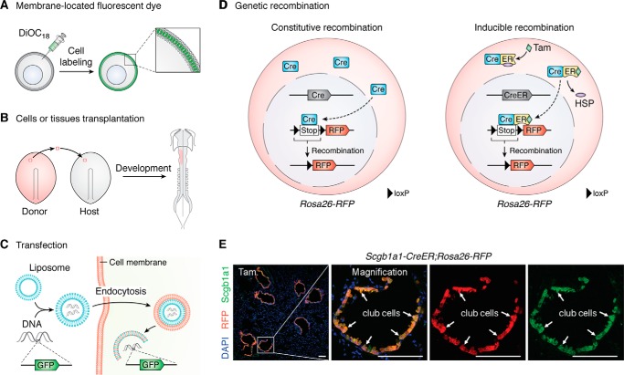
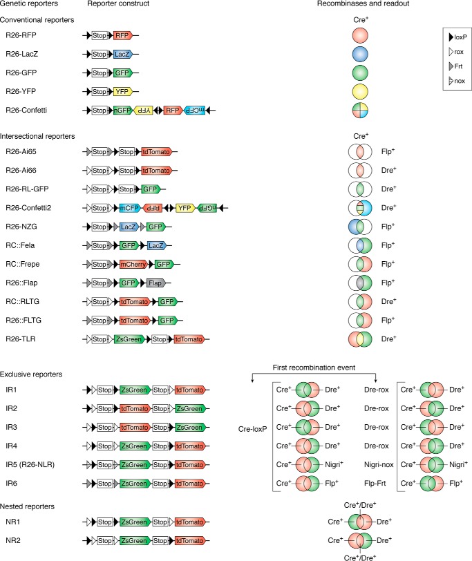
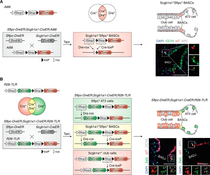
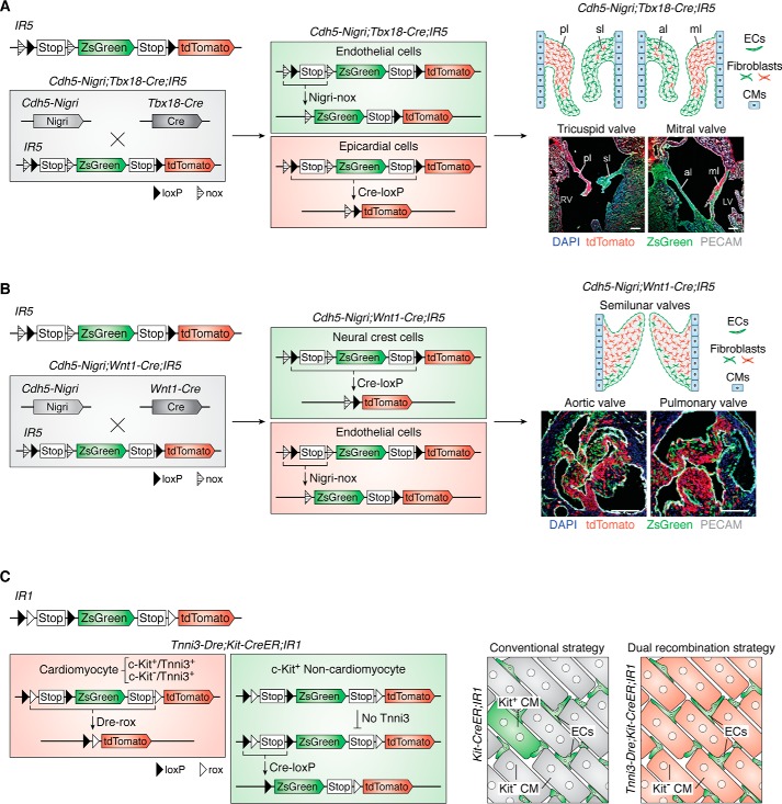
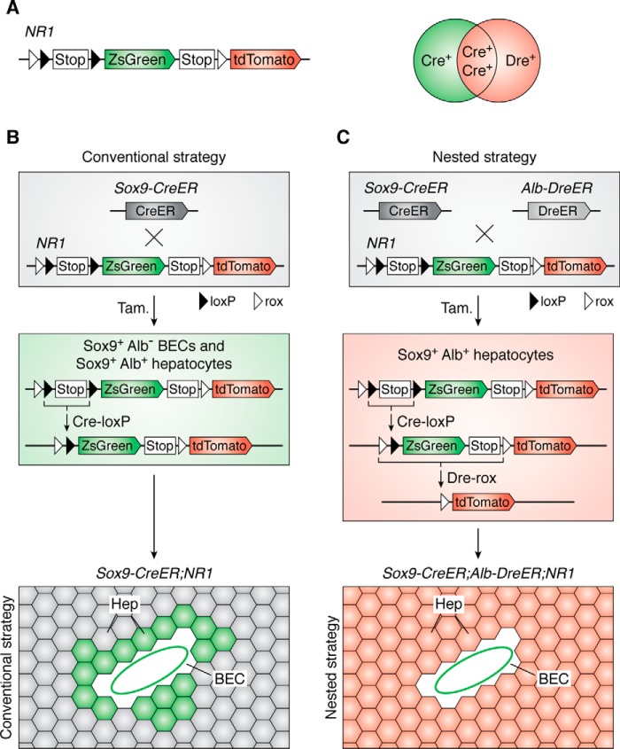
Site-specific recombinases, such as Cre, are a widely used tool for genetic lineage tracing in the fields of developmental biology, neural science, stem cell biology, and regenerative medicine. However, nonspecific cell labeling by some genetic Cre tools remains a technical limitation of this recombination system, which has resulted in data misinterpretation and led to many controversies in the scientific community. In the past decade, to enhance the specificity and precision of genetic targeting, researchers have used two or more orthogonal recombinases simultaneously for labeling cell lineages. Here, we review the history of cell-tracing strategies and then elaborate on the working principle and application of a recently developed dual genetic lineage-tracing approach for cell fate studies. We place an emphasis on discussing the technical strengths and caveats of different methods, with the goal to develop more specific and efficient tracing technologies for cell fate mapping. Our review also provides several examples for how to use different types of DNA recombinase-mediated lineage-tracing strategies to improve the resolution of the cell fate mapping in order to probe and explore cell fate-related biological phenomena in the life sciences.

Keywords: Cre-loxP; Dre-rox; cell fate mapping; development; dual recombination; gene expression; gene mapping; genetic recombination; genetics; lineage trace; organ regeneration; reporter gene; site-specific DNA recombinase; stem cell; tissue regeneration.


Conflict of interest statement

The authors declare that they have no conflicts of interest with the contents of this article

Figures

Figure 1.
Figure 1.
Lineage-tracing strategies. A, dyes for cell labeling. The lipid-soluble carbocyanine dyes could be embedded into lipid bilayer for cell tracing. B, transplantation of labeled cells or tissues from donor embryo to host embryo. The fate of labeled cells or tissues could be tracked during embryonic development. C, introduction of reporter genes such as GFP by transfection or viral infection has been used for cell labeling. D, cell fate tracing by DNA site–specific recombination. The genetic recombination includes constitutive recombination and inducible recombination. Take the widely used Cre-loxP system as an example. The loxP-flanked transcriptional stop cassette (Stop) was inserted before the RFP gene. In constitutive recombination, after the cell type–specific promoter drives the Cre recombinase expression in target cells, the loxP-flanked Stop cassette can be removed, and the reporter gene would be turned on. Because the excision of genomic level is permanent and heritable, all Cre+ cells and their progeny could be labeled by the reporter gene. However, in inducible recombination, binding to heat shock proteins (HSPs), the Cre-ER fusion protein is held in the cytoplasm. Only after the ligand (tamoxifen) enters the cell cytoplasm and binds to ER can the Cre-ER fusion protein release from the HSP and enter the cell nucleus for Cre-loxP recombination. E, immunostaining for RFP, DAPI, and club cell marker Scgb1a1 on Scgb1a1-CreER;Rosa26-RFP adult lung section after tamoxifen treatment. White arrows, RFP+Scgb1a1+ club cells. Tam, tamoxifen. Scale bar, 100 μm.
Figure 2.
Figure 2.
Genetic recombination systems. The first column indicates different reporter alleles, the second and third columns show the reporter construct and recombinases, and the last column shows the readout of related reporter alleles. The conventional reporter system includes the single-color reporter system and the multicolor reporter system, which are derived by one type recombination. The novel dual-reporter systems include intersectional reporters, exclusive reporters, and nested reporters. In exclusive reporters, the readout depends on the first recombination type. For example, in IR1, the first Cre-loxP will remove the Stop cassette and a rox site, which labels the Cre+ cells (including both Cre+Dre cells and Cre+Dre+ cells) as ZsGreen. The following Dre-rox recombination results in tdTomato expression of Dre+Cre cells. In contrast, if the first recombination event is Dre-rox, the Dre+ cells (including both Dre+Cre cells and Dre+Cre+ cells) are labeled as tdTomato, and the DreCre+ cells would be labeled as ZsGreen by the following Cre-loxP recombination.
Figure 3.
Figure 3.
Intersectional reporter systems. A, after dual Dre-rox and Cre-loxP recombination, the intersectional Ai66 reporter can specifically mark the Dre+Cre+ cells as tdTomato. In Sftpc-DreER;Scgb1a1-CreER;Ai66 triple-positive mouse, the Sftpc+Scgb1a1+ BASCs are labeled by tdTomato after tamoxifen treatment. The cartoon image (right) shows that BASCs are marked by red in the Sftpc-DreER;Scgb1a1-CreER;Ai66 triple-positive mouse. Yellow arrowheads in the sectional staining picture (right) indicate Sftpc+Scgb1a1+tdTomato+ BASCs. B, the R26-TLR is another intersectional system that could be used for tracing three cell populations simultaneously. Also, taking the lung epithelium as an example, in the Sftpc-DreER;Scgb1a1-CreER;R26-TLR triple-positive mouse, after Dre-rox recombination, the Sftpc+ AT2 cells are labeled as ZsGreen; after Cre-loxP recombination, the Scgb1a1+ are club cells labeled as tdTomato; and after both Dre-rox and Cre-loxP recombination, the Sftpc+Scgb1a1+ BASCs are labeled as ZsGreen and tdTomato, the readout for which is a yellow fluorescent color. A cartoon image (right) shows the club cells, AT2 cells, and BASCs marked as red, green, and yellow, respectively. Yellow arrowheads in the sectional staining picture (right) indicate ZsGreen+tdTomato+ BASCs. Scale bar, 100 μm.
Figure 4.
Figure 4.
Exclusive reporter systems. A, the exclusive IR5 reporter can be used to label distinct cell populations. In the Cdh5-Nigri;Tbx18-Cre;IR5 triple-positive mouse, Cdh5+ endothelial cells (including endocardial cells) are marked as ZsGreen by Nigri-nox recombination, and Tbx18+ epicardial cells are marked as tdTomato by Cre-loxP recombination. The image (right) shows the contribution of these two distinct cell populations for atrioventricular valve mesenchyme during heart development. B, in the Cdh5-Nigri;Wnt1-Cre;IR5 triple-positive mouse, Wnt1+ neural crest cells are marked as tdTomato by Cre-loxP recombination, and Cdh5+ endothelial cells (including endocardial cells) are marked as ZsGreen by Nigri-nox recombination. The image (right) shows the contribution of these two distinct cell populations for outflow track valve mesenchyme during heart development. C, the interleaved reporter 1 (IR1) can be used to label distinct cell populations. After Dre-rox recombination in Dre+ cells, all DNA sequences between these two rox sites would be removed, including a loxP site. So the next Cre-loxP recombination would not happen in Dre+ cells, and it can only happen in DreCre+ cells. Taking the c-kit as an example, because the c-kit ectopic expressed in a little cardiomyocytes, the Kit-CreER;IR1 mouse (conventional strategy) could label kit+ cardiomyocytes after Cre-loxP recombination. In Tnni3-Dre;Kit-CreER;IR1 triple-positive line, firstly, all Tnni3+ cardiomyocytes (including these unwanted c-kit+Tnni3+ cardiomyocytes) can be specific labeled by Dre-rox recombination. Then after tamoxifen induction, c-kit-CreER could specifically labeled all c-kit+ cells (excluding these unwanted c-kit+Tnni3+ cardiomyocytes). ECs, endothelial cells; CM, cardiomyocyte; LV, left ventricle; RV, right ventricle; pl, parietal leaflet; sl, septal leaflet; al, aortic leaflet; ml, mural leaflet. Scale bar, 100 μm.
Figure 5.
Figure 5.
Nested reporter systems. A, the nested NR1 reporter can be used to label distinct cell populations. The Cre+ cells are labeled as ZsGreen by Cre-loxP recombination. The Dre+Cre cells are marked as tdTomato by Cre-loxP recombination. The final readout of Dre+Cre+ cells is tdTomato because of the final Cre-loxP recombination. B, Sox9 is not a specific marker of BECs; it also targets some hepatocytes (Hep). The readout of Sox9-CreER;NR1 line includes both BECs and hepatocytes, which is same as conventional strategy. C, in the Sox9-CreER;Alb-CreER;NR1 triple-positive mouse (nested strategy), the Sox9+Alb+ hepatocytes are finally labeled as tdTomato. So the nested reporter system is valuable for labeling distinct cell populations.

References

    1. Kretzschmar K., and Watt F. M. (2012) Lineage tracing. Cell 148, 33–45 10.1016/j.cell.2012.01.002 - DOI - PubMed
    1. Fox D. T., Morris L. X., Nystul T., and Spradling A. C. (2008) Lineage analysis of stem cells. in StemBook, Harvard Stem Cell Institute, Cambridge, MA - PubMed
    1. Buckingham M. E., and Meilhac S. M. (2011) Tracing cells for tracking cell lineage and clonal behavior. Dev. Cell 21, 394–409 10.1016/j.devcel.2011.07.019 - DOI - PubMed
    1. Axelrod D. (1979) Carbocyanine dye orientation in red-cell membrane studied by microscopic fluorescence polarization. Biophys. J. 26, 557–573 10.1016/S0006-3495(79)85271-6 - DOI - PMC - PubMed
    1. Weisblat D. A., Sawyer R. T., and Stent G. S. (1978) Cell lineage analysis by intracellular injection of a tracer enzyme. Science 202, 1295–1298 10.1126/science.725606 - DOI - PubMed

Publication types

Substances