. 2020 Mar 10:15:1677-1691.
doi: 10.2147/IJN.S225807. eCollection 2020.

Multifunctional Immunoliposomes Combining Catalase and PD-L1 Antibodies Overcome Tumor Hypoxia and Enhance Immunotherapeutic Effects Against Melanoma

Affiliations

Multifunctional Immunoliposomes Combining Catalase and PD-L1 Antibodies Overcome Tumor Hypoxia and Enhance Immunotherapeutic Effects Against Melanoma

Yu Hei et al. Int J Nanomedicine. .

Abstract

Background: Immune checkpoint blockades (ICBs) are a promising treatment for cancers such as melanoma by blocking important inhibitory pathways that enable tumor cells to evade immune attack. Programmed death ligand 1 monoclonal antibodies (aPDL1s) can be used as an ICB to significantly enhance the effectiveness of tumor immunotherapy by blocking the PD-1/PD-L1 inhibitory pathway. However, the effectiveness of aPDL1s may be limited by low selectivity in vivo and immunosuppressed tumor microenvironment including hypoxia.

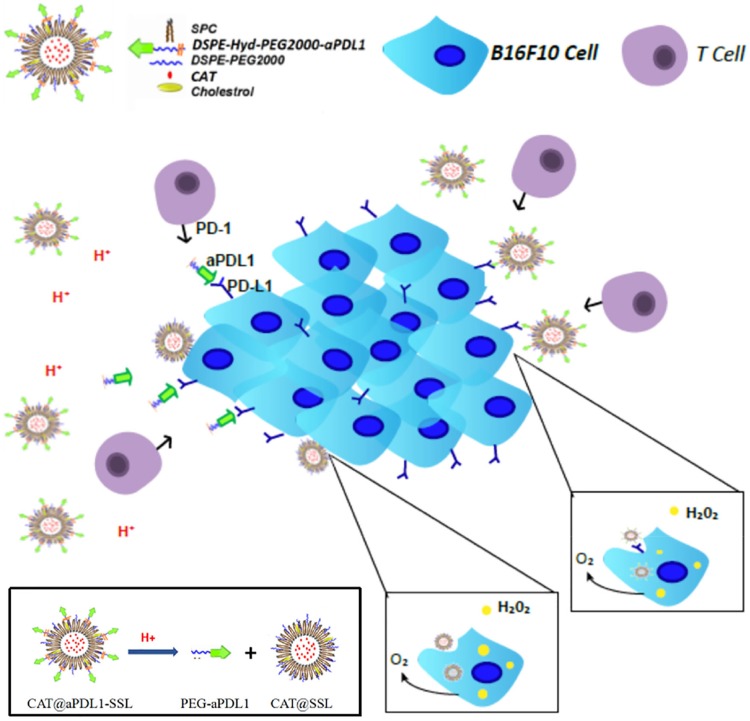
Purpose: To overcome the limitations, we develop a multifunctional immunoliposome, called CAT@aPDL1-SSL, with catalase (CAT) encapsulated inside to overcome tumor hypoxia and aPDL1s modified on the surface to enhance immunotherapeutic effects against melanoma.

Methods: The multifunctional immunoliposomes (CAT@aPDL1-SSLs) are prepared using the film dispersion/post-insertion method. The efficacy of CAT@aPDL1-SSLs is verified by multiple experiments in vivo and in vitro.

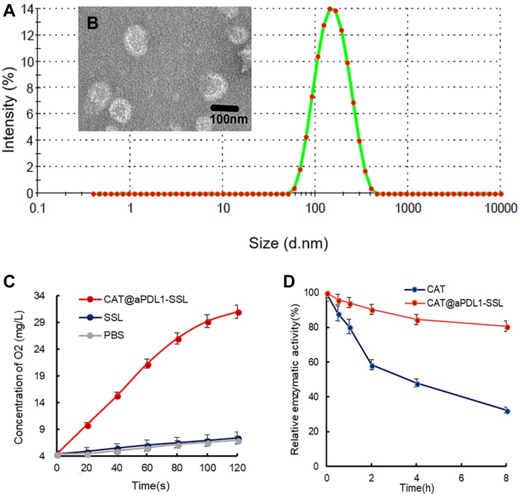
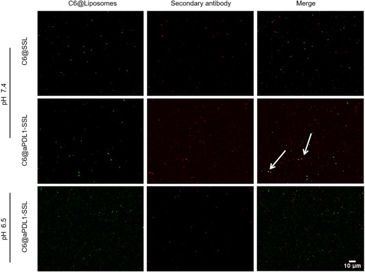
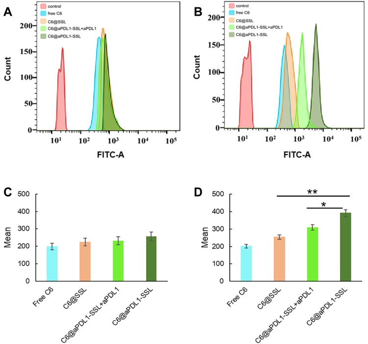
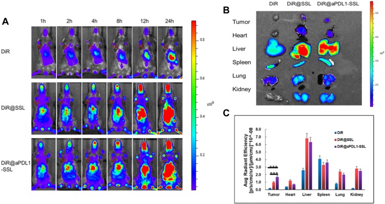
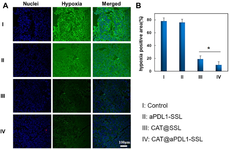
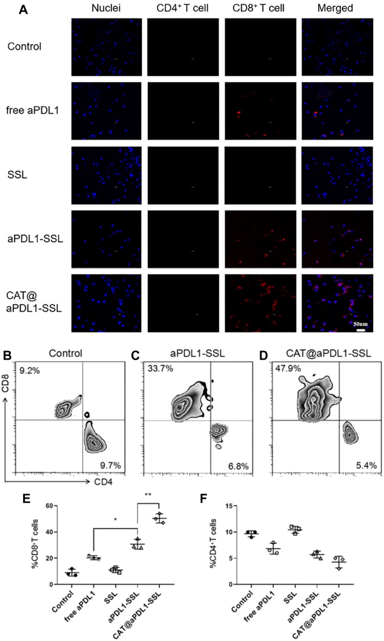
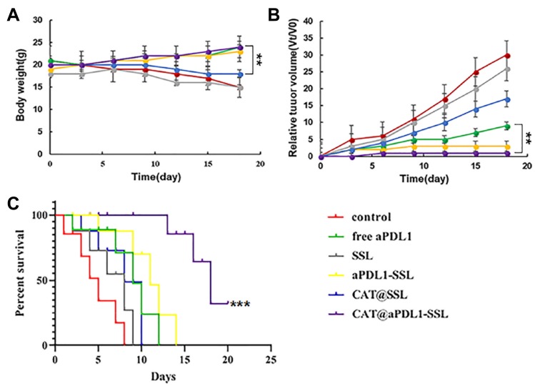
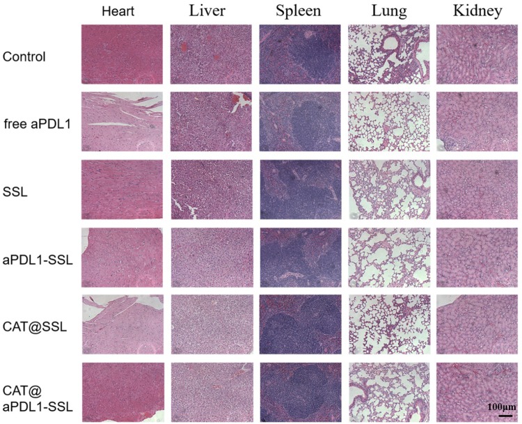
Results: The results of this study suggest that the multifunctional immunoliposomes preserve and protect the enzyme activity of CAT and ameliorate tumor hypoxia. Moreover, the enhanced cellular uptake of CAT@aPDL1-SSLs in vitro and their in vivo biodistribution suggest that CAT@aPDL1-SSLs have great targeting ability,resulting in improved delivery and accumulation of immunoliposomes in tumor tissue.Finally, by activating and increasing the infiltration of CD8+ T cells at the tumor site, CAT@aPDL1-SSLs inhibit the growth of tumor and prolong survival time of mice,with low systemic toxicity.

Conclusion: In conclusion, the multifunctional immunoliposomes developed and proposed in this study are a promising candidate for melanoma immunotherapy, and could potentially be combined with other cancer therapies like radiotherapy and chemotherapy to produce positive outcomes.

Keywords: aPDL1s; immunotherapy; liposomes; melanoma; programmed death ligand 1 monoclonal antibodies; tumor hypoxia.


Conflict of interest statement

The authors report no conflicts of interest in this work.

Figures

Figure 1
Figure 1
The schematic diagram of the structure and activating mechanism of CAT@aPDL1-SSL. Note: CAT@aPDL1-SSLs block the PD-1/PD-L1 pathway and deliver CAT into tumor cells to relieve hypoxia, promoting T lymphocyte infiltration and enhancing immunotherapeutic effects. Abbreviations: aPDL1, programmed death ligand 1 monoclonal antibody; CAT, catalase; SSL, sterically stabilized liposome; aPDL1-SSLs, aPDL1 modified immunoliposomes; CAT@aPDL1-SSLs, CAT-loaded and aPDL1 modified immunoliposomes; SPC, Lecithin (soy beans); DSPE-PEG2000, Distearoylphosphatidylethanoloamine-methoxy-polyethylene glycol MW:2000; Hyd, hydrazine; PD-1, programmed death receptor 1; PD-L1, programmed death ligand 1.
Figure 2
Figure 2
Characterization of CAT@aPDL1-SSLs. Notes: (A) The particle size distribution of CAT@aPDL1-SSLs determined by DLS. (B) Morphological images observed by TEM. Scale bar: 100 nm. (C) Concentrations of O2 in H2O2 solution detected by a portable dissolved oxygen meter after adding CAT@aPDL1-SSLs and SSLs. (D) Relative catalytic ability of free CAT and CAT@aPDL1-SSLs at different time points after protease K treatment (0.5 mg/mL). Abbreviations: CAT, catalase; CAT@aPDL1-SSLs,CAT-loaded immunoliposomes; DLS, dynamic light scattering; TEM, transmission electron microscopy.
Figure 3
Figure 3
Colocalization results of C6@SSLs, C6@aPDL1-SSLs (pH 7.4), and C6@aPDL1-SSLs (pH 6.5). Notes: The colocalization results were observed by CLSM. The green signals represented C6@SSLs, C6@aPDL1-SSLs (pH 7.4) or C6@aPDL1-SSLs (pH 6.5) and the red signals represented Alexa Fluor 647-labelled secondary antibodies. The white arrow indicated the colocalization area. Scale bar: 10 μm. Abbreviations: C6, Coumarin-6; C6@SSLs,C6-loaded liposomes; C6@aPDL1-SSLs, C6-loaded immunoliposomes; CLSM, confocal laser scanning microscopy.
Figure 4
Figure 4
Flow cytometry histograms and mean value of different formulations under different pH conditions. Notes: (A, C) pH 6.5. (B, D) pH 7.4. (n = 3, results are shown as means ± S.D. *P < 0.05, **P < 0.01.). Abbreviations: C6, Coumarin-6; C6@SSLs,C6-loaded liposomes; C6@aPDL1-SSLs, C6-loaded immunoliposomes.
Figure 5
Figure 5
Biodistribution of free DiR, DiR@SSLs, and DiR@aPDL1-SSLs. Notes: (A) In vivo fluorescence images of B16-F10 tumor-bearing mice after intravenous injection of the three formulations at 1, 2, 4, 8, 12, and 24 h (red circle represents the tumor area). (B) Ex vivo fluorescence images and (C) quantitative results of fluorescence efficiency of main organs and tumor tissues of the three groups 24 h after injection. (n = 3, results are shown as means ± S.D. ***P < 0.001.) Abbreviations: DiR, 1,1-dioctadecyl-3,3,3,3-tetramethylindotricarbocyanine; DiR@SSLs, DiR-loaded liposomes; DiR@aPDL1-SSLs,DiR- loaded immunoliposomes.
Figure 6
Figure 6
Effects of control (PBS), aPDL1-SSLs, CAT@SSLs, and CAT@aPDL1-SSLs on tumor hypoxia status. Notes: (A) Immunofluorescence images of tumor slices harvested from B16-F10 tumor-bearing mice following intravenous injection with four different formulations. The blue signals represent nuclei stained by DAPI and the green signals represent hypoxia areas stained by hypoxia probe, respectively. (B) The quantitative results of positive tumor hypoxia areas analyzed using Image J software based on the images shown in (A). (n = 3, results are means ± S.D., Scale bar: 100 μm,*P < 0.05) Abbreviations: aPDL1, programmed death ligand 1 monoclonal antibody; CAT, catalase; SSL, sterically stabilized liposome; aPDL1-SSLs, aPDL1 modified immunoliposomes; CAT@aPDL1-SSLs, CAT-loaded immunoliposomes.
Figure 7
Figure 7
Immunofluorescence of tumor slices and flow cytometry analysis showing CD4+ and CD8+ T cell infiltration. Notes: (A) Immunofluorescence of tumor slices.The blue signals represent nuclei stained with DAPI. The green signals represent CD4+ T cells stained with AF488-CD4+ antibodies and the red signals represent CD8+ T cells stained with AF647-CD8+ antibodies. (Scale bar: 50 μm). (B, C and D) Flow cytometry analysis and (E and F) their corresponding percentage of CD8+ and CD4+ tumor infiltrating T cells (n = 3, results are means ± S.D.,*P < 0.05,**P < 0.01). Abbreviations: aPDL1, programmed death ligand 1 monoclonal antibody; CAT, catalase; SSL, sterically stabilized liposome; aPDL1-SSLs, aPDL1 modified immunoliposomes; CAT@aPDL1-SSLs, CAT-loaded immunoliposomes.
Figure 8
Figure 8
Antitumor efficacy in vivo. Notes: (A) The body weights of B16-F10 tumor-bearing mice after injection with PBS, free aPDL1, SSLs, aPDL1-SSLs, CAT@SSLs, and CAT@aPDL1-SSLs. (B) The relative tumor volume of mice in each group. (C) Survival curves of mice after treatment with the formulations described above. (n = 6, results means ± S.D., **P < 0.01, ***P < 0.001) Abbreviations: aPDL1, programmed death ligand 1 monoclonal antibody; CAT, catalase; SSL, sterically stabilized liposome; aPDL1-SSLs, aPDL1 modified immunoliposomes; CAT@aPDL1-SSLs, CAT-loaded immunoliposomes.
Figure 9
Figure 9
Safety assessment of different formulations in vivo. Notes: The morphology of the main organs, including the heart, liver, spleen, lungs, and kidneys were observed using the H&E staining method after treating mice with PBS, free aPDL1, SSLs, aPDL1-SSLs, CAT@SSLs, or CAT@aPDL1-SSLs (Scale bar: 100 μm). Abbreviations: aPDL1, programmed death ligand 1 monoclonal antibody; CAT, catalase; SSL, sterically stabilized liposome; aPDL1-SSLs, aPDL1 modified immunoliposomes; CAT@aPDL1-SSLs, CAT-loaded immunoliposomes.

References

    1. Shergold AL, Millar R, Nibbs RJB. Understanding and overcoming the resistance of cancer to PD-1/PD-L1 blockade. Pharmacol Res. 2019;145:104–258. doi:10.1016/j.phrs.2019.104258 - DOI - PubMed
    1. Minn AL, Wherry EJ. Combination cancer therapies with immune checkpoint blockade: convergence on interferon signaling. Cell. 2016;165:272–275. doi:10.1016/j.cell.2016.03.031 - DOI - PubMed
    1. Gajewski TF, Schreiber H, Fu YX. Innate and adaptive immune cells in the tumor microenvironment. Nat Immunol. 2013;14:1014−1022. doi:10.1038/ni.2703 - DOI - PMC - PubMed
    1. Leisegang M, Kammertoens T, Uckert W, et al. Targeting human melanoma neoantigens by T cell receptor gene therapy. J Clin Invest. 2016;126:854–858. doi:10.1172/JCI83465 - DOI - PMC - PubMed
    1. Eroglu Z, Zaretsky JM, Hu-lieskovan S, et al. High response rate to PD-1 blockade in desmoplastic melanomas. Nature. 2018;553:347–350. doi:10.1038/nature25187 - DOI - PMC - PubMed

MeSH terms