. 2020 Jan 13:8:36-47.
doi: 10.1016/j.ibror.2019.12.004. eCollection 2020 Jun.

Dissociation of neonatal and adult mice brain for simultaneous analysis of microglia, astrocytes and infiltrating lymphocytes by flow cytometry

Affiliations

Dissociation of neonatal and adult mice brain for simultaneous analysis of microglia, astrocytes and infiltrating lymphocytes by flow cytometry

Belén Calvo et al. IBRO Rep. .

Abstract

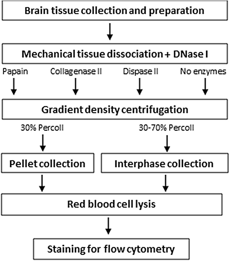
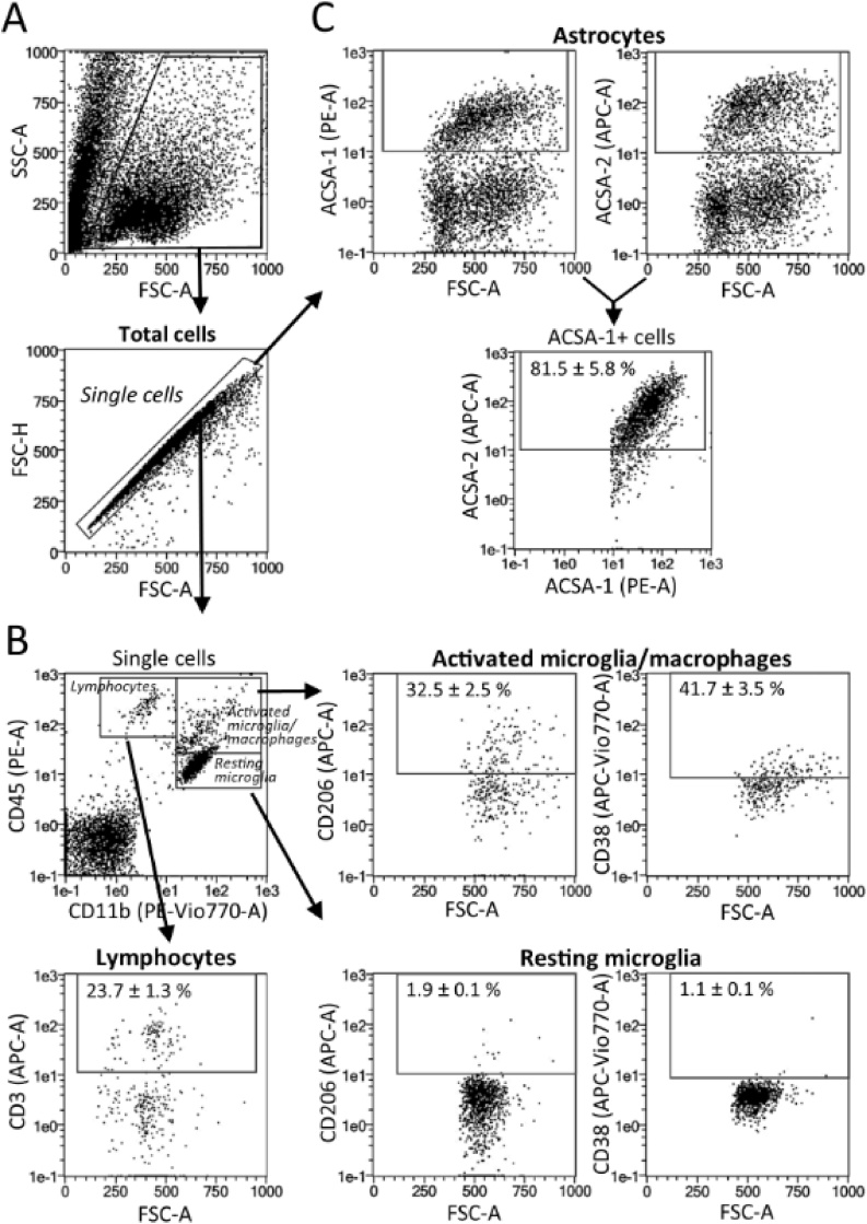
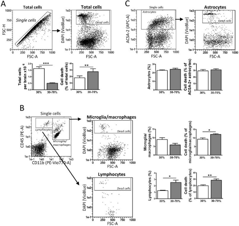
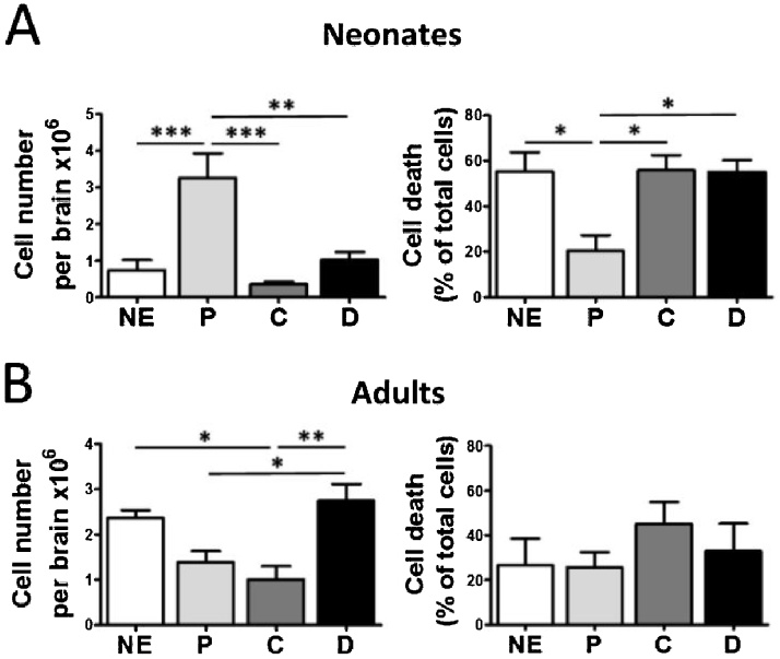
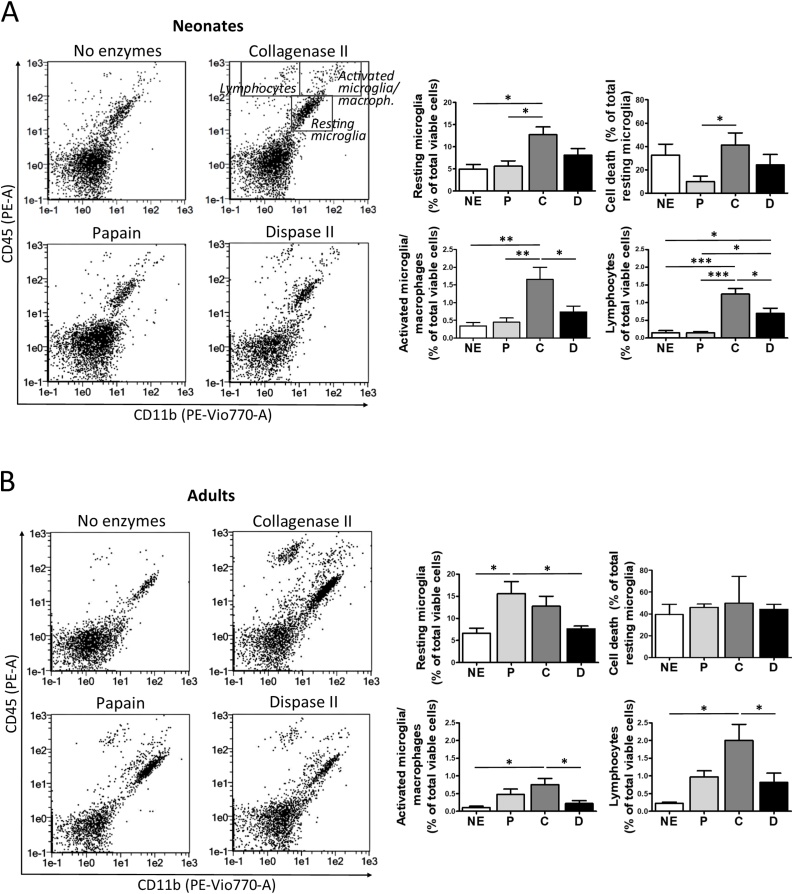
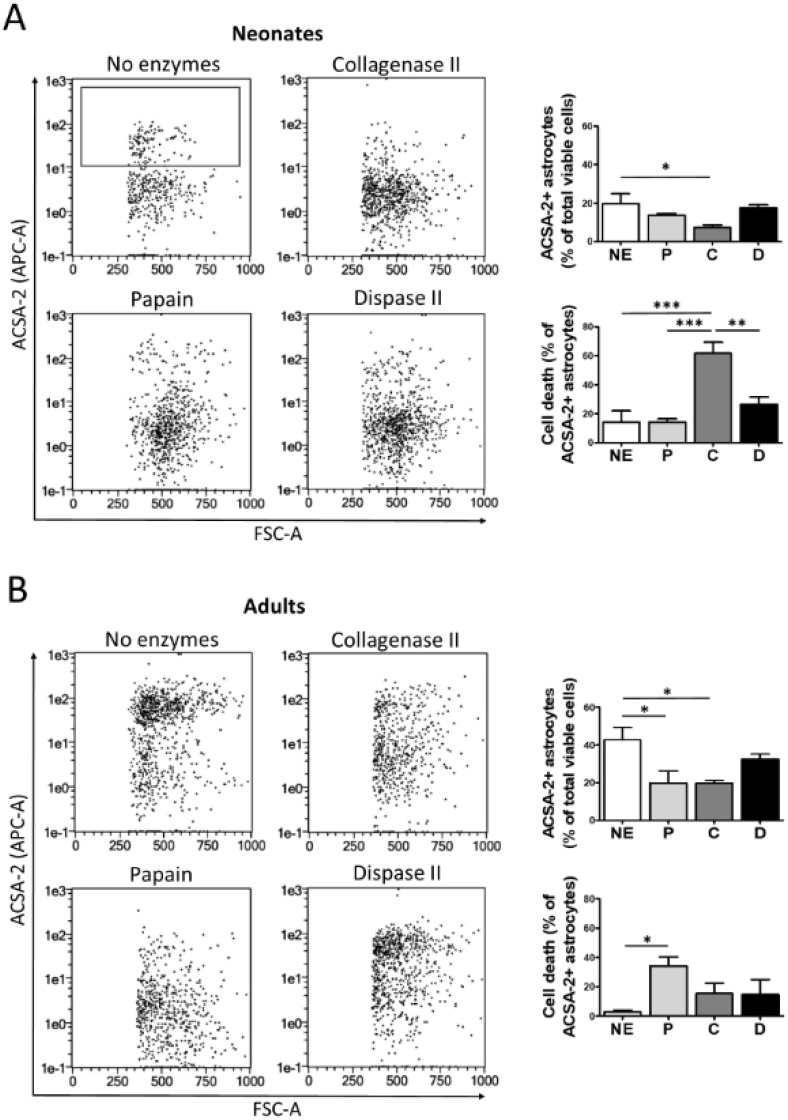
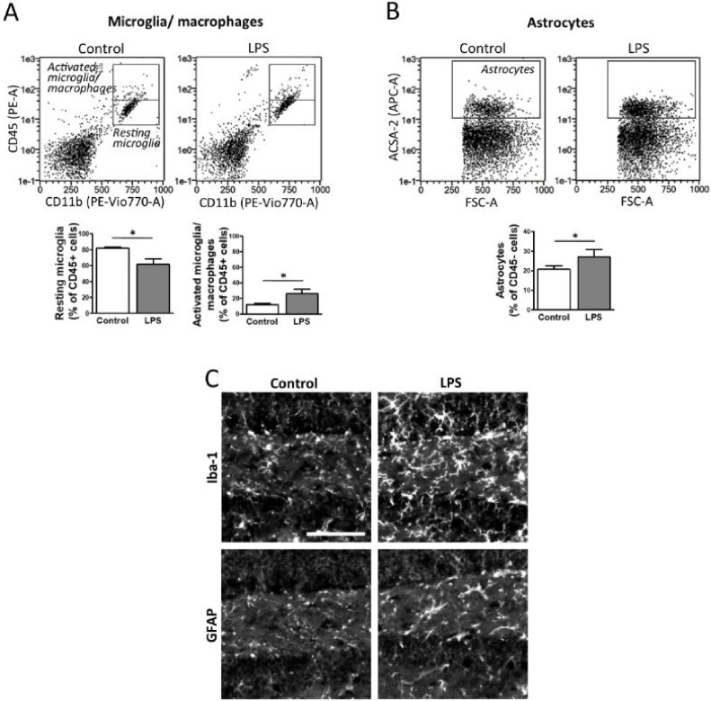
The technical difficulty to isolate microglia, astrocytes and infiltrating immune cells from mouse brain is nowadays a limiting factor in the study of neuroinflammation. Brain isolation requirements are cell-type and animal-age dependent, but current brain dissociation procedures are poorly standardized. This lack of comprehensive studies hampers the selection of optimized methodologies. Thus, we present here a comparative analysis of dissociation methods and Percoll-based separation to identify the most efficient procedure for the combined isolation of healthy microglia, astrocytes and infiltrated leukocytes; distinguishing neonatal and adult mouse brain. Gentle mechanical dissociation and DNase I incubation was supplemented with papain or collagenase II. Dispase II digestion was also used alone or in combination. In addition, cell separation efficiency of 30 % and 30-70 % Percoll gradients was compared. In these experiments, cell yield and integrity of freshly dissociated cells was measured by flow cytometry. We found that papain digestion in combination with dispase II followed by 30 % Percoll separation is the most balanced method to obtain a mixture of microglia, astrocytes and infiltrated immune cells; while addition of dispase II was not an advantage for neonatal brain. These dissociation conditions allowed flow cytometry detection of a slight glial activation triggered by sublethal LPS injection. In conclusion, the enzymes and Percoll density gradients tested here affected differently resting microglia, activated microglia/macrophages, astrocytes and infiltrated lymphocytes. Also, newborn and adult brain showed contrasting reactions to digestion. Our study highlights the strength of flow cytometry for the simultaneous analysis of neuroimmune cell populations once extraction is optimized.

Keywords: ANOVA, one-way analysis of variance; Astrocytes; CNS, Central Nervous System; CaCl2, calcium chloride; EBSS, Earle's Balanced Salt Solution; EDTA, ethylenediaminetetraacetic acid; FACS, Fluorescence-activated cell sorter; FSC, forward-scattered light; Flow cytometry; Glia reactivity; HBSS, Hank's Balanced Salt Solution; LD, lethal dose; LPS, lipopolysaccharide; Lymphocytes; MgCl2, magnesium chloride; MgSO4, magnesium sulfate; Microglia; Neuroimmunity; PBS, phosphate-buffered saline; RT, room temperature; SIP, stock solution of isotonic Percoll; SSC, side-scattered light; i.p, intraperitoneal injection.


Figures

Fig. 1
Fig. 1
Workflow outlining the different steps followed in this protocol.
Fig. 2
Fig. 2
Cell cytometry markers and gating strategy for the analysis of brain cell populations. Neural cells were isolated from mouse and further purified using 30 % Percoll. (A) Gates to exclude debris and cell aggregates in FSC-A/SSC-A and FSC-A/FSC-H plots. (B) Single cells from adult brain were obtained with papain and dispase II. Gates correspond to resting microglia (CD11b+/CD45low), activated microglia/macrophages (CD11b+/CD45high) and lymphocytes (CD11b-/CD45+). Activated microglia/ macrophage gating was further sustained by incubation with microglia/macrophage activation markers CD206 and CD38; while lymphocyte gating was verified by CD3 staining of T cells. (C) Gates for ACSA-1+ and ACSA-2+ astrocytes extracted from neonatal brain, showing extensive staining overlapping. Numerical values correspond to mean ± S.E.M (n = 4).
Fig. 3
Fig. 3
Comparison of Percoll density gradients for flow cytometry analysis of brain cell recovery. Neural cells were isolated from adult brain by enzymatic dissociation followed by comparison of 30 % and 30–70 % Percoll density gradients to separate cells from myelin and debris. DAPI staining of dead cells was applied to the analysis of total cells (A), CD11b+/CD45+ microglia/macrophages (B), CD11b-/CD45+ lymphocytes (B), and ACSA-2+ astrocytes (C). Graphs show cell recovery as well as the fraction of dead cells (%) for each brain population, comparing values after 30 % and 30–70 % Percoll purification. Data represent mean ± S.E.M. (n = 5–6). *, p < 0.05; **, p < 0.01; ***, p < 0.001.
Fig. 4
Fig. 4
Enzymatic digestion conditions neural cell recovery from neonate and adult brains. Neonatal (A) or adult brains (B) were digested with papain (P), collagenase II (C), dispase II (D) or non-enzymatically (NE), all in combination with DNase I and gentle mechanical dispersion. After enrichment using a 30 % Percoll gradient, the isolated cells were processed for flow cytometry analysis of total cell number. DAPI staining allowed evaluation of cell death. Data are expressed as mean ± S.E.M. (n = 4 for adult and n = 5–6 for neonate experiments). *, p < 0.05; **, p < 0.01; ***, p < 0.001.
Fig. 5
Fig. 5
Recovery of myeloid and lymphoid cells from newborn and adult mice brains. Tissue from neonatal (A) and adult brain (B) was mechanically dissociated while digested with papain (P), collagenase II (C), dispase II (D) or in the absence of enzymes (NE); and brain homogenates were stained for flow cytometry analysis. Representative plots for each isolation protocol are included. Gates drawn correspond to resting microglia (CD11b+/CD45low), activated microglia/macrophages (CD11b+/CD45high), and lymphocytes (CD11b-/CD45+). Cell death within resting microglia was measured after DAPI staining. Associated graphs correspond to % of each population and resting microglia death. Values in all graphs are expressed as percentage of total cells and represent mean ± S.E.M. (n = 4–5). *, p < 0.05; **, p < 0.01; ***, p < 0.001.
Fig. 6
Fig. 6
Dissociation procedure for the extraction of astrocytes from neonatal and adult brain. Tissue from neonatal (A) and adult (B) brain was mechanically dissociated in the presence of papain (P), collagenase II (C), dispase II (D) or no enzymes (NE); and further purified through a 30 % Percoll gradient. Representative flow cytometry plots are shown. Astrocytes were identified as ACSA-2+ cells (gate). Cell death was measured after DAPI staining. Associated graphs correspond to % of astrocytes and astrocyte death. Data are expressed as mean ± S.E.M. (n = 3–4). *, p < 0.05; **, p < 0.01; ***, p < 0.001.
Fig. 7
Fig. 7
Enzymatic isolation of brain cells with papain and dispase II to evaluate glia response to LPS. Animals were injected with LPS (0.5 mg/Kg, i.p.) to induce a mild inflammatory response. After 3 days, mice were sacrificed. Brain cells were isolated using papain and dispase II combined with gentle mechanical dissociation and DNase II incubation. Purification with 30 % Percoll was followed by flow cytometry analysis of (A) resting microglia (CD11b+/CD45low) and activated microglia/macrophages (CD11b+/CD45high), or (B) astrocytes (ACSA-2+/CD45-). Values are expressed as percentage of total CD45+ cells (A) or CD45-cells (B) and correspond to mean ± S.E.M. (n = 7–9). *, p < 0.05. (C) Another set of control and LPS-treated animals was fixed by perfusion, and brain sections stained for immunofluorescence detection of either Iba-1 or GFAP as markers of microglia and astrocytes, respectively. Representative images containing the dentate gyrus of hippocampus are shown (scale bar, 100 μm).

References

    1. Batiuk M.Y., de Vin F., Duque S.I., Li C., Saito T., Saido T., Fiers M., Belgard T.G., Holt M.G. An immunoaffinity-based method for isolating ultrapure adult astrocytes based on ATP1B2 targeting by the ACSA-2 antibody. J. Biol. Chem. 2017;292:8874–8891. - PMC - PubMed
    1. Bedi S.S., Smith P., Hetz R.A., Xue H., Cox C.S. Immunomagnetic enrichment and flow cytometric characterization of mouse microglia. J. Neurosci. Methods. 2013;219:176–182. - PMC - PubMed
    1. Ben-Gigi L., Sweetat S., Besser E., Fellig Y., Wiederhold T., Polakiewicz R.D., Behar O. Astrogliosis induced by brain injury is regulated by Sema4B phosphorylation. eNeuro. 2015;2:3. - PMC - PubMed
    1. Bergmann C.C., Altman J.D., Hinton D., Stohlman S.A. Inverted immunodominance and impaired cytolytic function of CD8+ T cells during viral persistence in the central nervous system. J. Immunol. 1999;163:3379–3387. doi: ji_v163n6p3379. - PubMed
    1. Boelen E., Stassen F.R., Steinbusch H.W., Borchelt D.R., Streit W.J. Ex vivo cultures of microglia from young and aged rodent brain reveal age-related changes in microglial function. Neurobiol. Aging. 2012;33:1. - PMC - PubMed