. 2020 Aug;30(8):4407-4416.
doi: 10.1007/s00330-020-06817-6. Epub 2020 Mar 25.

CT image visual quantitative evaluation and clinical classification of coronavirus disease (COVID-19)

Affiliations

CT image visual quantitative evaluation and clinical classification of coronavirus disease (COVID-19)

Kunwei Li et al. Eur Radiol. 2020 Aug.

Abstract

Objectives: To explore the relationship between the imaging manifestations and clinical classification of COVID-19.

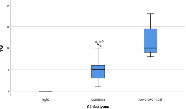
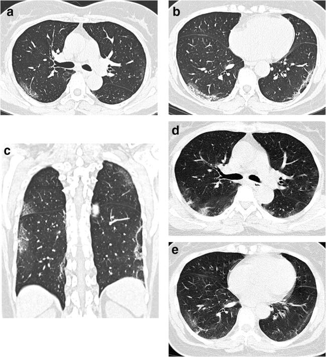
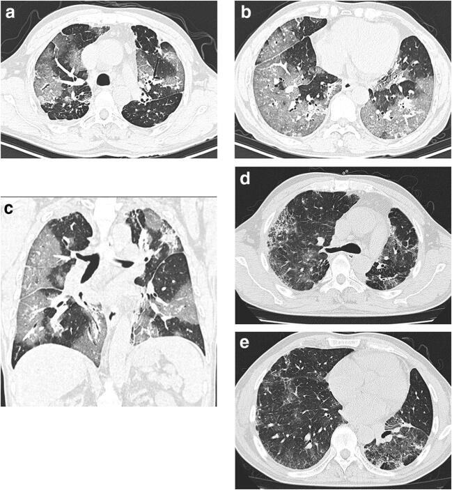
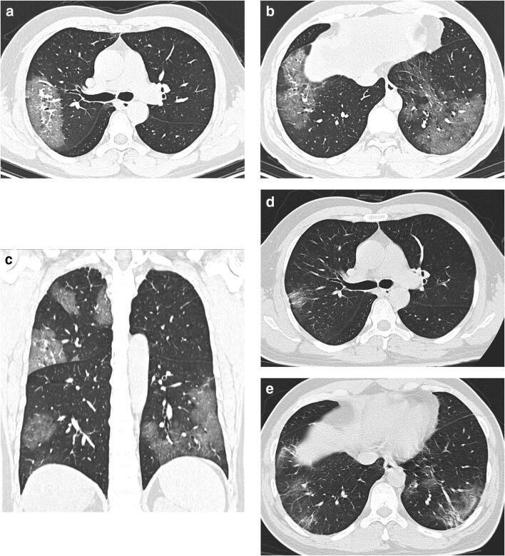
Methods: We conducted a retrospective single-center study on patients with COVID-19 from Jan. 18, 2020 to Feb. 7, 2020 in Zhuhai, China. Patients were divided into 3 types based on Chinese guideline: mild (patients with minimal symptoms and negative CT findings), common, and severe-critical (patients with positive CT findings and different extent of clinical manifestations). CT visual quantitative evaluation was based on summing up the acute lung inflammatory lesions involving each lobe, which was scored as 0 (0%), 1 (1-25%), 2 (26-50%), 3 (51-75%), or 4 (76-100%), respectively. The total severity score (TSS) was reached by summing the five lobe scores. The consistency of two observers was evaluated. The TSS was compared with the clinical classification. ROC was used to test the diagnosis ability of TSS for severe-critical type.

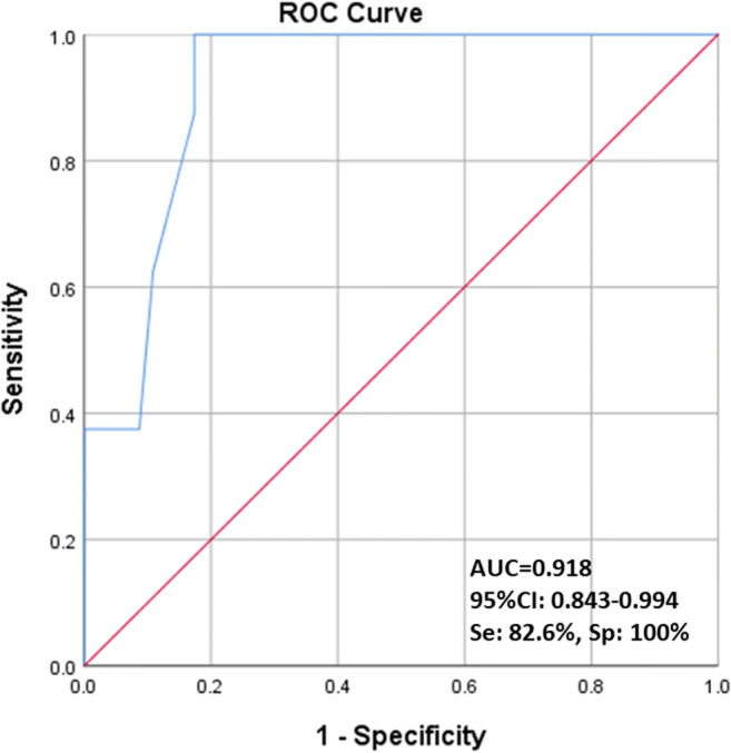
Results: This study included 78 patients, 38 males and 40 females. There were 24 mild (30.8%), 46 common (59.0%), and 8 severe-critical (10.2%) cases, respectively. The median TSS of severe-critical-type group was significantly higher than common type (p < 0.001). The ICC value of the two observers was 0.976 (95% CI 0.962-0.985). ROC analysis showed the area under the curve (AUC) of TSS for diagnosing severe-critical type was 0.918. The TSS cutoff of 7.5 had 82.6% sensitivity and 100% specificity.

Conclusions: The proportion of clinical mild-type patients with COVID-19 was relatively high; CT was not suitable for independent screening tool. The CT visual quantitative analysis has high consistency and can reflect the clinical classification of COVID-19.

Key points: • CT visual quantitative evaluation has high consistency (ICC value of 0.976) among the observers. The median TSS of severe-critical type group was significantly higher than common type (p < 0.001). • ROC analysis showed the area under the curve (AUC) of TSS for diagnosing severe-critical type was 0.918 (95% CI 0.843-0.994). The TSS cutoff of 7.5 had 82.6% sensitivity and 100% specificity. • The proportion of confirmed COVID-19 patients with normal chest CT was relatively high (30.8%); CT was not a suitable screening modality.

Keywords: COVID-19; Classification; Quantitative evaluation; Tomography, X-ray computed.


Conflict of interest statement

The authors of this manuscript declare no relationships with any companies, whose products or services may be related to the subject matter of the article.

Figures

Fig. 1
Fig. 1
The total severity score (TSS) of different clinical classifications. There were 24 cases of light type (31%), 46 cases of common type (60%), and 8 cases of severe-critical type (9%). The median TSS was 10 in severe-critical-type group (range 8–18), which was significantly higher than that of common type (median 5, ranged 1–11)
Fig. 2
Fig. 2
A 32-year-old female had fever, cough, and sputum with a body temperature of 38.8 °C for 5 days and admitted to the hospital on Jan. 27, 2020. The leukocytes and lymphocytes were decreased. She was living in Zhuhai and traveled to Wuhan on Jan. 21 and stayed there for 2 days. She was healthy and nonsmoker. Chest CT (images ac) on the 1st day after admission demonstrated bilateral peripheral ground-glass opacities with linear opacities. TSS was 5. The clinical type was common type. Follow-up CT (images d, e) on the 20th day after onset showed peripheral shrinking consolidation with ground-grass opacities in both lungs
Fig. 3
Fig. 3
A 60-year-old male was admitted to the hospital 5 days after fever and cough with a body temperature of 38 °C. The leukocytes were normal and lymphocytes were decreased. He was living in Wuhan and traveled to Zhuhai for the Spring Festival 5 days before the onset of the disease. He had tuberculosis. Chest CT (images ac) on the 2nd day after admission demonstrated bilateral peripheral ground-glass opacities with minimal consolidation. TSS was 17. The clinical type was severe-critical type. Follow-up CT (images d, e) on the 32nd day after onset showed bilateral fibrotic changes with ground-grass opacities with a left shift of mediastinum
Fig. 4
Fig. 4
TSS for diagnosing severe-critical COVID-19. Using ROC to test the differential diagnosis ability of TSS in common-type group and severe-critical-type group. ROC analysis showed the area under the curve (AUC) of TSS for diagnosing severe-critical type was 0.918 (95%CI 0.843–0.994). The TSS cutoff of 7.5 had 82.6% sensitivity and 100% specificity
Fig. 5
Fig. 5
A 44-year-old male was admitted to the hospital 1 day after fever and cough with a body temperature of 39 °C. The leukocytes were normal and lymphocytes were decreased. He was living in Zhuhai and traveled to Macao 12 days before the onset of the disease and stayed in Macao for 1 week. He was healthy and nonsmoker. Chest CT (images ac) on the 4th day after admission demonstrated bilateral peripheral ground-glass opacities without consolidation. TSS was 9. The clinical type was severe-critical type. Follow-up CT (images d, e) on the 22nd day after onset showed bilateral fibrotic changes with traction bronchiectasis and ground-grass opacities

References

    1. Huang C, Wang Y, Li X et al (2020) Clinical features of patients with 2019 novel coronavirus in Wuhan, China. Lancet. 10.1016/S0140-6736(20)30183-5 - PMC - PubMed
    1. Lu R, Zhao X, Li J et al (2020) Genomic characterization and epidemiology of 2019 novel coronavirus: implications of virus origins and receptor binding. Lancet. 10.1016/S0140-6736(20)30251-8 - PMC - PubMed
    1. Zhu N, Zhang D, Wang W et al (2020) A novel coronavirus from patients with pneumonia in China, 2019. N Engl J Med. 10.1056/NEJMoa2001017 - PubMed
    1. WHO main website. https://www.who.int. Accessed March 12th, 2020
    1. Phan LT, Nguyen TV, Luong QC et al (2020) Importation and human-to-human transmission of a novel coronavirus in Vietnam. N Engl J Med. 10.1056/NEJMc2001272 - PMC - PubMed

Publication types

LinkOut - more resources