. 2020 Mar 26;18(1):139.
doi: 10.1186/s12967-020-02310-2.

LTBP1 promotes esophageal squamous cell carcinoma progression through epithelial-mesenchymal transition and cancer-associated fibroblasts transformation

Affiliations

LTBP1 promotes esophageal squamous cell carcinoma progression through epithelial-mesenchymal transition and cancer-associated fibroblasts transformation

Rui Cai et al. J Transl Med. .

Abstract

Background: Esophageal squamous cell carcinoma (ESCC) is one of the most prevalent cancers worldwide. Due to its high morbidity and mortality rates, it is urgent to find a molecular target that contributes to esophageal carcinogenesis and progression. In this research, we aimed to investigate the functions of Latent transforming growth factor β binding protein 1(LTBP1) in ESCC progression and elucidate the underlying mechanisms.

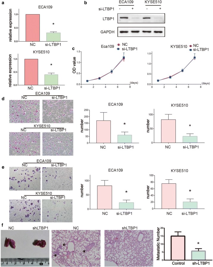
Methods: The tandem mass tag-based quantitative proteomic approach was applied to screen the differentially expressed proteins (DEPs) between 3 cases of ESCC tumor samples and paired normal tissues. Then the DEPs were validated in human ESCC tissues using western blot assays and GEPIA database respectively. The expression level of LTBP1 was detected in 152 cases of ESCC tissues and paired normal tissues. Loss-of-function assays were performed to detect the function of LTBP1 in vivo and in vitro. Immunofluorescence and Western blot assays were used to detect the expression of apoptosis, epithelial-mesenchymal transition (EMT) and cancer-associated fibroblasts (CAFs) markers.

Results: A total of 39 proteins were screened to be up-regulated (ratio > 2.0) in all three ESCC tissues. The results of immunohistochemistry assays indicated that the expression level of LTBP1 was higher in ESCC tissues than that in paired normal tissues (p < 0.001). Overexpression of LTBP1 was positively associated with lymphatic metastasis in ESCC (p = 0.002). Down-regulation of LTBP1 inhibited the invasion and migration as well as metastatic abilities in vitro and in vivo. It was also observed the down-regulation of LTBP1 not only decreased the mesenchymal phenotypes but also inhibited TGFβ-induced EMT in ESCC cells. We further found that down-regulation of LTBP1 enhanced ESCC cells' sensitivity to 5-FU treatment. Inhibition of LTBP1 expression could also attenuate induction of CAFs transformation and restrain fibroblast express fibronectin (FN1) in ESCC cells.

Conclusion: Overexpression of LTBP1 was associated with lymph node metastasis in ESCC. Our results indicated that LTBP1 not only increased the malignant behaviors of ESCC cells but also induced EMT and CAFs transformation. Our studies suggested an oncogenic role of LTBP1 in ESCC progression and it may serve as a potential therapeutic target for ESCC patients.

Keywords: Cancer-associated fibroblasts; Carcinogenesis; Epithelial–mesenchymal transition; Esophageal squamous cell carcinoma; FN1; LTBP1; Metastasis.


Conflict of interest statement

The authors declare no conflict of interest.

Figures

Fig. 1
Fig. 1
Identification and verification of DEPs in ESCC tissues. a Paired ESCC and adjacent normal esophageal tissues from three patients were analyzed by TMT. Twofold change was defined as the threshold for a significant change in expression. We detected 39 proteins up-regulated in cancer tissues compared with that in adjacent normal tissues (paired-samples t-test, p < 0.001). b Gene ontology enrichment analysis of identified DEPs which were classified into cellular component, molecular function, and biological process. c, d Validation of DEPs in ESCA. The expression of FN1, LTBP1, PLOD2, THBS1 and RCN3 were confirmed by GEPIA and western blot analysis. N non-tumorous tissues, T tumorous tissues. (one-way ANOVA and paired-samples t-test, *p < 0.05)
Fig. 2
Fig. 2
LTBP1 was up-regulated in ESCC and associated with lymphatic metastasis. a IHC staining of LTBP1 protein in ESCC and adjacent normal esophageal tissues. LTBP1 was localized in cytoplasm of cancer cells, but rarely expressed in normal esophageal tissues. b Frequency of negative, positive LTBP1 expression in ESCC and adjacent normal esophageal tissue. c Frequency of low/high LTBP1 expression in ESCC when categorized by lymphatic metastasis (Chi Square, *p < 0.05)
Fig. 3
Fig. 3
LTBP1 promoted ESCC cells migration, invasion and metastasis. a, b Efficiency of si-LTBP1 measured with qRT-PCR and Western blot. LTBP1 was reduced in si-LTBP1 groups compared with that in si-NC controls. c Cell proliferation capacity showed no significant changes in si-LTBP1 cells compared with the control cells, as measured by CCK8 assays. d Transwell migration assays of si-NC cells and si-LTBP1 cells (left). The number of cells that migrated after 24 h was counted in five randomly selected microscopic fields (right). e Transwell matrigel invasion assays of siNC cells and si-LTBP1 cells (left). The number of cells that invaded after 24 h was counted in five randomly selected microscopic fields (right). f ShLTBP1-ECA109, CON-ECA109 cells were injected into the tail vein of nude mouse. Lung tissues were autopsied 4 weeks after injection. Images of lung (left) and H&E staining (middle) of lung tissue sections were shown. Number of lung metastatic nodules (right) were significantly decreased in shLTBP1-ECA109 group. Bar graphs represent quantitative data from three independent experiments. Paired-samples t-test, *p < 0.05
Fig. 4
Fig. 4
LTBP1 promoted EMT and chemoresistant in ESCC cells. a Silencing LTBP1 inhibited hallmarks of the EMT, including loss of N-cadherin/vimentin and accumulation of E-cadherin in ESCC cells. b ESCC treated with si-NC or si-LTBP1 for 24 h, and treated with/without 20 ng/ml TGFβ for an additional 48 h. Western blot analysis of EMT markers in ESCC cells. c Morphological changes were observed under distinct treatment conditions. e BCL2 expression decreased and BAX expression increased over time in si-LTBP1 cells treated with 5-FU for 48 h. d, f Inhibition rate were detected by CCK8 assays and apoptosis rate was detected by flow cytometric analysis of ESCC cells after treatment with 5-FU for 48 h. Each experiment was performed in triplicate. Paired-samples t-test, *p < 0.05
Fig. 5
Fig. 5
LTBP1 contributed to CAFs transformation and expression of FN1 induced by ESCC cells. a The expression of LTBP1 and FN1 were positively correlated in the GEPIA database (Spearman correlation analysis, r = 0.6, p < 0.001). b, c The expression of LTBP1 and FN1 were detected in various cells by western blot and ELISA. d, f Western blot and immunofluorescent analysis of α-SMA and FN1 expression in fibroblasts which were con-cultured with si-NC, si-LTBP1 ESCC cells or negative for 96 h. e CCK8 assays comparing the effect of (± siLTBP1 ESCC cells or negative) conditioned medium on the activity of the fibroblasts. Each experiment was performed in triplicate. Paired-samples t-test, *p < 0.05
Fig. 6
Fig. 6
The expression of LTBP1 and FN1 showed positive correlation in ESCC tissues. IHC showed that LTBP1 was mainly observed in cancer cells while FN1 was mainly observed in the surrounding stroma. In LTBP1 positive cases, the expression of LTBP1 was mostly located in tumor parenchymal margin. E-cadherin was highly expressed in tumor parenchymal center and TGFβ was uniformly expressed in tumor parenchyma

References

    1. Bray F, Ferlay J, Soerjomataram I, Siegel RL, Torre LA, Jemal A. Global cancer statistics 2018: GLOBOCAN estimates of incidence and mortality worldwide for 36 cancers in 185 countries. CA Cancer J Clin. 2018;68(6):394–424. doi: 10.3322/caac.21492. - DOI - PubMed
    1. Chen W, Zheng R, Baade PD, Zhang S, Zeng H, Bray F, et al. Cancer statistics in China, 2015. Cancer J Clin. 2016;66(2):115–132. doi: 10.3322/caac.21338. - DOI - PubMed
    1. Pennathur A, Gibson MK, Jobe BA, Luketich JD. Oesophageal carcinoma. Lancet. 2013;381(9864):400–412. doi: 10.1016/S0140-6736(12)60643-6. - DOI - PubMed
    1. Cense HA, van Eijck CHJ, Tilanus HW. New insights in the lymphatic spread of oesophageal cancer and its implications for the extent of surgical resection. Best Pract Res Clin Gastroenterol. 2006;20(5):893–906. doi: 10.1016/j.bpg.2006.03.010. - DOI - PubMed
    1. Merkow RP, Bilimoria KY, Keswani RN, Chung J, Sherman KL, Knab LM, et al. Treatment trends, risk of lymph node metastasis, and outcomes for localized esophageal cancer. J Natl Cancer Inst. 2014;106:7. doi: 10.1093/jnci/dju133. - DOI - PubMed

Publication types

MeSH terms

Substances