Review
. 2020 Aug:38:100942.
doi: 10.1016/j.molmet.2020.01.006. Epub 2020 Feb 15.

Two genomes, one cell: Mitochondrial-nuclear coordination via epigenetic pathways

Affiliations
Review

Two genomes, one cell: Mitochondrial-nuclear coordination via epigenetic pathways

Meike Wiese et al. Mol Metab. 2020 Aug.

Abstract

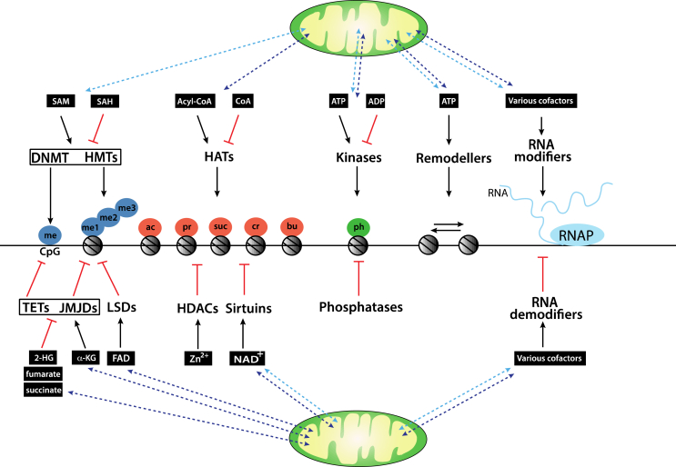
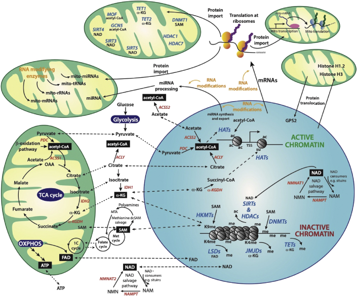
Background: Virtually all eukaryotic cells contain spatially distinct genomes, a single nuclear genome that harbours the vast majority of genes and much smaller genomes found in mitochondria present at thousands of copies per cell. To generate a coordinated gene response to various environmental cues, the genomes must communicate with each another. Much of this bi-directional crosstalk relies on epigenetic processes, including DNA, RNA, and histone modification pathways. Crucially, these pathways, in turn depend on many metabolites generated in specific pools throughout the cell, including the mitochondria. They also involve the transport of metabolites as well as the enzymes that catalyse these modifications between nuclear and mitochondrial genomes.

Scope of review: This study examines some of the molecular mechanisms by which metabolites influence the activity of epigenetic enzymes, ultimately affecting gene regulation in response to metabolic cues. We particularly focus on the subcellular localisation of metabolite pools and the crosstalk between mitochondrial and nuclear proteins and RNAs. We consider aspects of mitochondrial-nuclear communication involving histone proteins, and potentially their epigenetic marks, and discuss how nuclear-encoded enzymes regulate mitochondrial function through epitranscriptomic pathways involving various classes of RNA molecules within mitochondria.

Major conclusions: Epigenetic communication between nuclear and mitochondrial genomes occurs at multiple levels, ultimately ensuring a coordinated gene expression response between different genetic environments. Metabolic changes stimulated, for example, by environmental factors, such as diet or physical activity, alter the relative abundances of various metabolites, thereby directly affecting the epigenetic machinery. These pathways, coupled to regulated protein and RNA transport mechanisms, underpin the coordinated gene expression response. Their overall importance to the fitness of a cell is highlighted by the identification of many mutations in the pathways we discuss that have been linked to human disease including cancer.

Keywords: Chromatin; Enzymes; Epigenetics; Histones; Metabolites; Mitochondria; RNA modification.


Figures

Figure 1
Figure 1
Metabolic intermediates impact chromatin modifications. Epigenetic modifiers use metabolites as cofactors that are either directly produced within mitochondria (dark blue dotted arrows) or their production is indirectly affected by metabolic pathways within mitochondria (light blue dotted arrows). HMTs and DNMTs use SAM as a methyl donor to methylate (me) histones at lysine or arginine residues, or DNA, respectively. Both enzyme families are inhibited by the by-product of the methylation reaction, SAH. TET DNA dioxygenases and HDMs of the JMJD family use the tricarboxylic acid cycle intermediate α-KG as a cofactor and are inhibited by fumarate, succinate, and 2-HG. HDMs of the lysine-specific histone demethylase (LSDs) family use the cofactor FAD. HATs can utilise acyl-CoA derivatives acetyl (ac)-CoA, propionyl (pr)-CoA, succinyl (suc)-CoA, crotonyl (cr)-CoA, or butyryl (bu)-CoA as cofactors. Some HATs are inhibited by the acetylation by-product CoA. Histone acetylations are removed by HDACs. The sirtuin family of HDACs uses NAD+ as a cofactor, which is generated in the mitochondria, cytoplasm, and nucleus. Other HDACs depend on Zn2+, which is generated ubiquitously. Histones can be phosphorylated (ph) at serine, threonine, or tyrosine residues by ATP-dependent kinases that are inhibited by the by-product ADP. Dephosphorylation is mediated by phosphatases. Mitochondrially generated ATP is an essential cofactor for ATP-dependent remodelling activities that use the energy derived from ATP hydrolysis to move/slide nucleosomes. There are many families of RNA-modifying (and de-modifying) enzymes that require a wide variety of cofactors for their catalytic activity (see the text for details). Abbreviations: 2-HG: 2-hydroxyglutarate, α-KG: α-ketoglutarate, Acyl-CoA: acyl-coenzyme A, ADP: adenosine diphosphate, ATP: adenosine triphosphate, DNMT: DNA methyltransferase, FAD: flavin adenine dinucleotide, HAT: histone acetyltransferase, HDAC: histone lysine deacetylase, HDM: histone demethylase, HMT: histone methyltransferase, JMJD: Jumonji C domain-containing protein, LSD: lysine-specific histone demethylase, NAD+: nicotinamide adenine dinucleotide, SAH: S-adenosyl-homocysteine, SAM: S-adenosyl methionine, TET: ten-eleven translocation. (For interpretation of the references to colour in this figure legend, the reader is referred to the Web version of this article).
Figure 2
Figure 2
Epigenetic and epitranscriptomic pathways depend on metabolism. Crosstalk between the mitochondria and nucleus is regulated by the translocation of metabolic enzymes, metabolites, RNAs, and epigenetic regulators. Epigenetic gene regulation is established by enzymes that write, read, or erase post-translational modifications on histones, in particular acetylation (ac) and methylation (me), and regulate the methylation of DNA. In a similar fashion, modifications of RNA molecules have the potential to regulate all RNA-dependent functions and activities. Acetyl-CoA, the donor of acetyl groups used by HATs, is generated in distinct pools within the cell. Three main pathways in the mitochondria generate acetyl-CoA: (i) fatty acid breakdown via the β-oxidation pathway, (ii) PDC acting on pyruvate derived from glucose through glycolysis, and (iii) ACSS1 acting on acetate. Mitochondrial acetyl-CoA is subsequently used to acetylate mitochondrial proteins or channelled into the TCA cycle to generate energy. In the cytoplasm, two main pathways generate acetyl-CoA: (i) by ACLY acting on citrate, which in turn can be generated from the TCA cycle and shuttled between the mitochondria and cytoplasm and (ii) by ACSS2 on acetate. The three main precursors of acetyl-CoA (pyruvate, acetate, and citrate) can also diffuse into the nucleus, where they are processed into acetyl-CoA by nuclear pools of PDC, ACSS2, and ACLY, respectively. Distinct pools of acetyl-CoA regulate acetylation of mitochondrial, cytoplasmic, and nuclear proteins, including histones. HAT-mediated histone acetylation results in chromatin de-condensation and active transcription (active chromatin). Nuclear HATs MOF and GCN5 also localise to mitochondria. For more information on nuclear epigenetic factors localising to mitochondria (blue), see the main text and Table 1. α-KG is generated in the TCA cycle in mitochondria from isocitrate by IDH2. Isocitrate can diffuse into the cytosol where it is converted by IDH1 to α-KG. α-KG diffuses into the nucleus, where it is used as a cofactor by JMJD histone demethylases, TET DNA demethylases, or is converted by α-KGDH into succinyl-CoA, a cofactor for HATs. Mitochondria regulate the redox levels of FAD, a cofactor for LSDs. During β-oxidation and the TCA cycle, FAD is reduced to FADH2 and returned to its oxidised form during OXPHOS. FAD can then diffuse into the cytosol and nucleus. SAM is the donor of methyl groups utilised by HMTs and DNMTs in the nucleus. SAM is generated through the coupling of the folate and methionine cycles in the cytosol, which in turn is sustained by 1-C metabolism in mitochondria or is generated via the methionine salvage pathway. SAM can also enter the mitochondria. Methylation of DNA and lysine (K) 9 of histone H3 (K9me) together with histone deacetylation are associated with chromatin compaction and gene repression (inactive chromatin). Sirtuin histone deacetylates use NAD+ as a cofactor, which is generated de novo from the amino acid tryptophan or via the NAD+ salvage pathway, which uses NAD+ precursors such as NAM directly from the diet or recycled from intracellular reactions. A nuclear pool of NAD+ is either generated via a nuclear NAD+ salvage pathway or through passive diffusion of cytoplasmic NAD+ through nuclear pores. All classes of nuclear RNAs (mRNAs, miRNAs, lncRNAs, rRNAs, and tRNAs) are transcribed from active chromatin and actively transported into the cytoplasm. miRNAs are processed in the cytoplasm and can be transported into the mitochondria, while mRNAs are translated at cytoplasmic ribosomes. Transcription in the mitochondria of RNAs (mRNAs, rRNAs, tRNAs, and perhaps miRNAs) also occurs with translation of the mRNAs at mito-ribosomes. Throughout the cell, all classes of RNAs are targeted by specific RNA-modifying enzymes (orange). Proteins synthesised by cytoplasmic ribosomes either reside in the cytoplasm, are imported into mitochondria and/or into the nucleus. During mitochondrial stress, mitochondrial GPS2 rapidly translocates to the nucleus where it collaborates with H3K9 demethylases to activate gene expression facilitating mitochondrial stress response. Also, certain nuclear proteins translocate to the mitochondria only during cellular stress. For example, both histone H3 and H1.2 are released from chromatin and translocate to the mitochondria during apoptosis. This figure was adapted from [145]. Abbreviations: 1-C: one-carbon, Acetyl-CoA: acetyl-coenzyme A, ACLY: ATP-citrate lyase, ACSS1: acyl-CoA synthetase short-chain family member 1, α-KG: α-ketoglutarate, α-KGDH: α-ketoglutarate dehydrogenase, ATP: adenosine triphosphate, dcSAM: decarboxylated SAM, DNMT: DNA methyltransferase, FAD: flavin adenine dinucleotide, GCN5: lysine acetyltransferase 2A, GPS2: G-protein pathway suppressor 2, HAT: histone acetyltransferase, HDAC: histone lysine deacetylase, HMT: histone methyltransferase, IDH2/3: isocitrate dehydrogenase 2/3, JMJD: Jumonji C domain-containing histone demethylase, K9: lysine 9, LSDs: lysine-specific histone demethylases, MOF: lysine acetyltransferase 8, MTA: 5′-methylthioadenosine, NAD+: nicotinamide adenine dinucleotide, NAM: nicotinamide, NAMPT: nicotinamide phosphoribosyl transferase, NMN: nicotinamide mononucleotide, NMNAT1/2: nicotinamide mononucleotide adenylyltransferase 1/2/3, OAA: oxaloacetate, OXPHOS: oxidative phosphorylation, PDC: pyruvate dehydrogenase complex, SAM: S-adenosyl methionine, SIRT: sirtuin deacetylase, TCA: tricarboxylic acid, TET: ten-eleven translocation DNA demethylase. (For interpretation of the references to colour in this figure legend, the reader is referred to the Web version of this article).
Figure 3
Figure 3
Chemical structures of key metabolites regulating epigenetic enzymes. (A) Structures of metabolites affecting histone acylation. The acetyl group donor acetyl-CoA together with other acyl moieties is depicted on the left side and the deacetylase cofactor NAD+ on the right side. (B) Structures of metabolites affecting histone and DNA methylation. The methyl group donor SAM is shown (on the left side) together with the demethylase cofactors FAD (in the centre) and α-KG (on the right side).

References

    1. Roundtree I.A., Evans M.E., Pan T., He C. Dynamic RNA modifications in gene expression regulation. Cell. 2017;169(7):1187–1200. - PMC - PubMed
    1. Bannister A.J., Kouzarides T. Regulation of chromatin by histone modifications. Cell Research. 2011;21(3):381–395. - PMC - PubMed
    1. Xhemalce B., Dawson M.A., Bannister A.J. 2012. Epigenetic regulation and epigenomics in Wiley-Blackwell Advances in molecular biology and medicine; pp. 657–703.
    1. Lyko F. The DNA methyltransferase family: a versatile toolkit for epigenetic regulation. Nature Reviews Genetics. 2017;19:81. - PubMed
    1. Shi H., Wei J., He C. Where, when, and how: context-dependent functions of RNA methylation writers, readers, and erasers. Molecular Cell. 2019;74(4):640–650. - PMC - PubMed

Publication types