. 2020 May:175:113923.
doi: 10.1016/j.bcp.2020.113923. Epub 2020 Mar 23.

The inhibitory effect of ECG and EGCG dimeric procyanidins on colorectal cancer cells growth is associated with their actions at lipid rafts and the inhibition of the epidermal growth factor receptor signaling

Affiliations

The inhibitory effect of ECG and EGCG dimeric procyanidins on colorectal cancer cells growth is associated with their actions at lipid rafts and the inhibition of the epidermal growth factor receptor signaling

Wei Zhu et al. Biochem Pharmacol. 2020 May.

Abstract

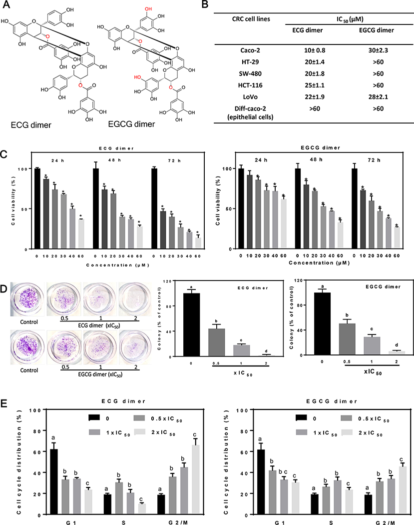
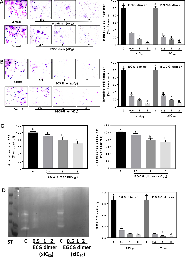
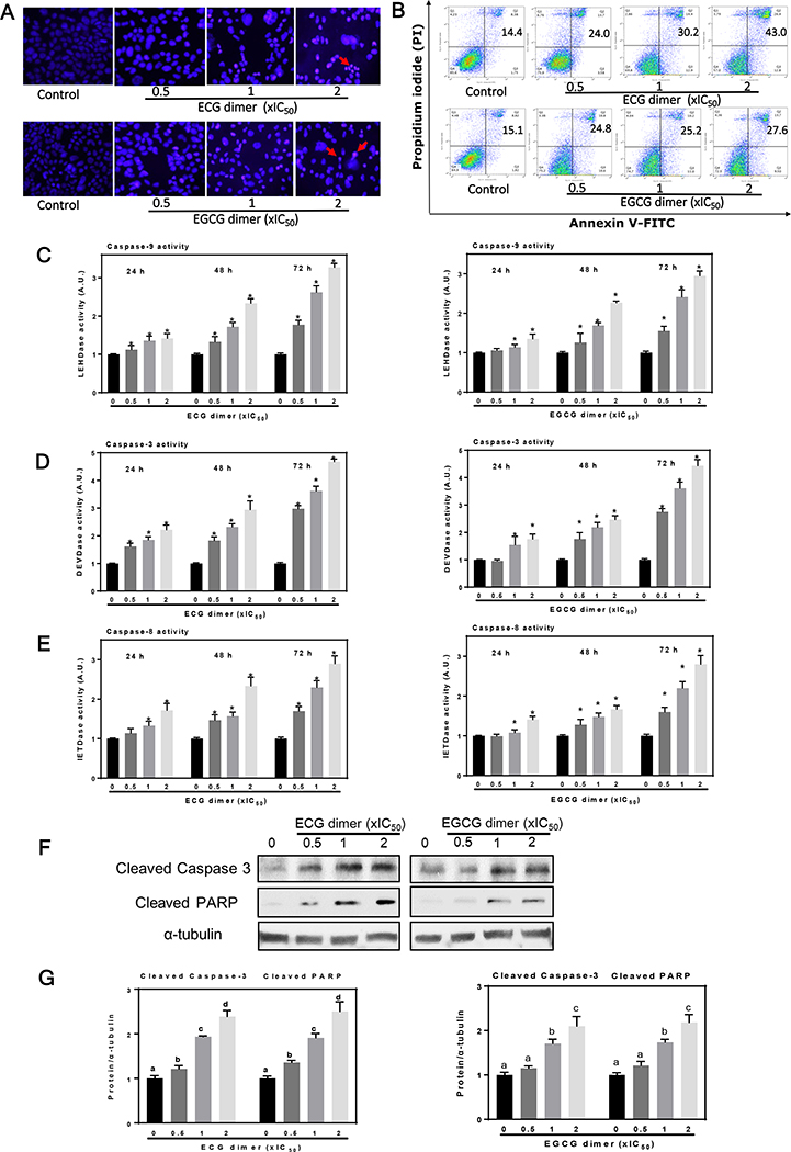
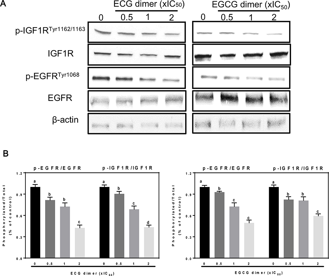
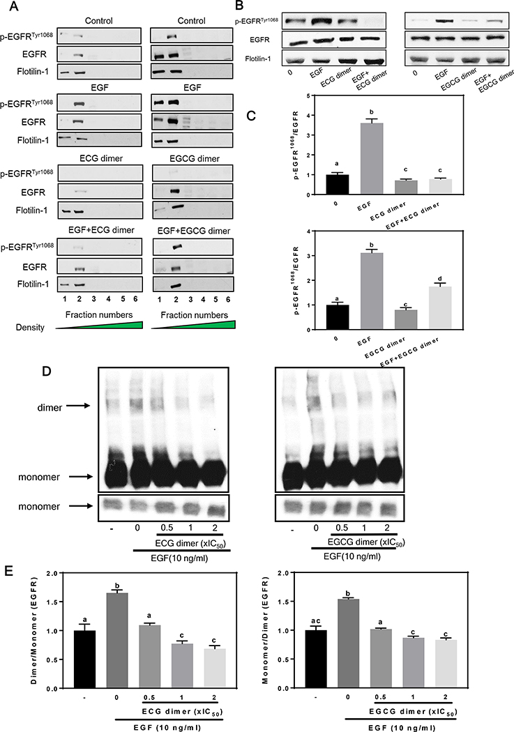
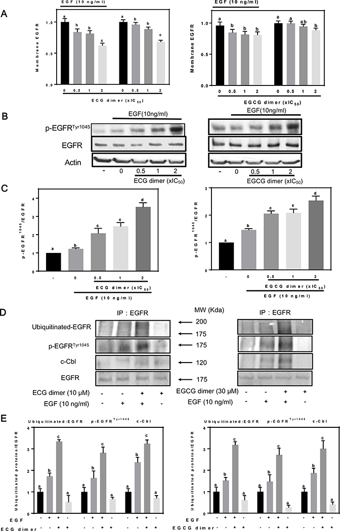
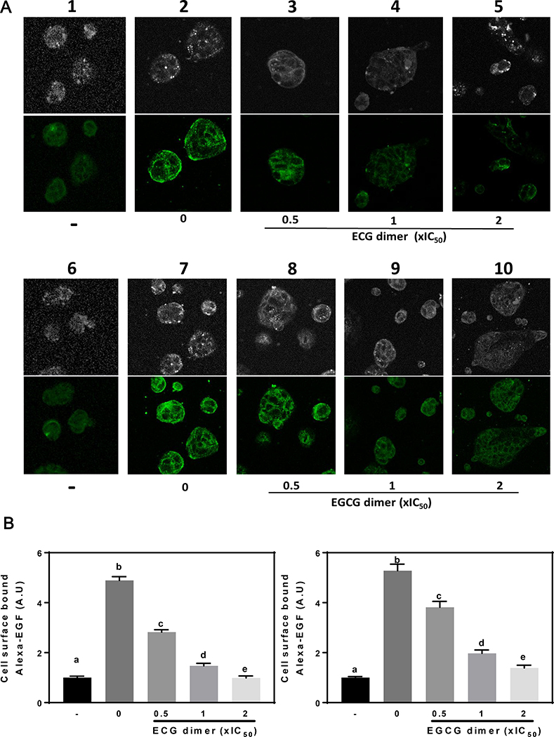
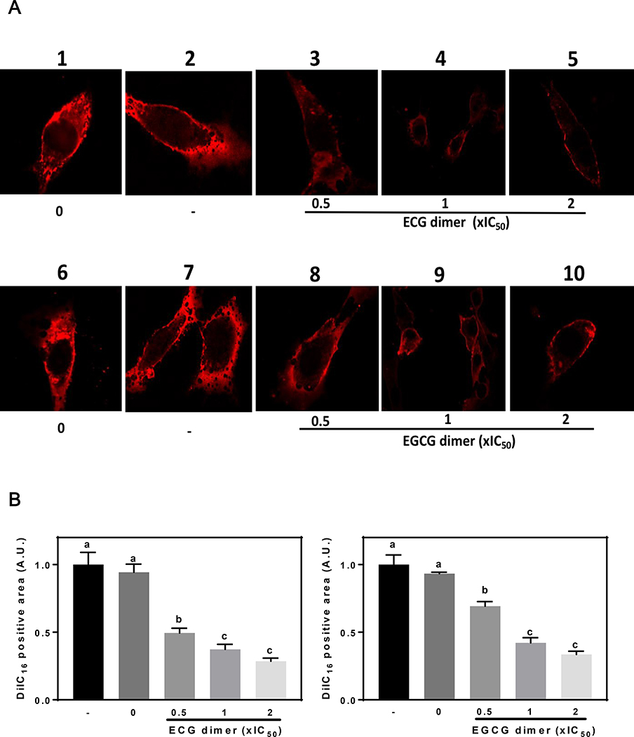
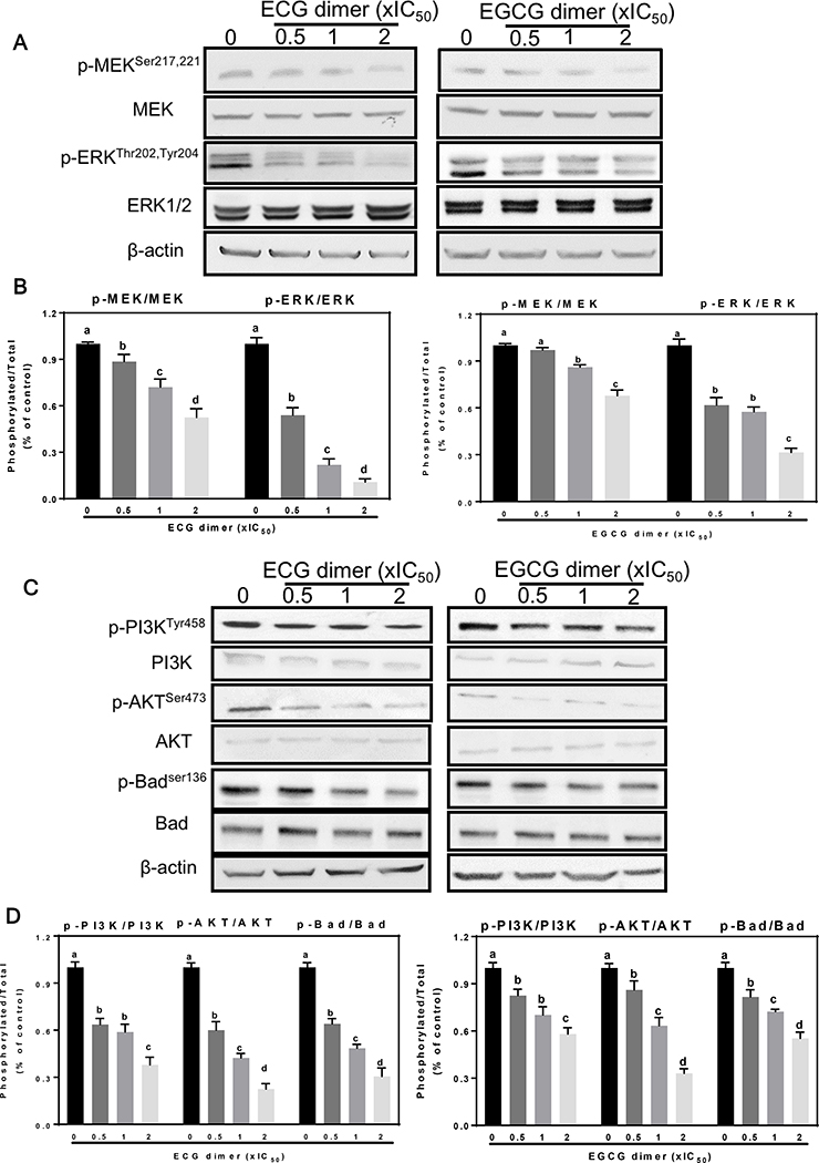
Colorectal cancer (CRC) is one of the most common cancers worldwide. Epidemiological studies indicate that consumption of fruits and vegetables containing procyanidins is associated with lower CRC risk. This study investigated the capacity of two dimeric procyanidins composed of epicatechin gallate (ECG) or epigallocatechin gallate (EGCG) isolated from persimmons, to inhibit CRC cell growth and promote apoptosis, characterizing the underlying mechanisms. ECG and EGCG dimers reduced the growth of five human CRC cell lines in a concentration (10-60 μM)- and time (24-72 h)-dependent manner, with a 72 h-IC50 value in Caco-2 cells of 10 and 30 μM, respectively. ECG and EGCG dimers inhibited Caco-2 cell proliferation by arresting the cell cycle in G2/M phase and by inducing apoptosis via the mitochondrial pathway. In addition, ECG and EGCG dimers inhibited cell migration, invasion, and adhesion, decreasing the activity of matrix metalloproteinases (MMP-2/9). Mechanistically, ECG and EGCG dimers inhibited the activation of lipid raft-associated epidermal growth factor (EGF) receptor (EGFR), without affecting its localization at lipid rafts. In particular, ECG and EGCG dimers reduced EGFR phosphorylation at Tyr1068 residue, prevented EGFR dimerization and activation upon stimulation, and induced EGFR internalization both in the absence and presence of EGF. Furthermore, ECG and EGCG dimers increased EGFR phosphorylation at Tyr1045 residue, providing a docking site for ubiquitin ligase c-Cbl and induced EGFR degradation by the proteasome. Downstream of EGFR, ECG and EGCG dimers inhibited the activation of the MEK/ERK1/2 and PI3K/AKT signaling pathways, downregulating proteins involved in the modulation of cell survival. In conclusion, ECG and EGCG dimers reduced CRC cell growth by inhibiting EGFR activation at multiple steps, including the disruption of lipid rafts integrity and promoting EGFR degradation. These results shed light on a potential molecular mechanism on how procyanidins-rich diets may lower CRC risk.

Keywords: Apoptosis; Colorectal cancer; ECG and EGCG dimers; EGFR signaling; Lipid rafts.


Conflict of interest statement

Declaration of Competing Interest The authors declare that they have no known competing financial interests or personal relationships that could have appeared to influence the work reported in this paper.

Figures

Fig. 1.
Fig. 1.
ECG and EGCG dimers decreased cell viability of human CRC cells and arrested the Caco-2 cell cycle in G2/M phase. (A) Chemical structures of ECG and EGCG dimers. (B) IC50 for ECG and EGCG dimers to inhibit, after 72h incubation, the growth of five different human CRC cell lines and Caco-2 cells differentiated into intestinal epithelial cells. Results are expressed as the mean ± SEM. (C) ECG and EGCG dimers concentration- and time-dependently decreased Caco-2 cell viability. (D) ECG and EGCG dimers inhibited Caco-2 cell colony formation measured at 24 h. (E) ECG and EGCG dimers blocked Caco-2 cell cycle progression after 72 h incubation. Values are shown as means ± SEM of 3–5 independent experiments. *indicated significantly different from untreated cells. Values having different superscripts are significantly different (p< 0.05, One-way ANOVA-test).
Fig. 2.
Fig. 2.
ECG and EGCG dimers inhibited the Caco-2 cell migration, invasion, and adhesion. The effects of ECG and EGCG dimers on Caco-2 cell migration, invasion, and MMPs activation were evaluated after 24 h incubation as described in the methods section. (A) ECG and EGCG dimers concentration-dependently decreased Caco-2 cell migration evaluated after 24 h treatment. (B) ECG and EGCG dimers concentration-dependently decreased Caco-2 cell invasion after 24 h treatment. (C) ECG and EGCG dimers concentration-dependently decreased Caco-2 cell adhesion. (D) ECG and EGCG dimers concentration-dependently decreased MMP2/9 activity measured in the cell culture medium after 24 h treatment. Results are shown as means ± SEM of 3–5 independent experiments. Values having different superscripts are significantly different (p< 0.05, One-way ANOVA-test).
Fig. 3.
Fig. 3.
ECG and EGCG dimers induced apoptotic cell death in Caco-2 cells. Cells were incubated for 48 h in the presence of the ECG and EGCG dimers. (A) Cell apoptosis was evaluated by staining with Hoechst (blue fluorescence)/PI (red fluorescence) and subsequent immunocytochemistry. The red arrows indicated apoptotic cells. (B) Early and late apoptosis was quantified by Annexin-V/PI dual staining and subsequent flow cytometry analysis (numbers in the top right quadrants indicated the percentage of cells undergoing apoptosis). (C-E) ECG and EGCG dimers promoted caspase 8, 9, and 3 activation in Caco-2 cells after 24, 48 and 72 h incubation. (F-G) ECG or EGCG dimers exposure for 48 h dose-dependently increased levels of cleaved caspase 3 and cleaved PARP as measured by Western blot. Results are shown as means ± SEM of 3–5 independent experiments. *indicated significantly different from untreated cells. Values having different superscripts are significantly different (p< 0.05, One-way ANOVA-test).
Fig. 4.
Fig. 4.
ECG and EGCG dimers inhibited both the EGFR and IGF1R activation in human Caco-2 cancer cells. Cells were incubated for 24 h in the absence or presence of 0.5X, 1X and 2XIC50 concentrations of ECG and EGCG dimers. EGFR phosphorylation (Tyr1068), or IGF1R phosphorylation (Tyr1162/1163) were evaluated by Western blot. Bands were quantified and values for phosphorylated proteins were referred to the respective total protein content. Results are shown as means ± SEM of 3–5 independent experiments. Values having different superscripts are significantly different (p< 0.05, One-way ANOVA-test).
Fig. 5.
Fig. 5.
ECG and EGCG dimers did not displace the EGFR from lipid rafts but inhibited EGF-induced phosphorylation and dimerization of the EGFR at lipid rafts. After 24 h starvation, cells were incubated in the absence (control) or presence of ECG or EGCG dimers for 30 min at 37°C, subsequently, cells were incubated without or with EGF (10 ng/ml) for further 15 min. Cells were harvested, and lipid rafts fractions were isolated as described in Methods. (A-C) EGFR phosphorylation in Tyr1068 was evaluated by Western blot. Bands were quantified and values for phosphorylated proteins were referred to the respective total protein content. (D-E) EGFR dimerization was measured as by crosslinking with (BS3) and subsequent Western blot, as described in Methods. Results are shown as means ± SEM of 3–5 independent experiments. Values having different superscripts are significantly different (p< 0.05, One-way ANOVA-test).
Fig. 6.
Fig. 6.
ECG and EGCG dimers promoted EGFR internalization and degradation. After 24 h starvation, cells were treated in the absence or presence of ECG or EGCG dimers for 30 min at 37 °C, and subsequently incubated without or with EGF (10 ng/ml) for 15 min. (A) The internalization of the EGFR was evaluated by measuring membrane EGFR content by cell-Elisa assay as described in Methods. (B-C) The effects of ECG and EGCG dimers on the phosphorylation of the EGFR at Tyr1045 was evaluated by Western blot. (D-E) ECG and EGCG dimers promoted degradation of the EGFR via the ubiquitin pathway. After immunoprecipitation of the EGFR, Western blots were done for (D) ubiquitinated-EGFR, p-EGFRTyr1045 and the ubiquitin ligase c-Cbl and (E) bands were quantified and values for phosphorylated proteins were referred to the respective total protein content. Results are shown as means ± SEM of 3–5 independent experiments. Values having different superscripts are significantly different (p< 0.05, One-way ANOVA-test).
Fig. 7.
Fig. 7.
ECG and EGCG dimers inhibited EGF binding to the EGFR. Caco-2 cells were treated with/without ECG or EGCG dimers of 0.5x, 1x, 2xIC50 for 30 min at 37 ° C. Then, cells were exposed to Alexa-EGF (10 ng/ml) for 1 h on ice. (A) Representative confocal microscopy images. (B) Images were quantified using Image J, and values referred to the total cell area that was positive for Alexa-EGF staining. Results are shown as means ± SEM of 3–5 independent experiments. Values having different superscripts are significantly different (p< 0.05, One-way ANOVA-test).
Fig. 8.
Fig. 8.
ECG and EGCG dimers decreased lipid rafts domains surface area. The lipid raft relative surface area was evaluated using the DiIC16 staining and cold 1% (v/v) Triton X-100 solubility assay as described in Methods. (A) representative confocal microscopy images of cells before (images 1, 6) or after (images 2–5, 7–10) cold 1% (v/v) Triton X-100 extraction. (B) Fluorescence intensity was quantified using ImageJ and was expressed as the percentage of the total cell area that was positive for DiIC16 staining. Results are shown as means ± SEM of 3–5 independent experiments. Values having different superscripts are significantly different (p< 0.05, One-way ANOVA-test).
Fig. 9.
Fig. 9.
ECG and EGCG dimers inhibited both the EGFR and IGF1R-associated activation of the MEK/ERK1/2 and PI3K/AKT pathways in human Caco-2 cancer cells. Cells were incubated for 24 h in the absence or presence of 0.5X, 1X and 2XIC50 concentrations of ECG or EGCG dimers. (A, B) MEK and ERK1/2 phosphorylation and (C, D) PI3K, AKT and Bad phosphorylation evaluated by Western blot. Bands were quantified and values for phosphorylated proteins were referred to the respective total protein content. Results are shown as means ± SEM of 3–5 independent experiments. Values having different superscripts are significantly different (p< 0.05, One-way ANOVA-test).

References

    1. Adachi S, et al. (2007). “The inhibitory effect of (−)-epigallocatechin gallate on activation of the epidermal growth factor receptor is associated with altered lipid order in HT29 colon cancer cells.” Cancer Research 67(13): 6493–6501. - PubMed
    1. Arnold M, et al. (2017). “Global patterns and trends in colorectal cancer incidence and mortality.” Gut 66(4): 683–691. - PubMed
    1. Aune D, et al. (2011). “Nonlinear Reduction in Risk for Colorectal Cancer by Fruit and Vegetable Intake Based on Meta-analysis of Prospective Studies.” Gastroenterology 141(1): 106–118. - PubMed
    1. Baena R and Salinas P (2015). “Diet and colorectal cancer.” Maturitas 80(3): 258–264. - PubMed
    1. Ben QW, et al. (2014). “Dietary Fiber Intake Reduces Risk for Colorectal Adenoma: A Meta-analysis.” Gastroenterology 146(3): 689–+. - PubMed

Publication types

MeSH terms