. 2020 May 8;295(19):6252-6262.
doi: 10.1074/jbc.RA120.012605. Epub 2020 Mar 26.

A single amino acid substitution uncouples catalysis and allostery in an essential biosynthetic enzyme in Mycobacterium tuberculosis

Affiliations

A single amino acid substitution uncouples catalysis and allostery in an essential biosynthetic enzyme in Mycobacterium tuberculosis

Wanting Jiao et al. J Biol Chem. .

Abstract

Allostery exploits the conformational dynamics of enzymes by triggering a shift in population ensembles toward functionally distinct conformational or dynamic states. Allostery extensively regulates the activities of key enzymes within biosynthetic pathways to meet metabolic demand for their end products. Here, we have examined a critical enzyme, 3-deoxy-d-arabino-heptulosonate 7-phosphate synthase (DAH7PS), at the gateway to aromatic amino acid biosynthesis in Mycobacterium tuberculosis, which shows extremely complex dynamic allostery: three distinct aromatic amino acids jointly communicate occupancy to the active site via subtle changes in dynamics, enabling exquisite fine-tuning of delivery of these essential metabolites. Furthermore, this allosteric mechanism is co-opted by pathway branchpoint enzyme chorismate mutase upon complex formation. In this study, using statistical coupling analysis, site-directed mutagenesis, isothermal calorimetry, small-angle X-ray scattering, and X-ray crystallography analyses, we have pinpointed a critical node within the complex dynamic communication network responsible for this sophisticated allosteric machinery. Through a facile Gly to Pro substitution, we have altered backbone dynamics, completely severing the allosteric signal yet remarkably, generating a nonallosteric enzyme that retains full catalytic activity. We also identified a second residue of prime importance to the inter-enzyme communication with chorismate mutase. Our results reveal that highly complex dynamic allostery is surprisingly vulnerable and provide further insights into the intimate link between catalysis and allostery.

Keywords: DAH7PS; allosteric regulation; aromatic amino acid biosynthesis; enzyme inhibitor; enzyme mutation; enzyme structure; molecular dynamics; protein sequence; shikimate pathway; statistical coupling analysis; tuberculosis.


Conflict of interest statement

The authors declare that they have no conflicts of interest with the contents of this article

Figures

Figure 1.
Figure 1.
Structures of DAH7PS from different subfamilies. A, crystal structure of E. coli DAH7PS (PDB 4UC5), representing type Iα. B, crystal structure of T. maritima DAH7PS (PDB 3PG9), representing type Iβ. C, crystal structure of MtuDAH7PS (PDB 3KGF), representing type II DAH7PS. D, tetrameric structure of MtuDAH7PS. Decoration structural elements associated with allostery are shown as orange cartoons, corresponding allosteric ligands are displayed as spheres with green carbon atoms, active site metal ions (cyan) and phosphate (sulfate) are displayed as spheres if present in the crystal structure.
Figure 2.
Figure 2.
Sector 1 identified from SCA. A, Sector 1 mapped onto crystal structure of MtuDAH7PS. Ligand-binding sites are indicated by displaying the Trp and Phe ligands as spheres with green carbon atoms. The active site Mn2+ (cyan) and the phosphate ions observed in the crystal are displayed as spheres to indicate location of the active site. B, residues in IC 7 mapped onto structure of MtuDAH7PS.
Figure 3.
Figure 3.
Sector 2 identified from SCA. A, Sector 2 residues mapped onto crystal structure of MtuDAH7PS. The active site Mn2+ (cyan) and the phosphate ions observed in the crystal are displayed as spheres to indicate location of the active site. B, residues in IC 2 mapped onto structure of MtuDAH7PS. Ligand-binding sites are indicated by displaying the Trp and Phe ligands as spheres with green carbon atoms. Crystal structure of a single chain MtuCM is shown in magenta cartoon representation to indicate location of the MtuDAH7PS-CM interface.
Figure 4.
Figure 4.
Position of Gly-190 on MtuDAH7PS. A, Gly-190 (yellow) on the monomer of MtuDAH7PS with other residues in IC 7 (dark blue spheres). Allosteric ligands Trp and Phe are shown as spheres with green carbon atoms. B, close-up view of the Trp-binding site relative to the position of Gly-190. Polar interactions formed between Trp and surrounding residues are shown as black dashed lines. Gly-190 is clearly not involved in any direct interactions with Trp.
Figure 5.
Figure 5.
A, allosteric response of MtuDAH7PSG190P to aromatic amino acids. Assay contained 50 mm BTP (pH 7.5), 1 mm TCEP, 100 μm Mn2+, 260 μm PEP, 160 μm E4P in the absence or presence of total concentration of 200 μm of aromatic amino acids, alone or in combinations. For the normalized data, the mean of each apo-dataset was defined as 100%. B, activity of MtuCM in the presence of MtuDAH7PSWT and MtuDAH7PSG190P in response to aromatic amino acids. Assay contained 7.5 nm MtuCM, 1500 nm MtuDAH7PS variants, 40 μm chorismate, and 200 μm total ligand. Data were generated from independent triplicate measurements, the mean values were plotted with error bars showing the S.D. C, SAXS scattering profiles of MtuDAH7PSWT (green dots) and MtuDAH7PSY131A (blue dots) and theoretical scattering of MtuDAH7PSWT (black line, PDB 3NV8). D, superimposed crystal structures of MtuDAH7PSG190P (blue, PDB 6PBJ) and MtuDAH7PSWT (gray, PDB 3NV8).
Figure 6.
Figure 6.
Position of Tyr-131 on MtuDAH7PS. A, Tyr-131 (yellow) on the monomer of MtuDAH7PS with other residues in IC 2 (dark red spheres). Allosteric ligands Trp and Phe are shown as spheres with green carbon atoms. Active site Mn2+ and phosphates are shown as spheres. B, close-up view of the Phe-binding site relative to the position of Tyr-131, showing this residue is not in any direct contact with Phe. Polar interactions formed between Tyr-131 and surrounding residues are shown as black dashed lines.
Figure 7.
Figure 7.
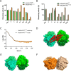
A, allosteric response of MtuDAH7PSY131A to aromatic amino acids. Assay contained 50 mm BTP (pH 7.5), 1 mm TCEP, 100 μm Mn2+, 260 μm PEP, 143 μm E4P in the absence or presence of total concentration of 200 μm of aromatic amino acids, alone or in combinations. For the normalized data, the mean of each apo-dataset was defined as 100%. B, activity of MtuCM in the presence of MtuDAH7PSWT and MtuDAH7PSY131A in response to aromatic amino acids. Assay contained 7.5 nm MtuCM, 1500 nm MtuDAH7PS variants, 40 μm chorismate, and 200 μm total ligand. Data were generated from independent triplicate measurements, the mean values were plotted with error bars showing the S.D. C, SAXS profile of MtuDAH7PSY131A (orange dots) and theoretical scattering from the rigid body model (black line). D, relative position of MtuCM in MtuCM-MtuDAH7PS complex shown in red cartoon (PDB 5HUD), aligned with PDB 3NV8 (surface). E, MtuDAH7PSWT shown in shades of green (PDB 3NV8). F, MtuDAH7PSY131A SAXS model shown in shades of orange.

References

    1. Goodey N. M., and Benkovic S. J. (2008) Allosteric regulation and catalysis emerge via a common route. Nat. Chem. Biol. 4, 474–482 10.1038/nchembio.98 - DOI - PubMed
    1. Walker G. E., Dunbar B., Hunter I. S., Nimmo H. G., and Coggins J. R. (1996) Evidence for a novel class of microbial 3-deoxy-d-arabino-heptulosonate-7-phosphate synthase in Streptomyces coelicolor A3(2), Streptomyces rimosus and Neurospora crassa. Microbiology 142, 1973–1982 - PubMed
    1. Shumilin I. A., Bauerle R., and Kretsinger R. H. (2003) The high-resolution structure of 3-deoxy-d-arabino-heptulosonate-7-phosphate synthase reveals a twist in the plane of bound phosphoenolpyruvate. Biochemistry 42, 3766–3776 10.1021/bi027257p - DOI - PubMed
    1. Wagner T., Shumilin I. A., Bauerle R., and Kretsinger R. H. (2000) Structure of 3-deoxy-d-arabino-heptulosonate-7-phosphate synthase from Escherichia coli: comparison of the Mn(2+)*2-phosphoglycolate and the Pb(2+)*2-phosphoenolpyruvate complexes and implications for catalysis. J. Mol. Biol. 301, 389–399 10.1006/jmbi.2000.3957 - DOI - PubMed
    1. Shumilin I. A., Kretsinger R. H., and Bauerle R. H. (1999) Crystal structure of phenylalanine-regulated 3-deoxy-d-arabino-heptulosonate-7-phosphate synthase from Escherichia coli. Structure 7, 865–875 10.1016/S0969-2126(99)80109-9 - DOI - PubMed

Publication types

MeSH terms

Substances

LinkOut - more resources