. 2020 Mar 25;12(4):359.
doi: 10.3390/v12040359.

Modelling Lyssavirus Infections in Human Stem Cell-Derived Neural Cultures

Affiliations

Modelling Lyssavirus Infections in Human Stem Cell-Derived Neural Cultures

Vinod Sundaramoorthy et al. Viruses. .

Abstract

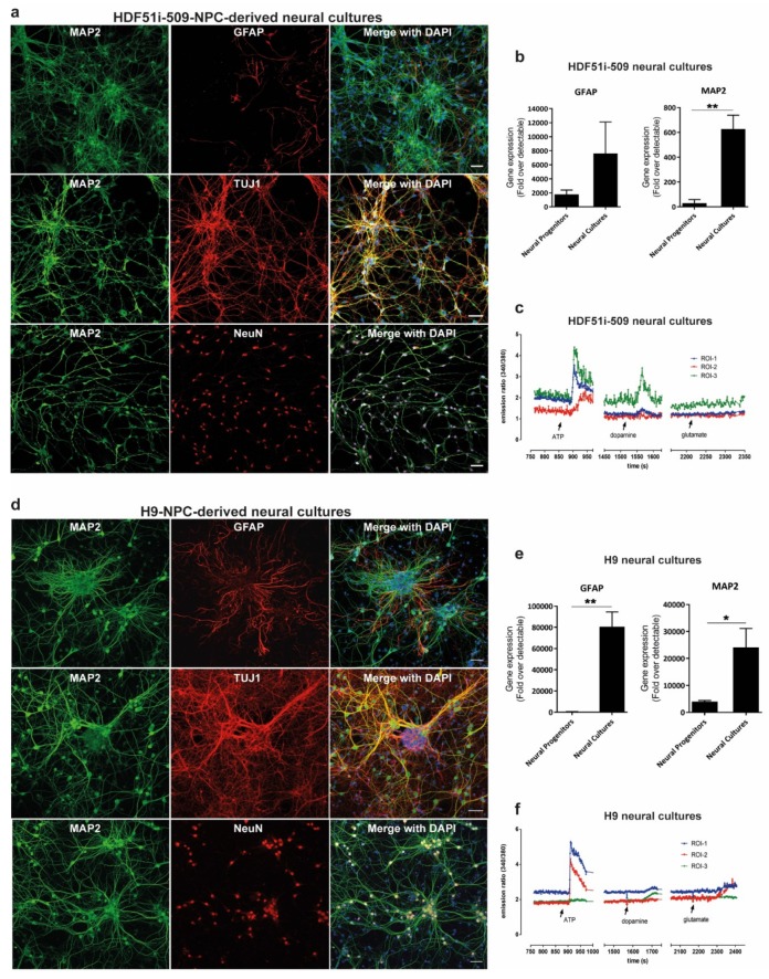
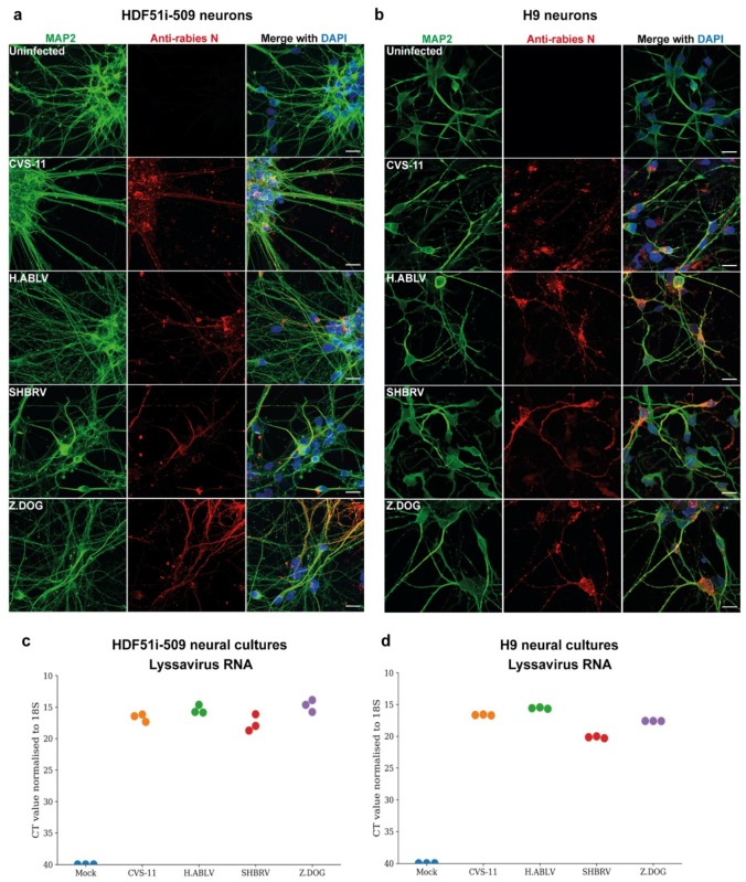
Rabies is a zoonotic neurological infection caused by lyssavirus that continues to result in devastating loss of human life. Many aspects of rabies pathogenesis in human neurons are not well understood. Lack of appropriate ex-vivo models for studying rabies infection in human neurons has contributed to this knowledge gap. In this study, we utilize advances in stem cell technology to characterize rabies infection in human stem cell-derived neurons. We show key cellular features of rabies infection in our human neural cultures, including upregulation of inflammatory chemokines, lack of neuronal apoptosis, and axonal transmission of viruses in neuronal networks. In addition, we highlight specific differences in cellular pathogenesis between laboratory-adapted and field strain lyssavirus. This study therefore defines the first stem cell-derived ex-vivo model system to study rabies pathogenesis in human neurons. This new model system demonstrates the potential for enabling an increased understanding of molecular mechanisms in human rabies, which could lead to improved control methods.

Keywords: chemokine and cytokine response; ex-vivo models; lyssavirus; neuronal apoptosis; rabies; stem cell-derived neurons; trans-synaptic axonal trafficking; viral pathogenesis.


Conflict of interest statement

The authors declare no conflict of interest.

Figures

Figure 1
Figure 1
Generation of forebrain type human neural lineage cell cultures from induced pluripotent stem cells (iPSC)- and embryonic stem cells (ESC)-derived neuronal precursor cells (hNPCs). (a) HDF51i-509-derived and (d) H9-derived hNPCs were differentiated into forebrain type human neural cell cultures for 24 days and 21 days, respectively. The cultures were then immunostained for the detection of mature neuron markers MAP2, beta tubulin III (TUJI), and NeuN, as well as astrocytes using anti-GFAP antibody. Images were taken using 20× objective. Scale bar 50 µm (images are maximum intensity projection of Z-stacks). Gene expression of neuronal (MAP2) and glial (GFAP), markers detected by qPCR in (b) HDF51i-509-derived and (e) H9-derived cultures following differentiation. ** p < 0.01, * p < 0.05 neural progenitors vs neural cultures, n = 3. (c) HDF51i-509-NPC and (f) H9-NPC-derived neural cultures were subjected to live calcium imaging following loading with the calcium binding dye, Fura-2 AM. Cells morphologically characteristic of neurons with axonal projections are identified as regions of interest (ROI) for Fura-2 intensity measurements. Graphs (c,f) show representative calcium influx (measured by ratio of emission intensities at 340 and 380 nm) in three different ROI in response to treatment with 100 µM ATP, 100 µM glutamate and 100 µM dopamine. Arrows indicate the timepoint at which stimulants were added.
Figure 2
Figure 2
Lyssavirus infection of stem cell-derived human neural cultures. (a) HDF51i-509-NPCs and (b) H9-NPCs were differentiated for 21 days and 29 days, respectively. Differentiated neural cultures were then infected with different lyssavirus strains (CVS-11, H.ABLV, SHBRV and Z.DOG) at a virus:cell ratio of 1 for 72 h. Representative images show infection of (a) HDF51i-509-NPC-derived and (b) H9-NPC-derived neural cultures immunostained for detection of MAP2-positive neurons (green) and rabies virus antigen by anti-nucleoprotein staining (red). Nuclei stained with DAPI (blue), shown as a merged image with red and green channels. Scale bar 50 µm (images are maximum intensity projection of Z-stacks). (c,d) Validation of viral infection shown by qPCR analysis of lyssavirus RNA in (c) HDF51i-509-NPC- and (d) H9-NPC-derived neural cultures, n = 3.
Figure 3
Figure 3
Lack of apoptosis in stem cell-derived human neural cultures infected with lyssavirus strains. HDF51i-509-NPCs and H9-NPCs were differentiated for 24 days and 29 days, respectively. These neural cultures were infected with different lyssavirus strains for 72 h at virus:cell ratio of 1 and examined for apoptosis by TUNEL staining (TMR-red). (a) Representatives images of HDF51i-509-NPC-derived neurons identified by anti-neurofilament immunofluorescence (NF, green) stained with TUNEL (red) and DAPI (blue) for nuclei. Infection with each lyssavirus strain was identified by anti-nucleoprotein staining (magenta). Images were taken with 20× objective and are maximum intensity projections of Z-stacks. Scale bar 50 µm. Quantification of apoptotic DNA fragmentation using TUNEL staining in (b) H9-NPC-derived neurons and (c) HDF51i-509-NPC-derived neurons. At least 500 neurons were quantified per sample, n = 3. No statistical significance in apoptotic cell death observed between uninfected and lyssavirus infected neurons. qPCR analysis of apoptotic (caspase9) and necrosis (RIP1) genes in total RNA from (d) H9-NPC-derived and (e) HDF51i-509-NPC-derived neural cell cultures. No statistical significance difference is observed in the expression of caspase9 and RIP1 genes in uninfected and lyssavirus infected neural cultures.
Figure 4
Figure 4
Gene expression profiles of chemokines and cytokines in H9- and HDF51i-509-derived human neural cultures infected with CVS-11 and Z.Dog rabies virus. H9-NPCs and HDF51i-509-NPCs were differentiated for 27 days and 28 days, respectively, before infection with rabies virus. qPCR analysis of chemokine and cytokine genes (29 genes selected from initial screen of 91 genes, Supplementary Materials Table S1) in H9-NPC and HDF51i-509-NPC-derived neural cultures infected with CVS-11 and Z.Dog rabies virus for 72 h at virus:cell ratio of 1. (a,b) Graphs show fold change (log2) of all 29 genes screened relative to mock infection in both H9-NPC-derived and HDF51i-509-NPC-derived neural cultures. (a) Infection with CVS-11 and (b) infection with Z.Dog strain. (c) Graphs show group of cytokines found to be significantly upregulated (p < 0.05) in both CVS-11 and Z.Dog infection, when compared with mock infection. (d) Graphs show IL5 and TNFSF13B/BAFF gene expression levels which were found to significantly regulated in CVS-11 infection but not with Z.Dog. (e) Graphs show group of cytokines including IFN-γ found to differently upregulated in Z.Dog infection in comparison to CVS-11 infection. * p < 0.05, ** p < 0.01, *** p < 0.001, n.s. = not significant. n = 3. Mean values are indicated by horizontal lines in the graph.
Figure 5
Figure 5
Stem cell-derived ex-vivo model of human neuronal network. (a) Representative DIC image taken using 10× objective of Xona microfluidic device seeded with HDF51i-509-NPCs at 5 days post-differentiation. (b) HDF51i-509-NPCs after 26 days of differentiation in microfluidic device were infected with CVS-11 rabies virus in the inoculated panel for 72 h. A unidirectional flow of media from the non-inoculated panel to the inoculated panel is maintained to prevent random diffusion of viral particles. Immunostaining with MAP2 (green) reveals extensive neuronal network between the panels after 26 days of differentiation. Immunostaining with rabies nucleoprotein antibody (red) shows equivalent viral antigen between both panels, indicating efficient spread of CVS-11 rabies infection within and between the neuronal networks of both panels. Confocal images were taken using 20× objective with tile function and stitched together. Scale bar 100 µm (images are maximum intensity projection of Z-stacks) (c) Representative high magnification (40×) confocal images shown in (b). Scale bar 10 µm. Images show spread of rabies antigen (red) in MAP2-neurites (green) within microchannels. (d) HDF51i-509-NPC-derived neural cultures in microfluidic device after 26 days of differentiation were infected with CVS-11 or Z.Dog rabies virus for 24 h. Confocal images show increased presence of rabies antigens (red) in the non-inoculated panel of CVS-11 infection when compared with Z.Dog infection. (e) Quantification of relative intensity of rabies nucleoprotein staining in the inoculated panel versus non-inoculated panel in CVS-11 and Z.Dog infection. Data show significantly reduced viral antigen staining in the non-inoculated panel of Z.Dog infection compared to CVS-11 infection. **** p < 0.0001 Z.Dog versus CVS-11 infection, n = 3.

References

    1. Delmas O., Holmes E.C., Talbi C., Larrous F., Dacheux L., Bouchier C., Bourhy H. Genomic diversity and evolution of the lyssaviruses. Plos One. 2008;3:e2057. doi: 10.1371/journal.pone.0002057. - DOI - PMC - PubMed
    1. Troupin C., Dacheux L., Tanguy M., Sabeta C., Blanc H., Bouchier C., Vignuzzi M., Duchene S., Holmes E.C., Bourhy H. Large-scale phylogenomic analysis reveals the complex evolutionary history of rabies virus in multiple carnivore hosts. Plos Pathog. 2016;12:e1006041. doi: 10.1371/journal.ppat.1006041. - DOI - PMC - PubMed
    1. Shipley R., Wright E., Selden D., Wu G., Aegerter J., Fooks A.R., Banyard A.C. Bats and viruses: Emergence of novel lyssaviruses and association of bats with viral zoonoses in the eu. Trop. Med. Infect. Dis. 2019;4:31. doi: 10.3390/tropicalmed4010031. - DOI - PMC - PubMed
    1. Rupprecht C., Kuzmin I., Meslin F. Lyssaviruses and rabies: Current conundrums, concerns, contradictions and controversies. F1000Research. 2017;6:184. doi: 10.12688/f1000research.10416.1. - DOI - PMC - PubMed
    1. Schnell M.J., McGettigan J.P., Wirblich C., Papaneri A. The cell biology of rabies virus: Using stealth to reach the brain. Nat. Rev. Microbiol. 2010;8:51. doi: 10.1038/nrmicro2260. - DOI - PubMed

Publication types

MeSH terms