Review
. 2020 Mar;87(Suppl 1):37-49.
doi: 10.1038/s41390-020-0781-1.

Preterm white matter injury: ultrasound diagnosis and classification

Collaborators, Affiliations
Review

Preterm white matter injury: ultrasound diagnosis and classification

Thais Agut et al. Pediatr Res. 2020 Mar.

Abstract

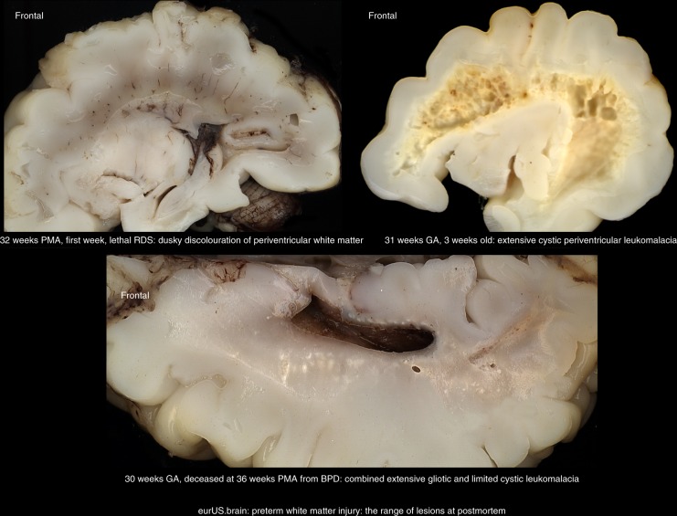
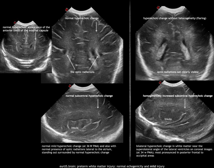
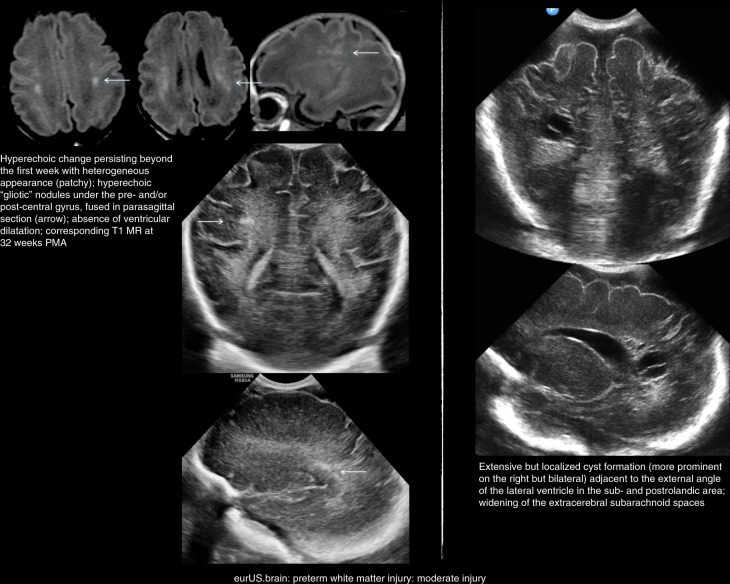
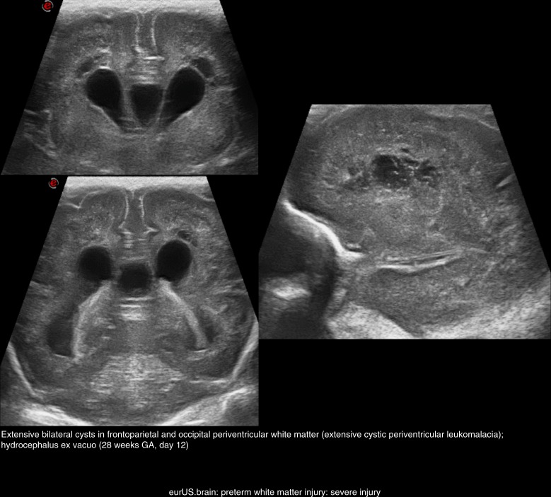
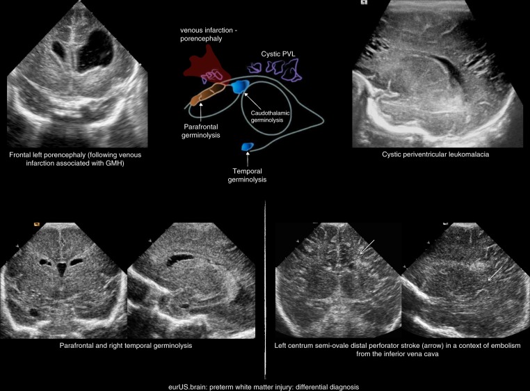
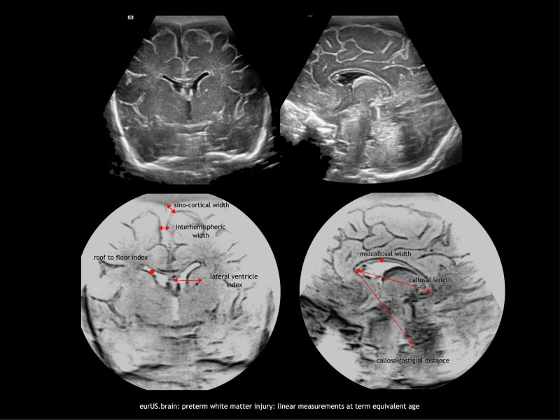
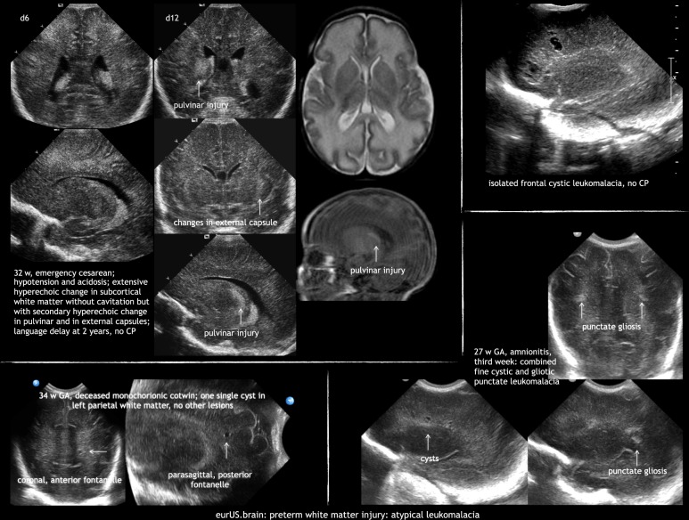
White matter injury (WMI) is the most frequent form of preterm brain injury. Cranial ultrasound (CUS) remains the preferred modality for initial and sequential neuroimaging in preterm infants, and is reliable for the diagnosis of cystic periventricular leukomalacia. Although magnetic resonance imaging is superior to CUS in detecting the diffuse and more subtle forms of WMI that prevail in very premature infants surviving nowadays, recent improvement in the quality of neonatal CUS imaging has broadened the spectrum of preterm white matter abnormalities that can be detected with this technique. We propose a structured CUS assessment of WMI of prematurity that seeks to account for both cystic and non-cystic changes, as well as signs of white matter loss and impaired brain growth and maturation, at or near term equivalent age. This novel assessment system aims to improve disease description in both routine clinical practice and clinical research. Whether this systematic assessment will improve prediction of outcome in preterm infants with WMI still needs to be evaluated in prospective studies.


Conflict of interest statement

The authors declare no competing interests.

Figures

Fig. 1
Fig. 1
Preterm white matter injury: the range of lesions at postmortem.
Fig. 2
Fig. 2
Preterm white matter injury: normal echogenicity and mild injury.
Fig. 3
Fig. 3
Preterm white matter injury: moderate injury.
Fig. 4
Fig. 4
Preterm white matter injury: severe injury.
Fig. 5
Fig. 5
Preterm white matter injury: differential diagnosis.
Fig. 6
Fig. 6
Preterm white matter injury: linear measurements at term equivalent age.
Fig. 7
Fig. 7
Preterm white matter injury: atypical leukomalacia.

References

    1. Volpe JJ. Cerebral white matter injury of the premature infant-more common than you think. Pediatrics. 2003;112:176–180. doi: 10.1542/peds.112.1.176. - DOI - PubMed
    1. Dyet LE, et al. Natural history of brain lesions in extremely preterm infants studied with serial magnetic resonance imaging from birth and neurodevelopmental assessment. Pediatrics. 2006;118:536–548. doi: 10.1542/peds.2005-1866. - DOI - PubMed
    1. Inder TE, et al. Defining the nature of the cerebral abnormalities in the premature infant: a qualitative magnetic resonance imaging study. J. Pediatrics. 2003;143:171–179. doi: 10.1067/S0022-3476(03)00357-3. - DOI - PubMed
    1. Horsch S, et al. Brain abnormalities in extremely low gestational age infants: a Swedish population based study. Acta Paediatr. 2007;96:979–984. doi: 10.1111/j.1651-2227.2007.00294.x. - DOI - PubMed
    1. Hamrick SEG, et al. Trends in severe brain injury and neurodevelopmental outcome in premature newborn infants: the role of cystic periventricular leukomalacia. J. Pediatr. 2004;145:593–599. doi: 10.1016/j.jpeds.2004.05.042. - DOI - PubMed

MeSH terms