Review
. 2020 Mar;87(Suppl 1):13-24.
doi: 10.1038/s41390-020-0780-2.

Cranial ultrasound findings in preterm germinal matrix haemorrhage, sequelae and outcome

Collaborators, Affiliations
Review

Cranial ultrasound findings in preterm germinal matrix haemorrhage, sequelae and outcome

Alessandro Parodi et al. Pediatr Res. 2020 Mar.

Abstract

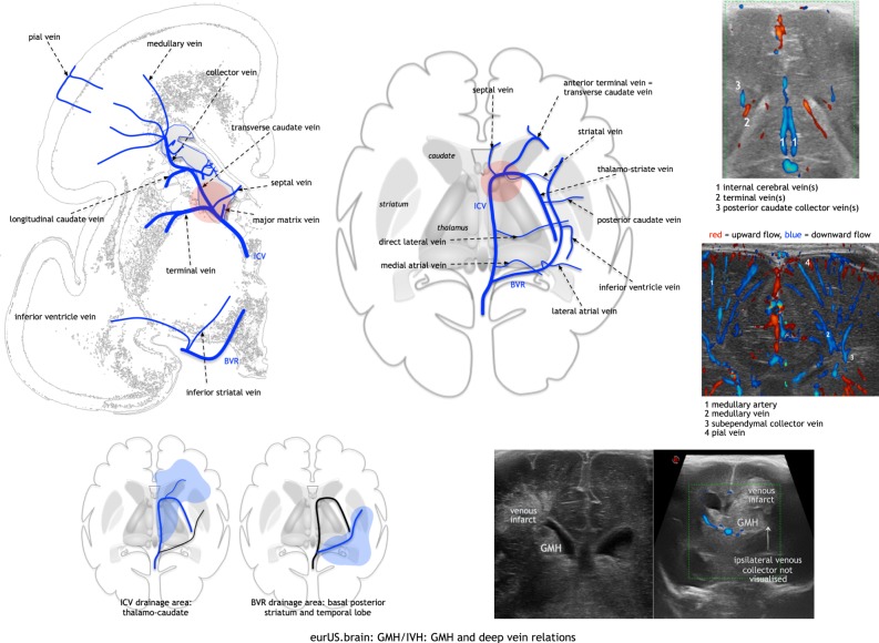
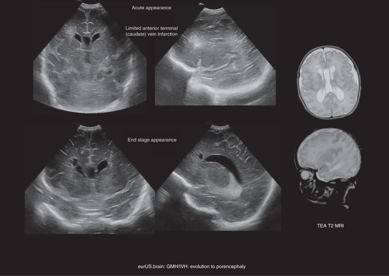
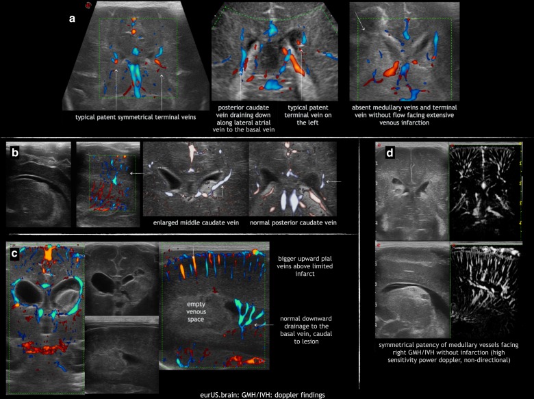
Germinal matrix-intraventricular haemorrhage (GMH-IVH), periventricular haemorrhagic infarction (PHI) and its complication, post-haemorrhagic ventricular dilatation (PHVD), are still common neonatal morbidities in preterm infants that are highly associated with adverse neurodevelopmental outcome. Typical cranial ultrasound (CUS) findings of GMH-IVH, PHI and PHVD, their anatomical substrates and underlying mechanisms are discussed in this paper. Furthermore, we propose a detailed descriptive classification of GMH-IVH and PHI that may improve quality of CUS reporting and prediction of outcome in infants suffering from GMH-IVH/PHI.


Conflict of interest statement

A.P. has received consulting fees from Shire HGT, Inc. The authors declare no competing interests.

Figures

Fig. 1
Fig. 1
GMH/IVH: typical postmortem examples.
Fig. 2
Fig. 2. GMH/IVH: origin and grading.
GMH starts in a venule that drains into lateral subependymal collector veins; it extends into white matter by virtue of venous compression and infarction; bottom row: T2-weighted MRI of GMH with limited IVH and limited venous infarct.
Fig. 3
Fig. 3. GMH/IVH: ultrasound grading.
CUS grading of GMH/IVH; arrowheads point to GMH, arrows to the presence of clot in the ventricle cavity; asterisk is choroid plexus.
Fig. 4
Fig. 4. GMH/IVH: indirect signs of IVH and unusual extension into cavum vergae.
Arrowhead points to GMH; asterisk represents clot in midline cavity.
Fig. 5
Fig. 5. GMH/IVH: GMH and deep vein relations.
Deep venous anatomy and some Doppler examples; in red circles the typical location of GMH near the caudo-thalamic groove; initially the GMH is often separate from a resulting venous infarct; the two may merge, and extensive lesions can be associated with absent terminal vein drainage.
Fig. 6
Fig. 6. GMH/IVH: examples of types of venous infarction.
Examples of different types of venous infarction: thalamostriate (terminal vein), anterior terminal vein (caudate), inferior ventricle vein, medial subependymal (midline) veins, compared with the image of typical posterior frontal developmental venous anomaly (DVA).
Fig. 7
Fig. 7
GMH/IVH: evolution to porencephaly.
Fig. 8
Fig. 8. GMH/IVH: post-haemorrhagic ventricular dilatation.
Measurements and inspection of dilated ventricles following extensive IVH (* third ventricle with extended protrusions).
Fig. 9
Fig. 9. GMH/IVH: Doppler findings.
a Asymmetry in terminal vein size, inversely related to size of ipsilateral direct lateral vein or lateral atrial vein; to be compared with absent terminal vein facing extensive venous infarction. b Apparent enlargement (congestion) of a vein by GMH. c Escape of blood from deep venous infarct along enlarged pial veins, above a Doppler-empty venous space. d Normal deep venous anatomy in GMH without infarction.

References

    1. Volpe, J. J. Neurology of the Newborn 5th edn, 517–588 (Saunders Elsevier, Philadelphia, PA, 2008).
    1. EXPRESS Group et al. One-year survival of extremely preterm infants after active perinatal care in Sweden. JAMA. 2009;301:2225–2233. doi: 10.1001/jama.2009.771. - DOI - PubMed
    1. Ancel PY, Goffinet F, the EPIPAGE-2 Writing Group. Epipage survival and morbidity of preterm children born at 22 through 34 weeks’ gestation in France in 2011 results of the EPIPAGE-2 Cohort Study. JAMA Pediatr. 2015;169:230–238. doi: 10.1001/jamapediatrics.2014.3351. - DOI - PubMed
    1. Stoll BJ, et al. Eunice Kennedy Shriver National Institute of Child Health and Human Development Neonatal Research Network. Neonatal outcomes of extremely preterm infants from the NICHD Neonatal Research Network. Pediatrics. 2010;126:443–456. doi: 10.1542/peds.2009-2959. - DOI - PMC - PubMed
    1. Ramenghi LA, et al. Cerebral venous thrombosis, intraventricular hemorrhage and white matter lesions in a preterm newborn with factor V (Leiden) mutation. Neuropediatrics. 2002;33:97–99. doi: 10.1055/s-2002-32370. - DOI - PubMed