Review
. 2020 Mar;87(Suppl 1):3-12.
doi: 10.1038/s41390-020-0776-y.

State-of-the-art neonatal cerebral ultrasound: technique and reporting

Collaborators, Affiliations
Review

State-of-the-art neonatal cerebral ultrasound: technique and reporting

Jeroen Dudink et al. Pediatr Res. 2020 Mar.

Abstract

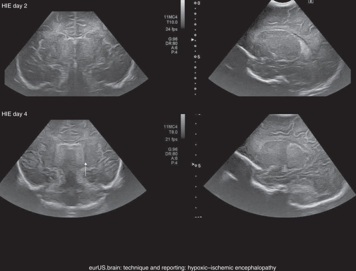
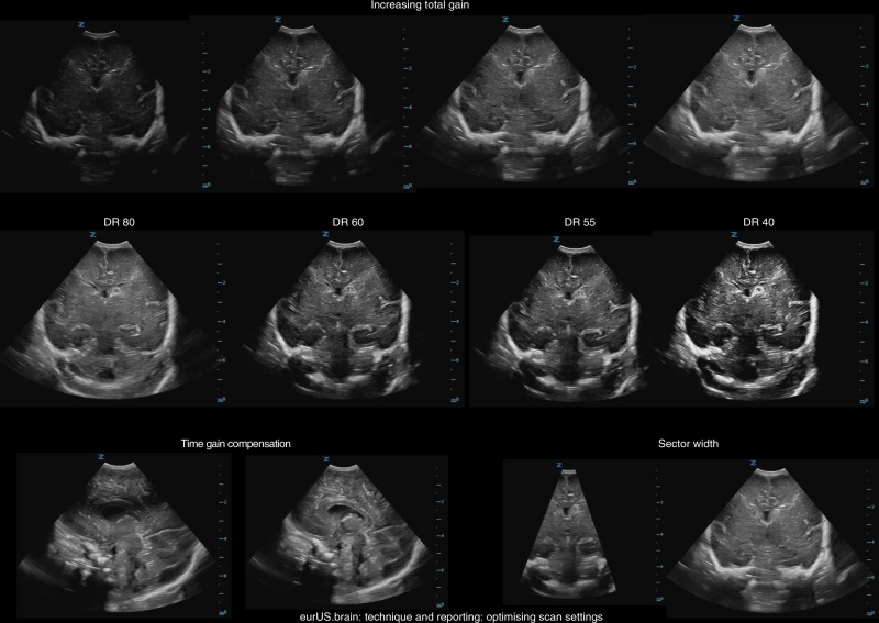
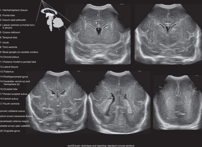
In the past three decades, cerebral ultrasound (CUS) has become a trusted technique to study the neonatal brain. It is a relatively cheap, non-invasive, bedside neuroimaging method available in nearly every hospital. Traditionally, CUS was used to detect major abnormalities, such as intraventricular hemorrhage (IVH), periventricular hemorrhagic infarction, post-hemorrhagic ventricular dilatation, and (cystic) periventricular leukomalacia (cPVL). The use of different acoustic windows, such as the mastoid and posterior fontanel, and ongoing technological developments, allows for recognizing other lesion patterns (e.g., cerebellar hemorrhage, perforator stroke, developmental venous anomaly). The CUS technique is still being improved with the use of higher transducer frequencies (7.5-18 MHz), 3D applications, advances in vascular imaging (e.g. ultrafast plane wave imaging), and improved B-mode image processing. Nevertheless, the helpfulness of CUS still highly depends on observer skills, knowledge, and experience. In this special article, we discuss how to perform a dedicated state-of-the-art neonatal CUS, and we provide suggestions for structured reporting and quality assessment.


Conflict of interest statement

The authors declare no competing interests.

Figures

Fig. 1
Fig. 1. Technique and reporting: hypoxic–ischemic encephalopathy.
Intrapartum asphyxia. Term infant, born at 41 weeks’ gestation with asphyxia and hypoxic–ischemic encephalopathy, treated with hypothermia. a, b Ultrasound on admission showing subtle increased echogenicity of the thalami on the coronal (a) but not on the sagittal (b) images. c, d Three days later, there is clearly abnormal increased echogenicity of the thalami in both planes (arrows), which are separated from more mildly echogenic basal ganglia by a band of low echogenicity, representing the posterior limb of the internal capsule (arrowhead).
Fig. 2
Fig. 2. Technique and reporting: arterial ischemic stroke.
Top: term infant with focal seizures on day 2: left posterior truncal MCA stroke; ultrasound and MRI (diffusion weighted and T2) on day 3. Bottom: vaginal breech delivery at 36 weeks’ GA, apnea, and tense fontanel at 24 h; pallor; and lowered consciousness: posterior cerebral artery stroke following uncal herniation due to right convexity subdural hematoma (left image on admission, other images on day 5) (arrow in the middle image: thalamic perforator stroke).
Fig. 3
Fig. 3. Technique and reporting: optimizing scan settings.
Top row: gradually increasing total gain; middle row: gradually decreasing dynamic range settings; bottom row, left: correction of wrong time gain compensation setting at 2 cm depth; bottom row, right: different sector widths.
Fig. 4
Fig. 4. Technique and reporting: standard coronal sections.
Standard coronal sections from anterior (top left) to posterior (bottom right); sectional planes in the scheme.
Fig. 5
Fig. 5. Technique and reporting: standard parasagittal sections.
Sections from midline (top left) to insula (bottom).
Fig. 6
Fig. 6. Technique and reporting: posterior fontanel images at 24 weeks’ GA.
Posterior fontanel section, top coronal (left anterior to right posterior), bottom sagittal and parasagittal.
Fig. 7
Fig. 7. Technique and reporting: temporo-squamosal section.
Top: sagittal section through brainstem and cerebellar vermis, compared with a sagittal scan of an infant of 27 weeks gestation, taken though the anterior fontanel with an 8.5 MHz scanhead; far right: sagittal 7.5 MHz ultrasound section of the area, taken through the posterior fontanel of an infant with cleidocranial dysplasia. Bottom: temporo-squamosal sections (parallel to the cantho-meatal line, indicated in red on the top scan): the echopoor mesencephalon looks like a butterfly; the cerebral peduncles and tectal lamina are surrounded by hyperechoic cisterns and parts of the tentorium; bright reflections in the posterior part of the brainstem coincide with the walls of the aqueduct; in the basal cisterns, the arteries of the circle of Willis show as short, pulsating lines; term MRI sections for comparison.

References

    1. Meijler, G. Neonatal Cranial Ultrasonography 2nd edn (Springer, Berlin, Heidelberg, 2012).
    1. Plaisier, A. et al. Serial cranial ultrasonography or early MRI for detecting preterm brain injury? Arch. Dis. Child. Fetal Neonatal Ed. 100, F293–F300 (2015). - PubMed
    1. Pape KE, et al. Ultrasound detection of brain damage in preterm infants. Lancet. 1979;1:1261–1264. doi: 10.1016/S0140-6736(79)92227-X. - DOI - PubMed
    1. Slovis TL, Kuhns LR. Real-time sonography of the brain through the anterior fontanel. AJR Am. J. Roentgenol. 1981;136:277–286. doi: 10.2214/ajr.136.2.277. - DOI - PubMed
    1. Thorburn RJ, et al. Accuracy of imaging of the brains of newborn infants by linear-array real-time ultrasound. Early Hum. Dev. 1982;6:31–46. doi: 10.1016/0378-3782(82)90055-X. - DOI - PubMed

MeSH terms