Review
. 2017;9(4):e371.
doi: 10.1038/am.2016.128. Epub 2017 Apr 7.

Engineering protein nanocages as carriers for biomedical applications

Affiliations
Review

Engineering protein nanocages as carriers for biomedical applications

Sathyamoorthy Bhaskar et al. NPG Asia Mater. 2017.

Abstract

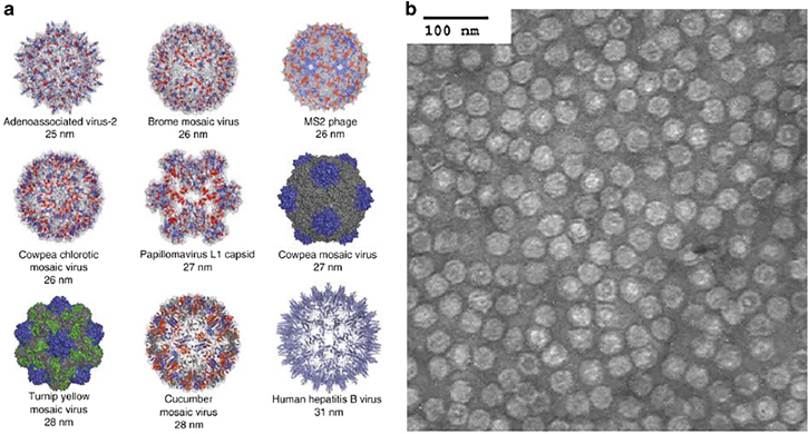
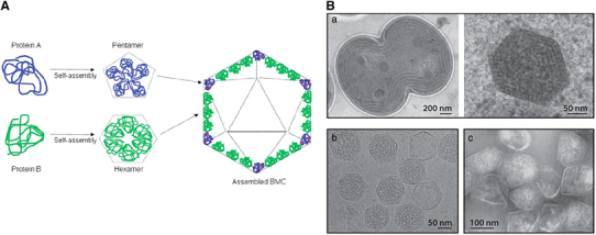
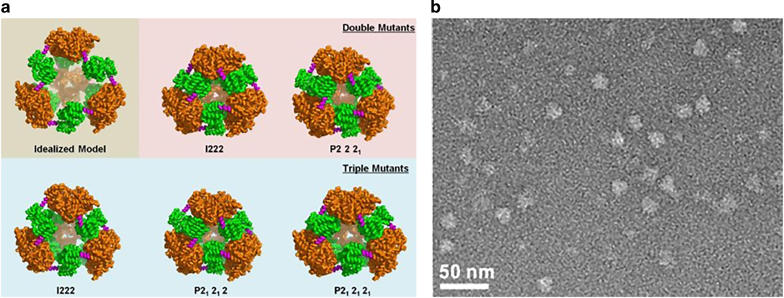
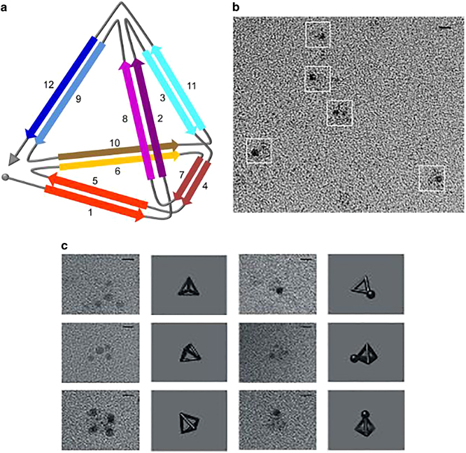
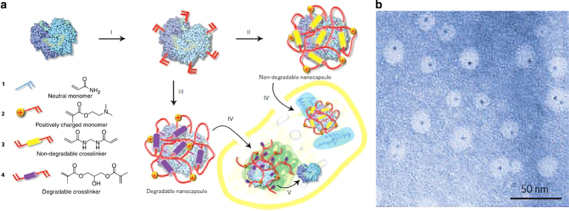
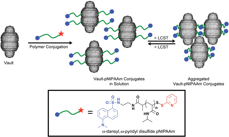
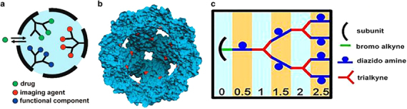
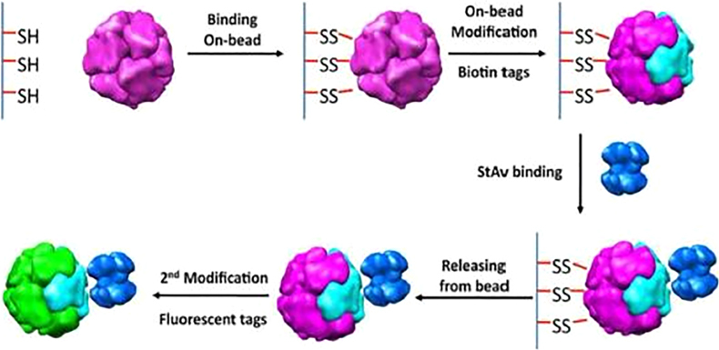
Protein nanocages have been explored as potential carriers in biomedicine. Formed by the self-assembly of protein subunits, the caged structure has three surfaces that can be engineered: the interior, the exterior and the intersubunit. Therapeutic and diagnostic molecules have been loaded in the interior of nanocages, while their external surfaces have been engineered to enhance their biocompatibility and targeting abilities. Modifications of the intersubunit interactions have been shown to modulate the self-assembly profile with implications for tuning the molecular release. We review natural and synthetic protein nanocages that have been modified using chemical and genetic engineering techniques to impart non-natural functions that are responsive to the complex cellular microenvironment of malignant cells while delivering molecular cargos with improved efficiencies and minimal toxicity.

Keywords: Biomaterials - proteins.


Figures

Figure 1
Figure 1
(a) Structural illustration of viruses used in bionanotechnology and biomedical applications. (b) Purified turnip yellow mosaic virus (TYMV) particles negatively stained with uranyl acetate, scale bar=100 nm (reprinted from Lee, L.A. & Wang, Q. Adaptations of nanoscale viruses and other protein cages for medical applications/virology. Nanomedicine 2, 137–149. Copyright (2006), with permission from Elsevier).
Figure 2
Figure 2
(a) PyMOL illustrations of the tetraeicosameric assembly observed in Archaeoglobus fulgidus ferritin viewed along different molecular symmetry axes. Left: One of four pores of the nanocage structure shown along the threefold noncrystallographic symmetry axis. Right: Engineered closed ferritin shell viewed along the axis the fourfold molecular symmetry axis (reprinted from Johnson, E., Cascio, D., Sawaya, M.R., Gingery, M. & Schroder, I. Crystal structures of a tetrahedral open pore ferritin from the hyperthermophilic archaeon Archaeoglobus fulgidus. Structure 13/4, 637–648. Copyright (2005), with permission from Elsevier). (b) Transmission electron micrograph of engineered A. fulgidus ferritin, scale bar=50 nm (reprinted from Sana, B., Johnson, E., Sheah, K., Poh, C.L. & Lim, S. Iron-based ferritin nanocore as a contrast agent. Biointerphases 5/3, FA48–FA52. Copyright (2010), with permission from Springer).
Figure 3
Figure 3
(a) E1-E2–E3 complex of Geobacillus stearothermophilus active site coupling model. Tetramers of E1 (purple) and dimers of E3 (yellow) are shown attached to the outer surface of icosahedral E2 protein (gray) formed by 60 subunits (reprinted from Milne, J.L. et al. Molecular structure of a 9 MDa icosahedral pyruvate dehydrogenase subcomplex containing the E2 and E3 enzymes using cryoelectron microscopy. J. Biol. Chem. 281, 4364–4370. Copyright (2006). with permission from The American Society for Biochemistry and Molecular Biology). (b) Electron micrograph of wild-type E2 protein, scale bar=50 nm (reprinted from Dalmau, M., Lim, S., Chen, H.C., Ruiz, C. & Wang, S.W. Thermostability and molecular encapsulation within an engineered caged protein scaffold. Biotechnology and Bioengineering 101, 654–664. Copyright (2008), with permission from John Wiley and Sons).
Figure 4
Figure 4
(A) Schematic representation of vault nanocages engineered by inclusion of additional amino acids at the N and C termini of major vault protein (MVP). Peptide extensions at the N termini (shown in red) and C termini (shown in blue) from the waist region (light red) and the extreme ends of the caps, respectively (Aa and b). By fusing an exogenous protein at INT binding site (Ac) and additional peptide at the N terminus (Ad), a composite assembly with multifunctional units could be formed (Ae) (reprinted (adapted) Han, M., Kickhoefer, V.A., Nemerow, G.R. & Rome, L.H. Targeted vault nanoparticles engineered with an endosomolytic peptide deliver biomolecules to the cytoplasm. ACS Nano 5, 6128–6137. Copyright (2011), with permission from American Chemical Society). (B) Transmission electron microscopy (TEM) image of recombinant vault particles, scale bar=100 nm (reprinted (adapted) from Kickhoefer, V.A. et al. Targeting vault nanoparticles to specific cell surface receptors. ACS Nano 3, 27–36. Copyright (2009), with permission from American Chemical Society).
Figure 5
Figure 5
(A) Structural illustration of the formation of icosahedral bacterial microcompartments (BMCs). The BMCs are formed by hexameric (green) and pentameric (blue) folding proteins. The hexamers assemble side by side to form a flat molecular layer of the icosahedron; the vertices of the same structure are formed by pentameric proteins (reprinted from Ferrer-Miralles, N. et al. Engineering protein self-assembling in protein-based nanomedicines for drug delivery and gene therapy. Crit. Rev. Biotechnol. 1–13, 1549–7801. Copyright (2013), with permission from Taylor & Francis Ltd). (Ba) TEM images showing the division of a cyanobacterial cell (left) and the structure of a carboxysomes (right). (Bb) TEM image showing purified carboxysomes derived from Halothiobacillus neopolitanus. (Bc) Purified Pdu microcompartments derived from Salmonella enterica (reprinted from Yeates, T.O., Crowley, C.S. & Tanaka, S. Bacterial microcompartment organelles: protein shell structure and evolution. Annu. Rev. Biophys. 39, 185–205. Copyright (2010), with permission from Annual Reviews).
Figure 6
Figure 6
(a) Crystal structures of double and triple mutant-type protein nanocages formed by the oligomer fusion strategy. The trimeric domains are shown in orange, and dimeric domains are shown in green. (b) TEM image of the triple mutant form of the designed protein nanocage, scale bar=50 nm35 (reprinted (adapted) Lai, Y.T. et al. Structure and flexibility of nanoscale protein cages designed by symmetric self-assembly. J. Am. Chem. Soc. 135, 7738–7743. Copyright (2013), with permission from American Chemical Society).
Figure 7
Figure 7
(a) Illustration of tetrahedron-shaped polypeptide structure. (b) Coil-forming elements in the polypeptide self-assembly are denoted by arrow marks. Transmission electron microscopy (TEM) image of TET12 tetrahedral polypeptide highlighted in white boxes. TEM images of TET12 structure (left) stained with 1.8 nm Ni-NTA nanogold beads followed by uranyl acetate staining, scale bar=5 nm. (c) On the right, TEM images of TET12 polypeptide from the white boxes shown in (b) (reprinted from Gradisar, H. et al. Design of a single-chain polypeptide tetrahedron assembled from coiled-coil segments. Nat. Chem. Biol. 9, 362–366. Copyright (2013), with permission from Macmillan Publishers Ltd.)
Figure 8
Figure 8
(a) Schematic representation of nanocapsule synthesis by in situ copolymerization of degradable and non-degradable nanocapsules from their constituent crosslinked subunits (I, II and III), followed by the cellular uptake of the formed nanocapsules via endocytosis (IV); eventually, the loaded protein cargoes are released via shell degradation in the intracellular environment (V). (b) TEM image of nanocages containing one 1.4-nm gold quantum dot-labeled horse radish peroxidase core within the nanoscale architecture, scale bar=50 nm (adapted from Yan, M. et al. A novel intracellular protein delivery platform based on single-protein nanocapsules. Nat. Nanotechnol. 5, 48–53. Copyright (2010), with permission from Macmillan Publishers Ltd).
Figure 9
Figure 9
Synthesis of thermoresponsive vault conjugates. The recombinant vault images are cryo-EM reconstructed (reprinted (adapted) from Matsumoto, N.M. et al. Smart vaults: thermally-responsive protein nanocapsules. ACS Nano 7, 867–874. Copyright (2013), with permission from American Chemical Society).
Figure 10
Figure 10
Protein cage modifications at distinct interfaces (reprinted from Uchida, M., Kang, S., Reichhardt, C., Harlen, K. & Douglas, T. The ferritin superfamily: supramolecular templates for materials synthesis/ferritin: structures, properties and applications. Biochim. Biophys. Acta 1800(8), 834–845. Copyright (2010), with permission from Elsevier).
Figure 11
Figure 11
Design of protein nanocage-polymer hybrids. (a) Representation of protein cage with polymer branches in the interior cavity with attachment sites for drugs or imaging agents. (b) A cross-sectional view of HSPG41C nanocage showing cysteine residues (red) in the interior cavity. (c) Scheme for the production of dendritic structure with the generation units indicated at the bottom (reprinted (adapted) from Abedin, M.J. et al. Synthesis of a cross-linked branched polymer network in the interior of a protein cage. J. Am. Chem. Soc. 131, 4346–4354. Copyright (2009), with permission from American Chemical Society).
Figure 12
Figure 12
Toposelective modification of Listeria innocua DNA-binding protein nanocage using masking/unmasking method using a solid bead support (reprinted from Uchida, M., Kang, S., Reichhardt, C., Harlen, K. & Douglas, T. The ferritin superfamily: supramolecular templates for materials synthesis/ferritin: structures, properties and applications. Biochim. Biophys. Acta 1800(8), 834–845. Copyright (2010), with permission from Elsevier).
Figure 13
Figure 13
Different methods for asymmetric ligand display on cowpea chlorotic mottle virus (CCMV) nanocage. (A) Dissociation of differentially engineered CCMV cages into subunits and reassembly. (B) Symmetrical dissociation of a CCMV mutant A163C. (Ba) Thiol modification of the viral nanocage by activated resin. (Bb) Neutralizing the unbound cysteines using IAA (Bc) Separation of symmetry broken mutant nanocage reduction (reprinted from Uchida, M., Klem, M.T., Allen, M., Suci, P., Flenniken, M., Gillitzer, E. et al. Biological containers: protein cages as multifunctional nanoplatforms. Adv. Mater. 19, 1025–1042. Copyright (2007), with permission from John Wiley and Sons).
Figure 14
Figure 14
TEM image of ferritin nanoparticles (a), hemagglutinin (HA) ligands displayed on ferritin nanocages (b) and comparison (c) of symmetry between computational models and TEM images of the engineered octahedral nanoparticles displaying HA spikes (numbered) (reprinted from Kanekiyo, M. et al. Self-assembling influenza nanoparticle vaccines elicit broadly neutralizing H1N1 antibodies. Nature 499, 102–106. Copyright (2013), with permission from Macmillan Publishers Ltd.).

References

    1. Ferrer-Miralles N, Rodríguez-Carmona E, Corchero JL, García-Fruitós E, Vázquez E, Villaverde A. Engineering protein self-assembling in protein-based nanomedicines for drug delivery and gene therapy. Crit. Rev. Biotechnol. 2013;35:209–221. - PubMed
    1. Yan M, Du J, Gu Z, Liang M, Hu Y, Zhang W, Priceman S, Wu L, Hong Z, Zhou H, Liu Z, Segura T, Tang Y, Lu Y. A novel intracellular protein delivery platform based on single-protein nanocapsules. Nat. Nanotechnol. 2010;5:48–53. - PubMed
    1. Lee LA, Wang Q. Adaptations of nanoscale viruses and other protein cages for medical applications. Nanomedicine. 2006;2:137–149. - PubMed
    1. Flenniken ML, Uchida M, Liepold LO, Kang S, Young MJ, Douglas T. A library of protein cage architectures as nanomaterials. Curr. Top. Microbiol. 2009;327:71–93. - PubMed
    1. Uchida M, Klem MT, Allen M, Suci P, Flenniken M, Gillitzer E, Carpness Z, Liepold LO, Young M, Douglas T. Biological containers: protein cages as multifunctional nanoplatforms. Adv. Mater. 2007;19:1025–1042.